
Pterostichus malkini
|
publication ID |
https://doi.org/ 10.11646/zootaxa.3682.4.7 |
|
publication LSID |
lsid:zoobank.org:pub:B22DA403-BAEE-4EED-86F5-D79AE60D8175 |
|
DOI |
https://doi.org/10.5281/zenodo.6148972 |
|
persistent identifier |
https://treatment.plazi.org/id/405F87B3-FFC9-FF92-FF74-8A31B2C089BA |
|
treatment provided by |
Plazi |
|
scientific name |
Pterostichus malkini |
| status |
|
Distinguishing Pterostichus malkini View in CoL from other species of P. ( Anilloferonia )
Four characters readily distinguish P. malkini from P. diana and P. testacea : the number of supraorbital setae, the shape of the male metafemur, the form of the last visible abdominal ventrite of the male just anterior of the apical margin, and the form of the median lobe of the aedeagus. There are other distinguishing characters but these are subtle, are often variable, and sometimes cannot be accurately assessed without access to reliably identified reference specimens. They will not be addressed herein as that would be more appropriate in a broader treatment of Anilloferonia .
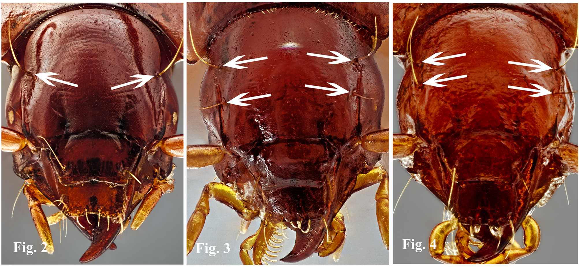
All known specimens (6) of P. malkini have only one pair of supraorbital setae ( Figure 2 View FIGURES 2 – 4 ). All specimens of P. diana (111) ( Figure 3 View FIGURES 2 – 4 ) and P. t e s t a c e a (21) ( Figure 4 View FIGURES 2 – 4 ) I examined have two pairs of supraorbital setae, with the exception of one specimen of P. diana with two setae on one side and three on the other.
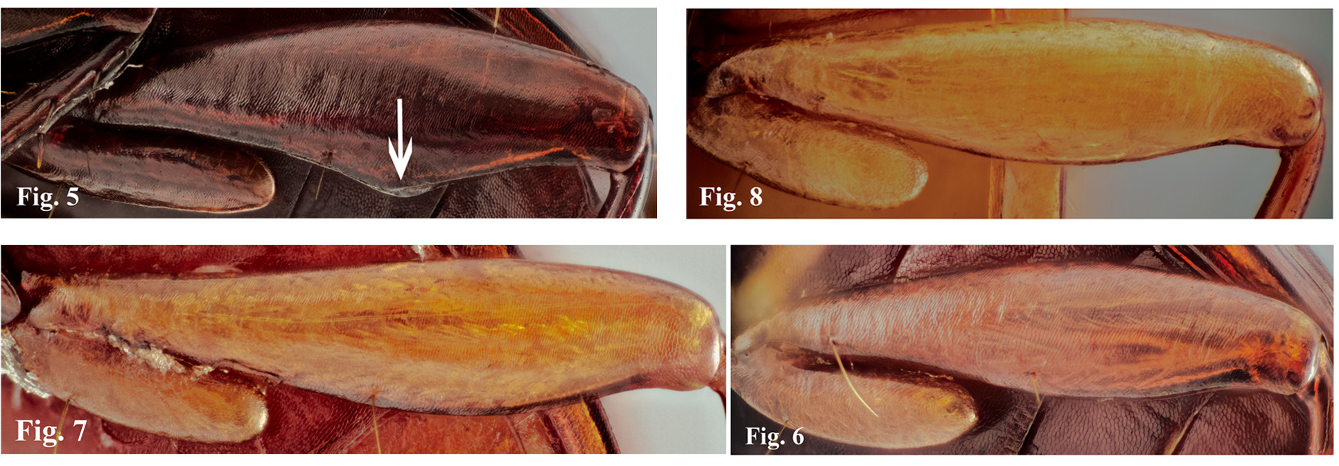
The median posterior margins of the metafemora of the three male P. malkini examined are obliquely angulate and the ventral faces of the metafemora are broader at that point than at either end ( Figure 5 View FIGURES 5 – 8 ). The posterior margins of the metafemora of female P. malkini ( Figure 6 View FIGURES 5 – 8 ) and both females and males of P. diana (male, Figure 7 View FIGURES 5 – 8 ) and P. testacea (male, Figure 8 View FIGURES 5 – 8 ) are shallowly, smoothly arcuate throughout and the metafemora are more or less narrowly cylindrical throughout.
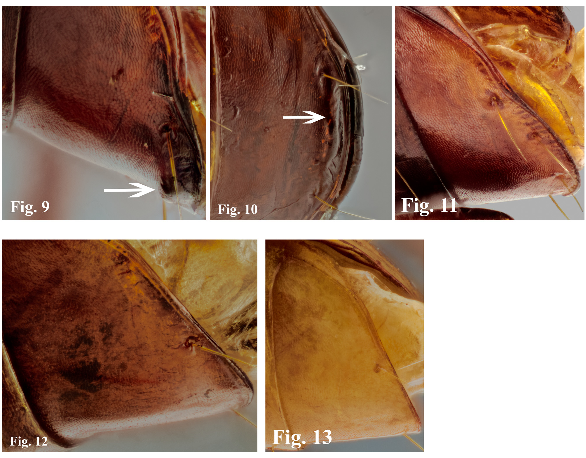
The last abdominal ventrite in the three available specimens of male P. malkini has a small, ventrally projecting ridge between the pair of paramedial preapical setae ( Figures 9–10 View FIGURES 9 – 13 ). Female P. malkini lack this ridge ( Figure 11 View FIGURES 9 – 13 ), although a small preapical protuberance is present. The last abdominal ventrites of females and males of P. diana (male, Figure 12 View FIGURES 9 – 13 ) and P. testacea (male, Figure 13 View FIGURES 9 – 13 ) lack preapical ridges or protuberances.
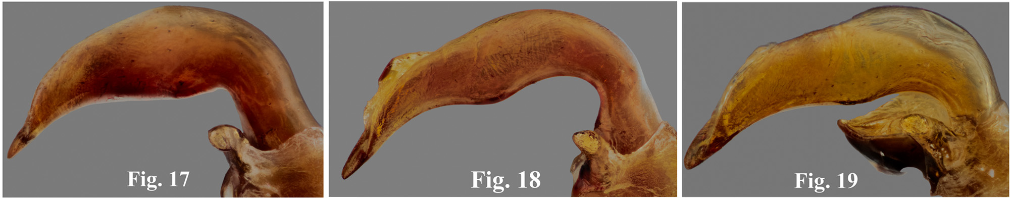
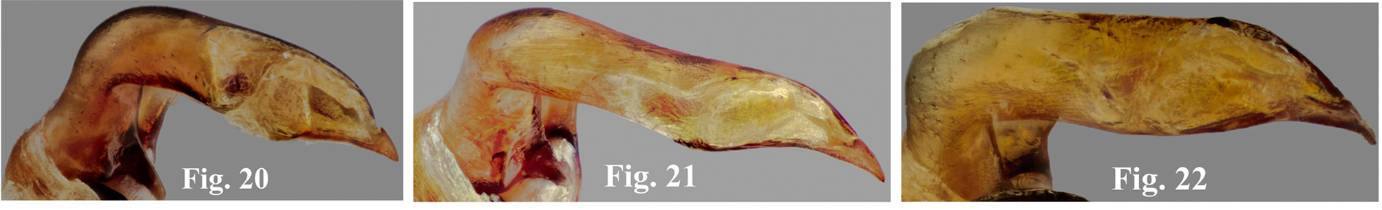
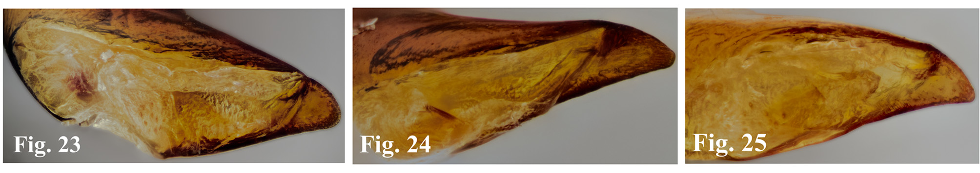
The shape of the median lobe of the aedeagus of P. malkini differs from those of P. diana and P. t e s t a c e a as follows: the median lobe is shorter and less sinuate in lateral view ( Figures 14–19 View FIGURES 14 – 16 View FIGURES 17 – 19 ) and dorsal view ( Figures 20– 22 View FIGURES 20 – 22 ), the right ventrolateral tumidity is much smaller and is very close to the base of the median lobe (versus large and more medially located) ( Figures 17–19 View FIGURES 17 – 19 ), the meeting of the shaft of the median lobe and the base is narrowly perpendicular in right lateral perspective ( Figure 17 View FIGURES 17 – 19 ) versus being arcuate and broader ( Figures 18–19 View FIGURES 17 – 19 ), the apex is shorter in both lateral and dorsal perspectives ( Figures 14–22 View FIGURES 14 – 16 View FIGURES 17 – 19 View FIGURES 20 – 22 ) and in dorsal perspective, the apex is less twisted to the anatomical left and the tip of the apex is more rounded ( Figures 23–25 View FIGURES 23 - 25 ).

No known copyright restrictions apply. See Agosti, D., Egloff, W., 2009. Taxonomic information exchange and copyright: the Plazi approach. BMC Research Notes 2009, 2:53 for further explanation.
|
Kingdom |
|
|
Phylum |
|
|
Class |
|
|
Order |
|
|
Family |
|
|
Genus |