
Ptilorhynchia mclachlani, Sandy & Hryniewicz & Hammer & Nakrem & Little, 2014
|
publication ID |
https://doi.org/ 10.11646/zootaxa.3884.6.1 |
|
publication LSID |
lsid:zoobank.org:pub:BD4F285D-358C-4350-88EA-7FA94513D930 |
|
DOI |
https://doi.org/10.5281/zenodo.5236100 |
|
persistent identifier |
https://treatment.plazi.org/id/466F6C33-6251-FFB3-9685-4766FBEE3486 |
|
treatment provided by |
Felipe |
|
scientific name |
Ptilorhynchia mclachlani |
| status |
sp. nov. |
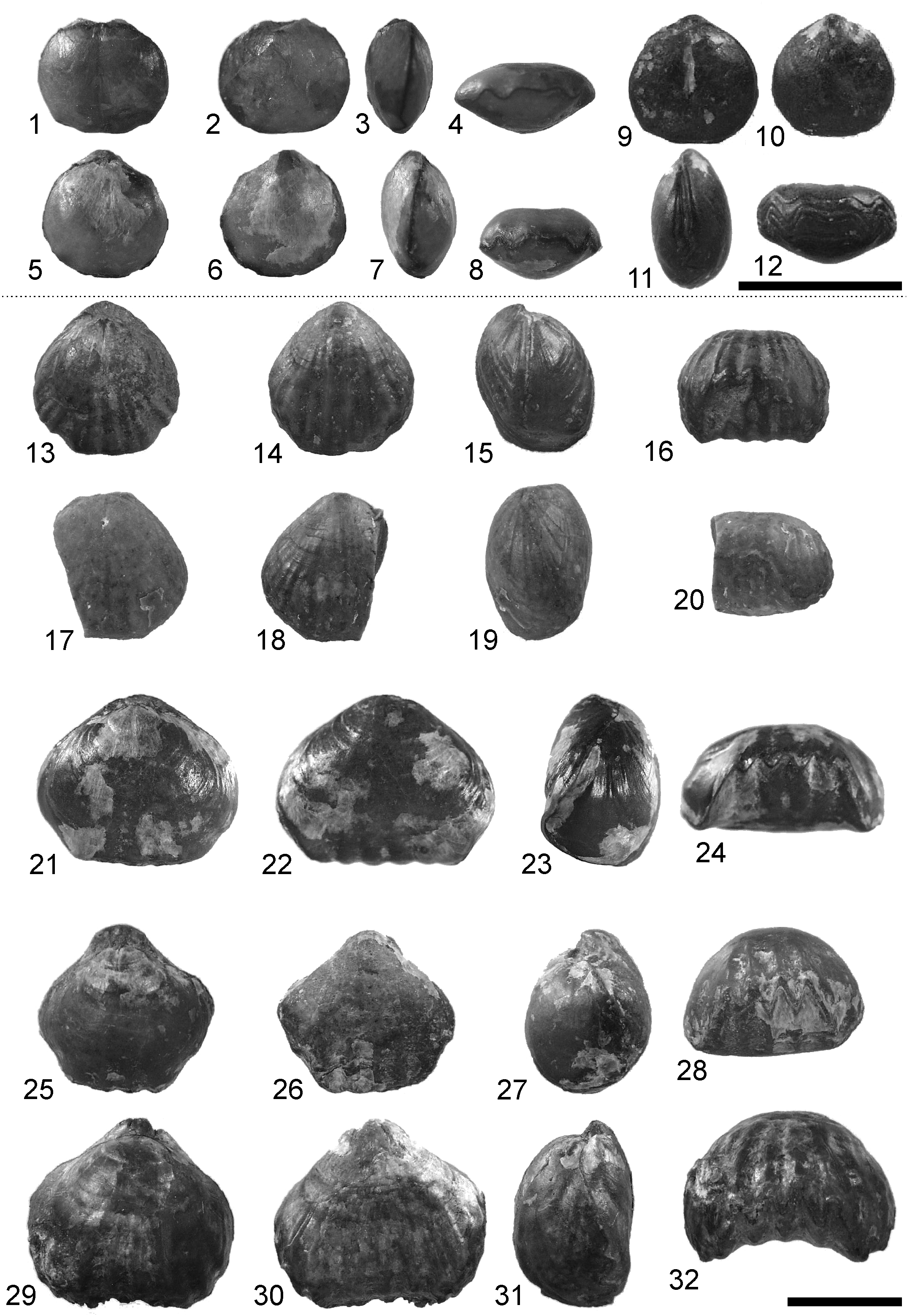
Ptilorhynchia mclachlani sp. nov.
Figs. 3.13–3.32 View FIGURE 3 , 5 View FIGURE 5
2011 v. partim ‘ Lacunosella ’ sp.—Hammer et al., p. 21, table 2, fig. 7R.
2011 v. partim ‘ Monticlarella ’ sp.—Hammer et al., p. 21, table 2.
Material and occurrence. Holotype: PMO 224.882; paratypes: PMO 227.431, PMO 227.432, PMO 224.871–873, PMO 224.875, PMO 224.883, PMO 217.199, PMO 224.914 (crushed). Juvenile specimens: PMO 224.876–879, PMO 227.433. All specimens from seep 9.
Type locality. Knorringfjellet , central Spitsbergen, N78° 18’ 49.9” E16° 10’ 58.9” GoogleMaps .
Etymology. For the late Mr. Archie McLachlan, Department of Geology, Queen Mary College, University of London (see Middlemiss 1997, p. 169).
Dimensions of the holotype. PMO 224.882—Length 17.1 mm, width 18.9 mm, thickness 11.6 mm.
Diagnosis. Subtriangular in outline, generally evenly biconvex in profile. Valve surface smooth except for ornament of costae which may be traceable over most of the length of the valves but are most strongly developed at the anterior commissure. Number of costae on valves variable, with typically 3–4 costae in the sulcus of the dorsal valve and 4–5 on the corresponding fold of the ventral valve. Dental lamellae present in ventral valve. Welldeveloped septalium in dorsal valve, outer hinge plates horizontal from which inner socket ridges are deflected. Crura develop at top of Y-shape of septalium, where inner and outer hinge plates meet. Anteriorly crura arch ventrally.
Discussion. The beak of the ventral valve is frequently damaged/incomplete in the specimens. As a result, the dental lamellae are usually clearly visible in the umbo of the ventral valve. Juvenile specimens have costae developed over both valves, some larger specimens have costae developed primarily in the sulcus of the ventral valve and at the anterior margin.
The material from Spitsbergen shows variability in outline and number of costae. The costae are traceable over most of the length of the valves. Owen (1972) described a variable species that he referred to Ptilorhynchia jeletzkyi from the latest Jurassic of British Columbia which is very similar to the material from Spitsbergen.
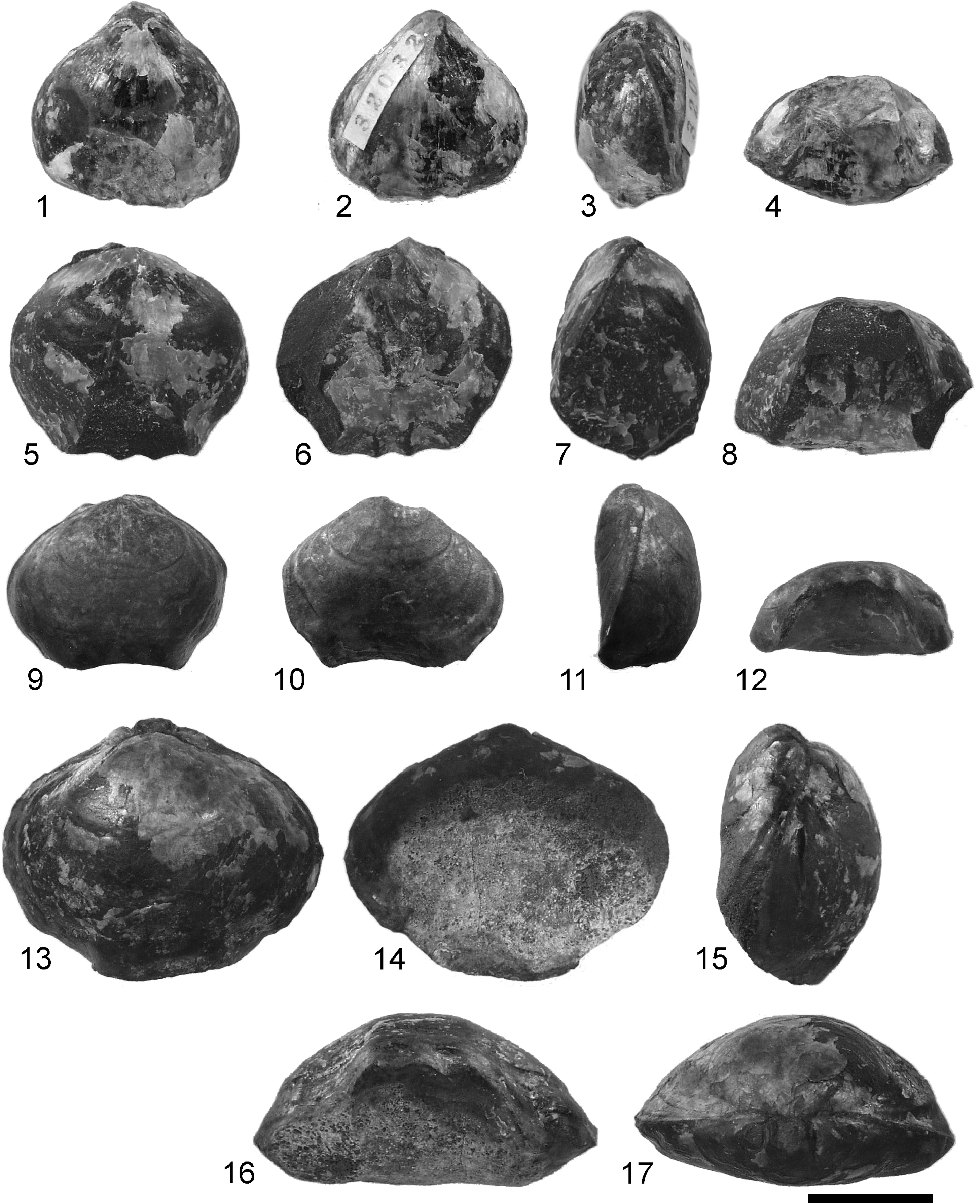
Owen (1972, p. 2) comments on Ptilorhynchia jeletzkyi “The shell surface is smooth, exhibiting no sign of striae, but often developing strong marginal plicae which originate midway between the umbones and anterior commissure. These plicae number from one to four...”. However, these strong plicae are not obvious in figured specimens ( Owen 1972, pl. 1, figs. 1–4) or a specimen referred herein to Ptilorhynchia jeletzkyi from the Upper Jurassic (Kimmeridgian–Tithonian?) of Alaska ( Fig. 6.5–6.8 View FIGURE 6 ). This specimen is slightly damaged at the anterior commissure and the lateral margin, but does show the generally smooth shell ornament and the development of marginal costae (3 costae on the dorsal fold, 2 in the ventral sulcus). The number of costae in the sulcus (at the anterior commissure) in Ptilorhynchia jeletzkyi ranges from one to 4 or 5 ( Owen 1972, fig. 4, pl. 1, figs. 1c, 2–4). Ptilorhynchia jeletzkyi is reported from the Buchia fisheriana and B. piochii zones (i.e., Upper Tithonian, in part equivalent to the Upper Volgian).
In Ptilorhynchia mclachlani , the specimens may show well-marked costae which may extend across most of the length of the specimen (e.g. Fig. 3.29 View FIGURE 3 ). This costate shell ornament is not typical of Ptilorhynchia , which tends to show costation developed at the shell margin (e.g. species figured by Dagys 1968; Owen 1972; Owen & Manceñido 2002; and P. jeletzkyi herein, Fig. 6.5–6.8 View FIGURE 6 , and a specimen identified as P. aff. plumasensis from the Middle Jurassic of Alaska, Fig. 6.1–6.4 View FIGURE 6 close to the type species from the Middle Jurassic Hinchman Formation (Callovian) of California ( Crickmay 1933; see also Owen 1972). MacFarlan et al. (2011) claim the type horizon for P. plumasensis to be the Upper Jurassic Knoxville Formation, but their reason for this revised assignation was not given). Most of the specimens from Spitsbergen display four costae on the dorsal fold and three costae in the ventral sulcus. Due to the distinctive nature of the costation it is considered desirable to refer the Spitsbergen material to a new species rather than to the morphologically similar P. jeletzyki .
The weakly costate Ptilorhynchia mclachlani resembles some species referred to Ptilorhynchia by Dagys (1968). Of these, the Spitsbergen specimens are closest to P. lenaensis in the nature of the costation of the anterior commissure but the overall size of the latter is smaller, and the number of costae on the fold and sulcus fewer than in the Spitsbergen specimens. Matching closely in outline and costation is P. anadyrensis Dagys (1968 , pl. 4, figs. 3, 4) although the nature of the costation appears more angular at the anterior commissure when compared to Ptilorhynchia mclachlani . The specimens of these species figured by Dagys appear to all be decorticated. The age assignment of P. lenaensis is Valanginian ( Dagys 1968) so it is slightly younger in age than the Spitsbergen specimens while P. anadyrensis was given as of uncertain age, referred questionably to the Bathonian–Callovian ( Dagys 1968, table 1, p. 145). A single rhynchonellide specimen (internal cast of a ventral valve, approximately 8 mm x 8 mm) from undifferentiated Berriasian–Valanginian of the Barents Sea ( Århus et al. 1990) identified as Ptilorhynchia sp. , was tentatively referred to this genus. MacFarlan et al. (2011) described Ptilorhynchia pugnaciformis from the Lower Cretaceous of the Misool Archipelago, eastern Indonesia. Like other species of Ptilorhynchia already discussed, the number of costae for this species is considered to be variable (between 3–6 per valve; MacFarlan et al., table 1). The posterior profile of the dorsal valve in P. pugnaciformis is not as rounded as it is in P. mclachlani or P. jeletzkyi ; in pugnaciformis the profile may be cynocephalous and therefore resembling, at least in profile, Homoeorhynchia Buckman.
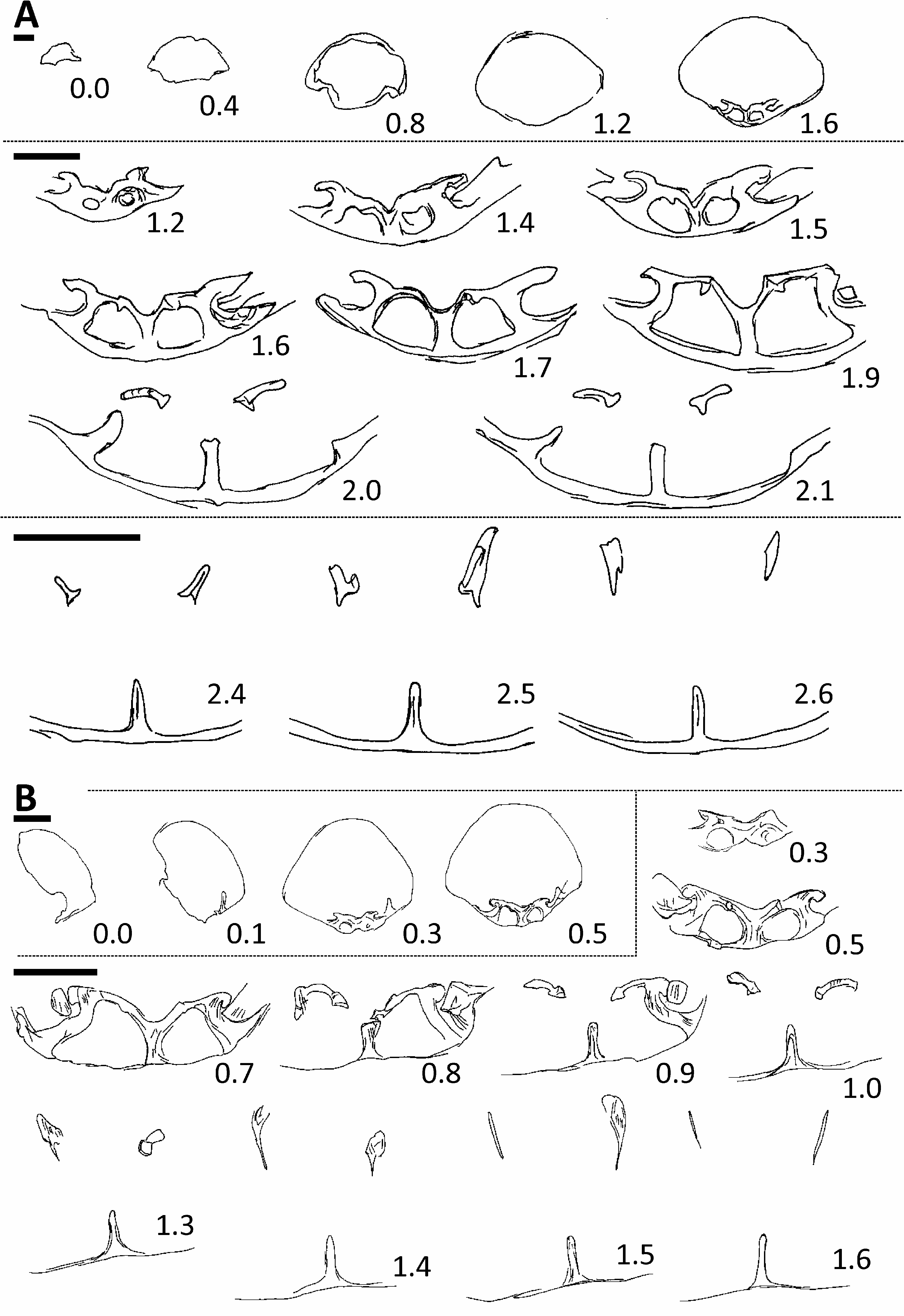
Small multicostate specimens are considered juveniles of Ptilorhynchia mclachlani . These specimens had been tentatively referred to Monticlarella by Sandy in Hammer et al. (2011); they have a similar outline to Monticlarella triloboides (Quenstedt) in Childs (1969, pl. I, figs. 5–7). However, the nature of the costation is consistent with Ptilorhynchia mclachlani and the broad uniplicate fold develops more strongly as the brachiopod approaches maturity, with a concomitant deflection of the lateral commissure ( Fig. 3.21–3.24, 3.29–3.32 View FIGURE 3 cf. for juveniles, Fig. 3.13–3.20, 3.25–3.28 View FIGURE 3 ). Dental lamellae and a median septum can be readily discerned in the majority of the smaller specimens; characters that if present in Monticlarella are typically poorly developed (e.g. Owen 1968; Manceñido et al. 2002, p. 1316). (From the palaeobiographic point of view, Monticlarella has not been recorded from Boreal faunas of the Russian Platform or Siberia, whereas several species have been referred to Ptilorhynchia from these regions.) Serial sections were taken of one of the smaller-sized specimens of Ptilorhynchia mclachlani to clarify the relationships among the material ( Fig. 5B View FIGURE 5 ). The serial sections show remnants of a dental lamella on the right side of the specimen ( Fig. 5B View FIGURE 5 , section 0.1), a well-developed septalium (0.3–0.7) and crura that arch ventrally towards the anterior of the brachiopod (1.0–1.6). The crura develop as discrete structures at the junction between the outer hinge plates and the inner hinge plates of the septalial structure (0.5–1.0). These serial sections are very similar in overall aspect to those taken from the larger specimen ( Fig. 5A View FIGURE 5 ) and both are considered Ptilorhynchia mclachlani .
In comparing the serial sections of Ptilorhynchia mclachlani ( Fig. 5 View FIGURE 5 ) with Ptilorhynchia jeletzkyi ( Owen 1972, fig. 1) there are similarities in the development of the septalium and the crura on the inner margins of the hinge plates, and the crura then arch ventrally anteriorly. The larger Spitsbergen specimen ( Fig. 5A View FIGURE 5 ) did not capture the dental lamellae of the ventral valve, and this is also true when comparing the serial sections of species referred to Ptilorhynchia by Dagys (1968). The presence of well-developed dental lamellae in the ventral valves of the Canadian ( Owen 1972), Siberian ( Dagys 1968) and Indonesian ( MacFarlan et al. 2011) material is a difference from the Spitsbergen material. The ventral umbo of the larger specimen selected for serial sectioning ( Fig. 3.21–3.24 View FIGURE 3 ) was damaged, with the outline of dental lamellae visible, and their traces can also be determined in the duplicate cast taken of this specimen. In addition, dental lamellae are clearly visible in the damaged ventral umbones of other specimens. Traces of dental lamellae were not seen in the serial sections and acetate peels (for specimen in Fig. 5A View FIGURE 5 ). This specimen was decorticated and the calcite shell of the dental lamellae apparently lost through decortication or weathering in the umbonal region of the ventral valve. Most of the external shell of the specimen was absent too. Of the serial sections of species described by Dagys, P. lenaensis Dagys (1968 , fig. 43) and P. anadyrensis Dagys (1968 , fig. 39) appear closest to those of Ptilorhynchia mclachlani ( Fig. 5 View FIGURE 5 ) based on the development of the septalium, hinge plates, and derivation of the crura from the inner margins of the hinge plates. It should be noted that plates 2 and 5 in Dagys 1968 were printed in the wrong order. The taxonomic descriptions and caption for plate 5 refers to P. glabra , P. lenaensis and P. obscuricostata . However, these species are figured on plate 2.
Serial sections of the Spitsbergen material ( Fig. 5 View FIGURE 5 ) lack the falciform crura characteristic of the genus Lacunosella recorded from Greenland ( Owen 1976, fig. 3).
Stratigraphic and geographic distribution. Upper Ryazanian of Spitsbergen.
No known copyright restrictions apply. See Agosti, D., Egloff, W., 2009. Taxonomic information exchange and copyright: the Plazi approach. BMC Research Notes 2009, 2:53 for further explanation.
|
Kingdom |
|
|
Phylum |
|
|
Class |
|
|
Order |
|
|
SuperFamily |
Rhynchonelloidea |
|
Family |
|
|
SubFamily |
Piarorhynchiinae |
|
Genus |