
Leptopilos Levy, 2009
|
publication ID |
https://doi.org/10.11646/zootaxa.5194.1.1 |
|
publication LSID |
lsid:zoobank.org:pub:E66D4948-BF8A-414A-9AB5-389AEF9D951B |
|
DOI |
https://doi.org/10.5281/zenodo.7141924 |
|
persistent identifier |
https://treatment.plazi.org/id/475287B4-FFD0-2B20-FF2E-FC0FFCDAF7D6 |
|
treatment provided by |
Plazi |
|
scientific name |
Leptopilos Levy, 2009 |
| status |
|
Genus Leptopilos Levy, 2009 View in CoL
Type species. Drassus tenerrimus O. Pickard-Cambridge, 1872 , by original designation.
Diagnosis. Leptopilos can be recognised from other Leptodrassinae by the presence of an anterior median hood in the female epigyne, and a male palp armed with closely grouped, often pointed distal laminae (tegular processes), and a single retrolateral tibial apophysis ( Levy 2009).
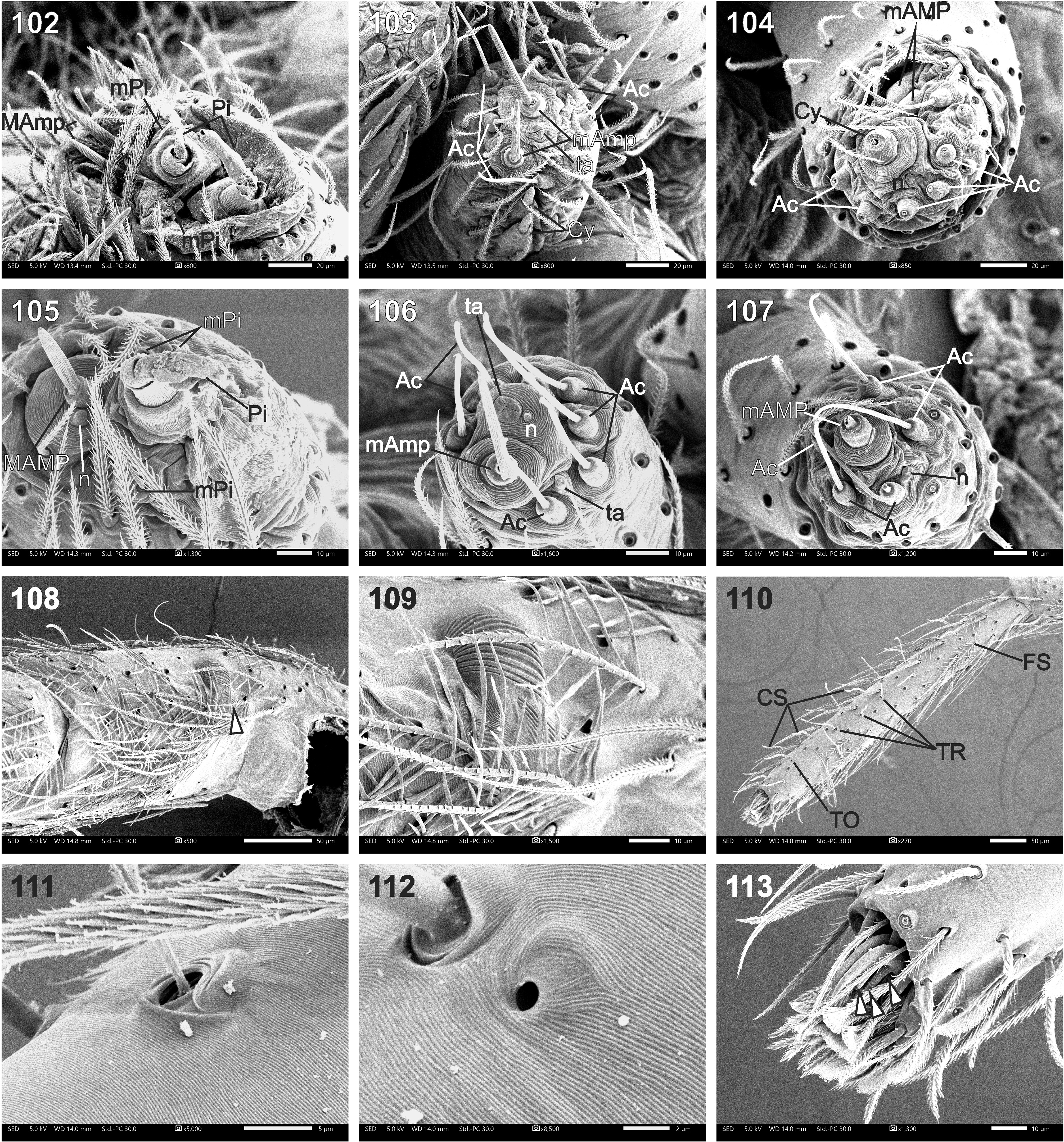
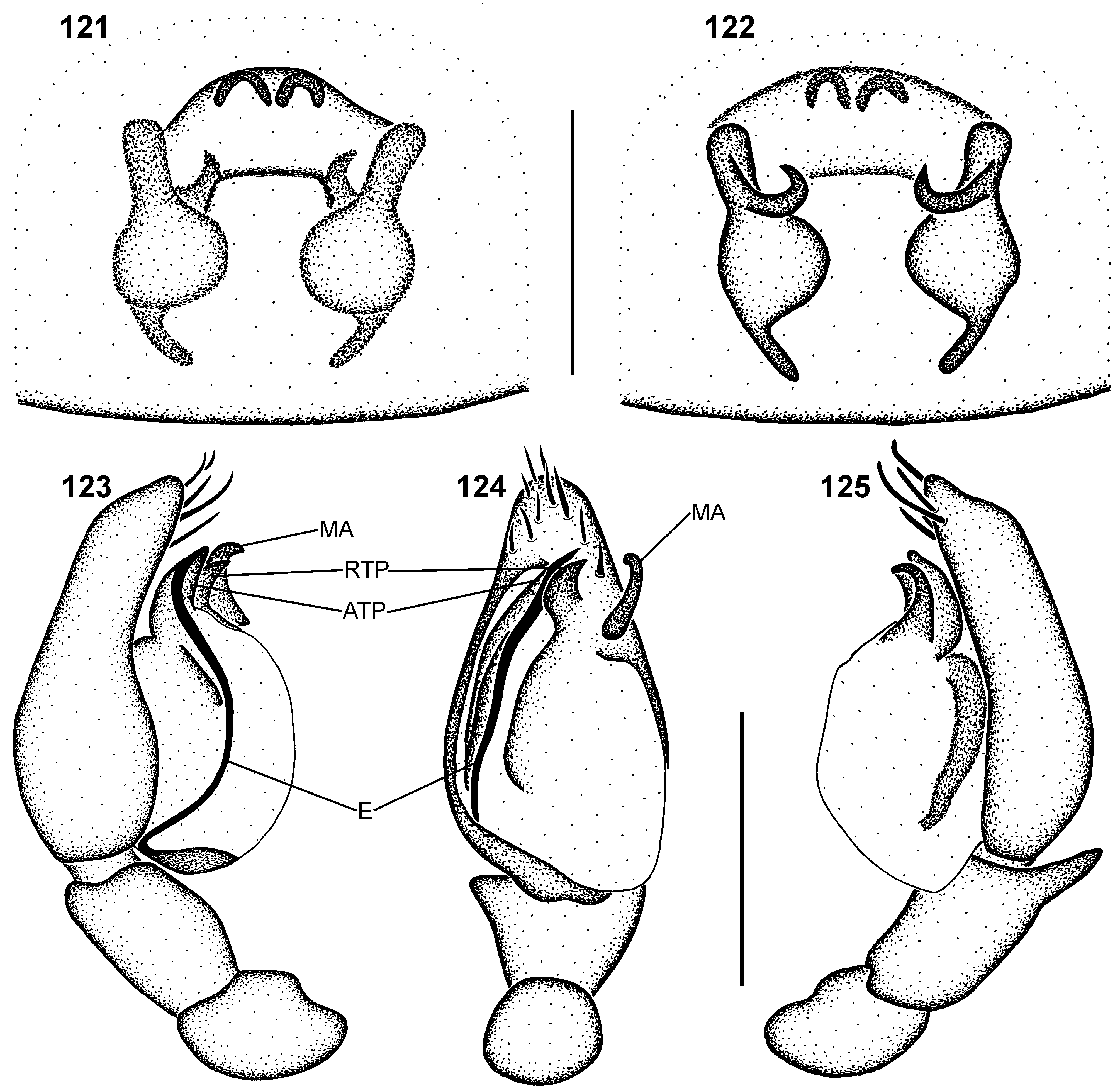
Description. Small pale spiders ( Figs 5, 6 View FIGURES 1–6 , 87–92 View FIGURES 87–92 ), females 2.15–3.80 mm and males 1.77–2.98 mm in length; carapace creamy-white to yellow; carapace oval, eye region narrow, broadest between coxae II and III, without fovea ( Fig. 93 View FIGURES 93–101 ); posterior margin slightly concave; carapace gradually elevated from eye region, highest at approximately 3/4 its length, with steep posterior slope; carapace smooth and matte, densely covered in feathery setae ( Figs 93, 94 View FIGURES 93–101 ), with long straight setae in eye region. All eyes surrounded by black rings, pigment continuous between anterior eyes ( Figs 87–92 View FIGURES 87–92 ); AER procurved in anterior view, slightly recurved in dorsal view ( Fig. 94 View FIGURES 93–101 ); clypeus height slightly larger than AME diameter; AME largest, separated by approximately 1/2 their diameter, separated from ALE by 1/8 ALE diameter; PER strongly procurved in dorsal view ( Fig. 94 View FIGURES 93–101 ); PME oval and flattened, PLE round, PME slightly larger than PLE; PME separated from each other by approximately 3/4 their diameter, from PLE by approximately 1/2 PME diameter; ALE and PLE almost touching ( Fig. 94 View FIGURES 93–101 ); MOQ slightly narrower posteriorly than anteriorly, anterior width slightly larger than MOQ length. Chelicerae: with promarginal escort seta and rake setae, and single retromarginal escort seta ( Fig. 96 View FIGURES 93–101 ); cheliceral dentition (southern African species): promargin with one tooth; retromargin with two teeth, proximal tooth larger than distal ( Figs 97, 98 View FIGURES 93–101 ); endites with slightly depressed lateral margins, distal margins rounded, with distinct serrula and maxillar hair tuft; serrula teeth with weakly undulating lateral margins ( Fig. 99 View FIGURES 93–101 ); labium trapezoid, slightly longer than wide, with rounded anterior margin. Pleural bars weakly sclerotised, isolated; sternum shield-shaped, approximately 1¼ times longer than broad, broadest at coxa II, surface smooth, sparsely covered in straight setae ( Fig. 100 View FIGURES 93–101 ); precoxal triangles present, intercoxal sclerites present between all coxal pairs. Abdomen oval, slightly broader than carapace, dorsal scutum absent in both sexes ( Figs 87–92 View FIGURES 87–92 ); dorsum with single pair of indistinct sigilla; dorsum and sides densely covered in feathery setae, with scattered fine plumose setae ( Fig. 101 View FIGURES 93–101 ), venter only with fine plumose setae. Spinnerets (only observed in L. digitus sp. nov.): ALS of female with two major ampullate gland spigots anteriorly, two large piriform gland spigots mesally, and two slender modified piriform gland spigots, one anterior and one posterior to anterior piriform gland spigot ( Fig. 102 View FIGURES 102–113 ); PMS of female with two large minor ampullate gland spigots mesally, two small cylindrical gland spigots posteriorly, single mesal tartipore, and eight small aciniform gland spigots peripherally ( Fig. 103 View FIGURES 102–113 ); PLS of female with two small minor ampullate gland spigots anteriorly, one large cylindrical gland spigot mesally, single mesal nubbin, and nine aciniform gland spigots peripherally ( Fig. 104 View FIGURES 102–113 ); ALS of male with one large major ampullate gland spigot with adjacent nubbin anteromesally, one large piriform gland spigot mesally, and three slender modified piriform gland spigots, two anterior and one posterior to anterior piriform gland spigot ( Fig. 105 View FIGURES 102–113 ); PMS of male with one posterior minor ampullate gland spigot, one tartipore and nubbin anterior to it, one posterior tartipore, and seven peripheral aciniform gland spigots ( Fig. 106 View FIGURES 102–113 ); PLS of male with single large anterior minor ampullate gland spigot, one posterior nubbin, and five aciniform gland spigots peripherally ( Fig. 107 View FIGURES 102–113 ). Leg formula 4123; legs densely covered in feathery setae, with scattered straight plumose setae between them, feathery setae sparse on tarsi ( Figs 108–110 View FIGURES 102–113 ); patellae with narrow indentation and lyriform organ on retrolateral side ( Figs 108, 109 View FIGURES 102–113 ), with single distal erect long seta dorsally on all patellae; metatarsi with well-developed dorsal stopper distally ( Fig. 110 View FIGURES 102–113 ); tarsi with sparse chemosensory setae, three pro- and retrolateral dorsal trichobothria in alternating arrangement, oval tarsal organ and dense claw tufts ( Figs 110–113 View FIGURES 102–113 ); tarsal claws with three ventral teeth ( Fig. 113 View FIGURES 102–113 ). Female epigyne with median anterior hood ( Figs 121 View FIGURES 121–125 , 131 View FIGURES 131–135 ) or pair of anterior ridges ( Fig. 126 View FIGURES 126–130 ), with atria frequently filled with secretory plugs ( Fig. 114 View FIGURES 114–120 ); internally with short copulatory ducts, initially directed laterally, then looping anteriorly and mesally, entering teardrop-shaped spermathecae anteriorly, with posteriorly-directed fertilization ducts. Male palpal femur and patella without apophyses; palpal patella with retrolateral lyriform organ ( Fig. 117 View FIGURES 114–120 ); palpal tibia with single retrolateral apophysis ( Figs 115, 116, 118 View FIGURES 114–120 ); cymbium ovoid, with dense setae distally on dorsal surface ( Fig. 115 View FIGURES 114–120 ); tegulum generally ovoid, with very slender embolus originating proximally, entering prolateral groove in subtegulum, leading embolus to distal apical tegular process, with embolus tip in narrow retrolateral groove ( Figs 119, 120 View FIGURES 114–120 ); retrolateral tegular process closely associated with apical tegular process ( Figs 119 View FIGURES 114–120 , 124 View FIGURES 121–125 , 129 View FIGURES 126–130 , 134 View FIGURES 131–135 ); median apophysis hook-like, originating retrolaterally ( Fig. 119 View FIGURES 114–120 ).


No known copyright restrictions apply. See Agosti, D., Egloff, W., 2009. Taxonomic information exchange and copyright: the Plazi approach. BMC Research Notes 2009, 2:53 for further explanation.
|
Kingdom |
|
|
Phylum |
|
|
Class |
|
|
Order |
|
|
Family |