
Moribaetis maculipennis ( Flowers 1979 )
|
publication ID |
https://doi.org/ 10.11646/zootaxa.4521.2.5 |
|
publication LSID |
lsid:zoobank.org:pub:F992EA3F-2AD0-49F8-9230-266B4D4C3ACE |
|
DOI |
https://doi.org/10.5281/zenodo.5952426 |
|
persistent identifier |
https://treatment.plazi.org/id/4A1E8797-FF67-AC65-FF48-8199FB6E9231 |
|
treatment provided by |
Plazi |
|
scientific name |
Moribaetis maculipennis ( Flowers 1979 ) |
| status |
|
Moribaetis maculipennis ( Flowers 1979) View in CoL
( Figs 1–61 View FIGURES 1–15 View FIGURES 16–21 View FIGURES 22–24 View FIGURES 25–33 View FIGURES 34–43 View FIGURES 44–47 View FIGURES 48–52 View FIGURES 53–55 View FIGURES 56–61 )
Baetis maculipennis Flowers 1979: 187 (male imago, larva). Moribaetis maculipennis: Waltz & McCafferty 1985: 245 View in CoL , figs 5, 26–27 (larva) (non fig. 32); McCafferty & Lugo-Ortiz 1998: 120 (key to male imagoes).
Material examined. PANAMA, provincia de Bocas del Toro, bosque protector Palo Seco, Altos del Valle , río Buris (8°47'37''N, 82°11'35''W), 24–29.I.2018, coll. N. Kluge & L. Sheyko: 4 L-S-I ♂ GoogleMaps , 5 L-S ♂, 11 L-S-I ♀, 2 L-S ♀, 99 larvae ( ZIN) ; provincia de Chiriquí, reserva forestal Fortuna, quebrada Honda (8°45'2.15''N, 82°14'18.68''W), 11.XI.2017, coll. J. Bernal & T. Ríos: 5 larvae ( UNACHI) GoogleMaps ; Alambique, río Fonseca (8°22'28.15''N, 82°4'28.91''W; 8°22'1.46''N, 82°4'28.65''W), 7.XII.2016, coll. T. Ríos: 9 larvae ( UNACHI) GoogleMaps .
Diagnosis. Larva. Described by Flowers (1979) and Waltz & McCafferty (1985). Differs from M. latipennis sp. n. by following characters.
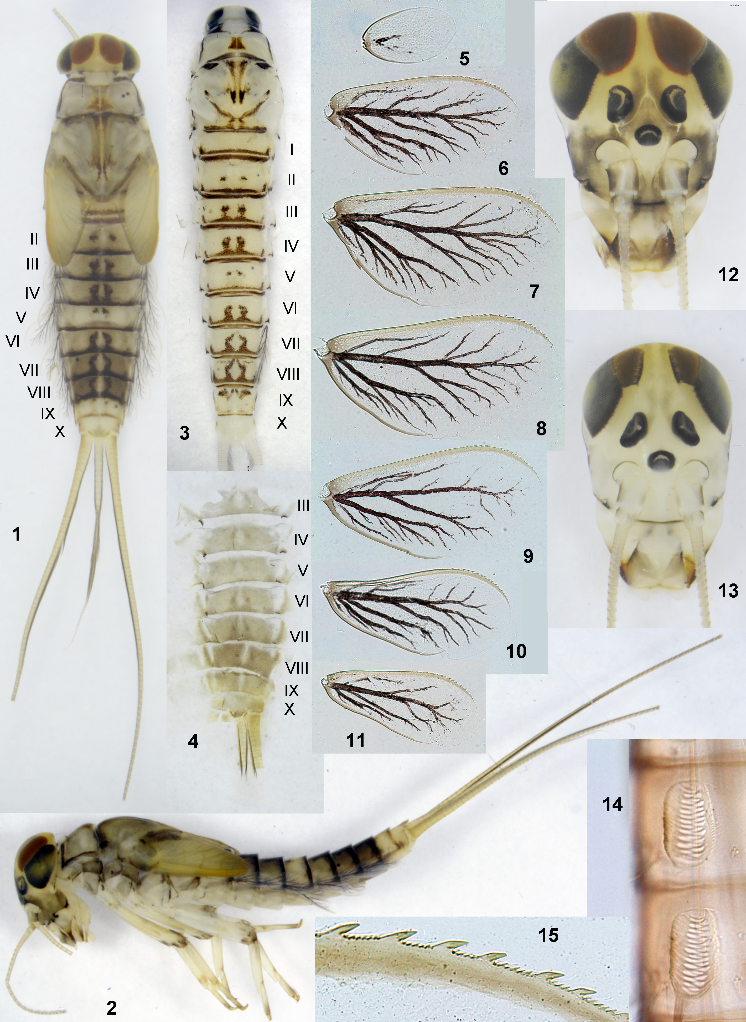
(1) Larval head elongated and narrowed, with antennae bases brought together, frons narrow and keel-shaped ( Figs 12–13 View FIGURES 1–15 ). In specimens with intensively developed cuticular coloration, cuticle of frons in last larval instar brown ( Fig. 12 View FIGURES 1–15 ), in previous instars colorless ( Fig. 13 View FIGURES 1–15 ) [other cuticular coloration as characterized for Moribaetis (18)].
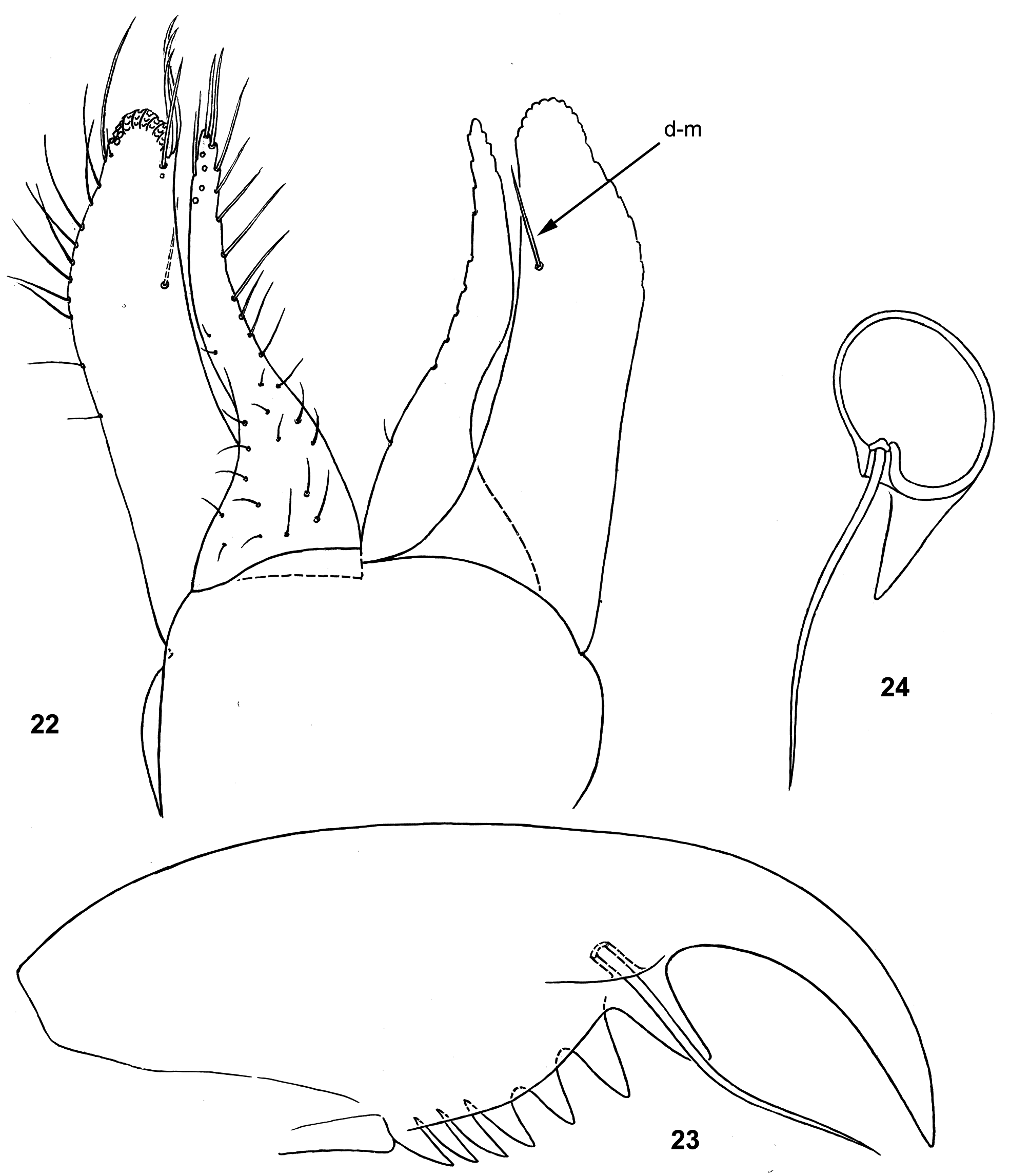
(2) Both left and right mandibles with incisor [blade-like – see Moribaetis (2)] and kinetodontium completely fused together and lacking denticles; prosthecae of both mandibles equally diminished and very slender [see Moribaetis (3)] ( Figs 18–21 View FIGURES 16–21 ).
(3) Femur of fore leg parallel-sided ( Fig. 25 View FIGURES 25–33 ); stout setae on its inner side pointed, occupy wide area ( Fig. 33 View FIGURES 25–33 ).
(4) Dark brown submedian hypodermal stripes on abdominal terga VI–VIII equally narrow, stripes on terga III–IV also not widened ( Figs 1, 3 View FIGURES 1–15 ).
(5) Posterior margins of abdominal sterna I–IV without denticles, that of sterna V–IX with regular pointed denticles [II–X terga with larger denticles – see Moribaetis (14)].
(6) Paraproct with denticles on inner and apical margins sharply differentiated: minute on inner margin and significantly larger on apical margin ( Flowers 1979: fig. 16).
(7) Larval cerci and their swimming setae unicolor ( Fig. 1 View FIGURES 1–15 ).
Male imago. Described by Flowers (1979). Differs from M. latipennis sp. n. and M. salvini by following characters.
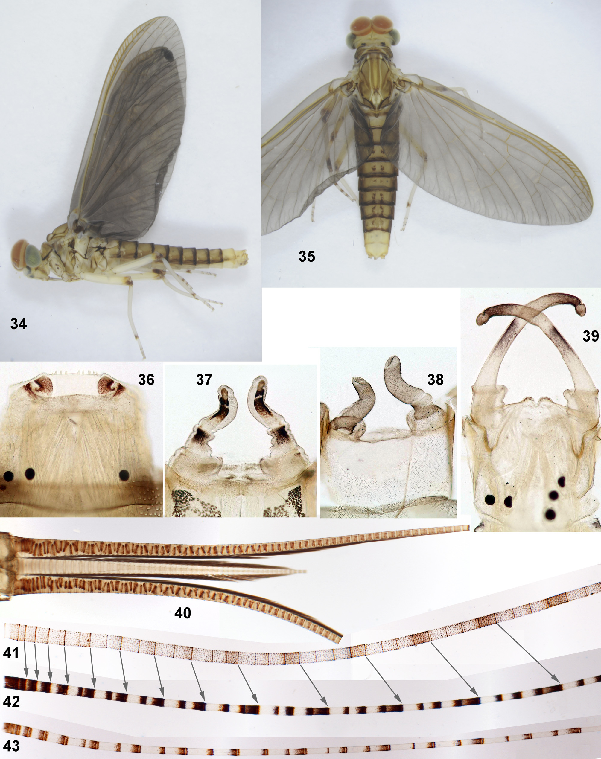
(1) Turbinate eyes low and wide, contiguous both in imago and in subimago ( Figs 34–35 View FIGURES 34–43 , 48–50 View FIGURES 48–52 ) (in contrast to narrow in M. latipennis ).
(2) On fore wing most cross veins light yellow, lighter than adjacent brown maculae (in contrast to dark brown in M. salvini ); brown maculae present all over wing, forming nearly regular bands ( Figs 48–50 View FIGURES 48–52 ). Hind wing often also with brown maculae, variably located ( Fig. 55 View FIGURES 53–55 ).
(3) Dark brown submedian hypodermal stripes on abdominal terga VI–VIII equally narrow ( Fig. 48 View FIGURES 48–52 ) (in contrast to M. latipennis ).
(4) Femur with small contrasting brown spot on posterior side ( Fig. 50 View FIGURES 48–52 ) (in contrast to wide brown band in M. salvini – Fig. 102 View FIGURES 102–105 ).
Female imago. Coloration of body, wings, legs and cerci as in male ( Figs 53–54 View FIGURES 53–55 ). Fore tarsus with apical spines on 2nd and 3rd segments (as in Fig. 31 View FIGURES 25–33 ) (as on middle and hind tarsi – as in Fig. 32 View FIGURES 25–33 ).
Eggs. Oval, covered with thick coat ( Fig. 56 View FIGURES 56–61 ). Outer surface of coat forms folds and regularly arranged round protuberances, each protuberance with concavity at middle ( Figs 57–59 View FIGURES 56–61 ). Chorion under the coat with dense fine concavities ( Figs 60–61 View FIGURES 56–61 ).
Dimensions. Fore wing length 7.5–9.2 mm (7.5–8.5 in our specimens, 9.0–9.2 according to Flowers 1979).
Distribution. Costa Rica and Panama.
Comparison. Imago of Moribaetis maculipennis differs from the earlier described M. salvini by coloration of wings, which in M. maculipennis have brown markings all over fore wing and cross veins yellow, lighter than adjacent brown markings ( Figs 48–50 View FIGURES 48–52 ), while in M. salvini brown markings are present only on anterior part of fore wing, and cross veins are brown, darker than adjacent brown markings ( Fig. 102 View FIGURES 102–105 ). The formerly reported difference between these species in coloration of cerci is wrong (see «Comments»).
Comments. Originally, male imagoes and larvae of this species were reliably associated based on one male imago reared from larva (Flowers 1989). Recent rearing of imagoes of both sexes allows to add here description of female and egg.
In the original description tergalius IV is figured greatly widened and angulate ( Flowers 1979: fig. 14), that somewhat differs from our specimens ( Fig. 8 View FIGURES 1–15 ).
The original description ( Flowers 1979), does not provide comparison of the new species B. maculipennis with the earlier described species B. salvini Eaton 1885 . Comparison of these two species was given by Waltz & McCafferty (1985), who reexamined the lectotype of B. salvini . These authors did not add any new characteristics for male imago of Baetis salvini and reported only that it was «Adequately characterized by Eaton (1885)» ( Waltz & McCafferty 1985: 247). In their diagnosis and the key to male imagoes, M. maculipennis is distinguished from M. salvini by three characters only: number of intercalaries of hind wing, «the abdominal color pattern» and coloration of cerci. Actually, no one of these characters allows to distinguish these two species:
Hind wing of Moribaetis normally has one intercalary between branches of the second vein ( Flowers 1979: fig. 3). Hind wing of M. salvini was characterized as «usually with two distinct intercalaries and two or three minor intercalaries (fig. 44)». The figures of fore and hind wing of M. salvini ( Waltz & McCafferty 1985: figs 43–44) were redrawn from the figures of the right fore and hind wings of the lectotype in the original description (Eaton 1885: pl. 16: figs 29a); no other winged specimens reliably attributed to M. salvini have been ever examined. Additional intercalary is present on individual hind wings of M. maculipennis ( Fig. 55 View FIGURES 53–55 ), so M. maculipennis and M. salvini have no difference by this character. McCafferty & Lugo-Ortiz (1998) wrote «It is possible that the number of marginal intercalaries between the forks of the second elongate vein in the hindwings is variable within M. macaferti , ... and thus we do not recommend attempting to identify adults of species of Moribaetis on that basis».
Waltz & McCafferty (1985) did not report, which feature of «the abdominal color pattern» distinguishes M. maculipennis from M. salvini ; the imaginal color pattern, being hypodermal, should be the same in imago and larva; the figure of larva, which illustrates this color pattern for M. maculipennis ( Waltz & McCafferty 1985: fig. 32), actually belongs to M. latipennis sp. n., whose larva was wrongly attributed to M. salvini (see below).
Waltz & McCafferty (1985) stated that imago of M. maculipennis differs from M. salvini by coloration of cerci, which in M. maculipennis are «(fig. 33) banded in alternating pattern of one white segment with three darkened segments» and in M. salvini are «(fig. 42) banded in alternating pattern of one lightly darkened segment with three white segments». This feature of M. maculipennis was rewritten from the original description (Flowers 1 979: 188), but illustrated differently: cercus is figured as having alternating two darkened segments and two white segments ( Waltz & McCafferty 1985: fig. 33); according to the original description, «all segments darker brown at apex», while on this figure apices of segments are not darkened. The paratype, from which this figure was done, was not reared, so its systematic position is unknown. All our specimens reared from larvae have apex of each cercal segment contrastingly darkened, and each 4th segment is more or less darker than others ( Figs 42–43 View FIGURES 34–43 ). This coloration is the same as in the lectotype of M. salvini ( Fig. 103 View FIGURES 102–105 ; Waltz & McCafferty 1985: fig. 42).
Redescription and drawings of M. maculipennis made by Waltz & McCafferty (1985) belong to this species, except for the drawing of larval thorax and abdomen ( Waltz & McCafferty 1985: fig. 32); judging by its color pattern, the larva drawn on this figure belongs not to B. maculipennis , but to M. latipennis sp. n., which in the same paper was described as « M. salvini » (see below). The authors of this paper wrote that «Larvae of M. maculipennis may be easily separated from other species of this genus by the distinctive abdominal color pattern (fig. 32)»; actually the abdominal color pattern shown on this figure is hypodermal, so it is the same in larva and imago. Abdominal color pattern of imago of M. maculipennis is adequately figured in the original description ( Flowers 1979: fig. 5); in contrast to M. latipennis sp. n., its paired stripes on abdominal tergum VI are not widened, being similar to the stripes on abdominal tergum VII ( Figs 1, 3 View FIGURES 1–15 ).
Waltz & McCafferty (1985) stated that larva of M. maculipennis differs from « M. salvini » (actually M. latipennis sp. n.) by having «frons with distinct medial pigmented area between ocelli» ( Waltz & McCafferty 1985: 250, fig. 5). Actually, pigmentation of larval frons in both these species is present only within cuticle, but not in underlying tissues, so it is absent at the beginning of each instar; in M. maculipennis cuticle of frons becomes dark only in the last larval instar ( Fig. 12 View FIGURES 1–15 ), being colorless during in all previous instars ( Fig. 13 View FIGURES 1–15 ).


| ZIN |
Russian Academy of Sciences, Zoological Institute, Zoological Museum |
No known copyright restrictions apply. See Agosti, D., Egloff, W., 2009. Taxonomic information exchange and copyright: the Plazi approach. BMC Research Notes 2009, 2:53 for further explanation.
|
Kingdom |
|
|
Phylum |
|
|
Class |
|
|
Order |
|
|
Family |
|
|
Genus |
Moribaetis maculipennis ( Flowers 1979 )
| Kluge, Nikita J. & Bernal Vega, Juan A. 2018 |
Baetis maculipennis
| McCafferty, W. P. & Lugo-Ortiz, C. R. 1998: 120 |
| Waltz, R. D. & McCafferty, W. P. 1985: 245 |
| Flowers, R. W. 1979: 187 |