
Amphibolips zacatecaensis Melika & Pujade-Villar
|
publication ID |
https://doi.org/10.5281/zenodo.279218 |
|
DOI |
https://doi.org/10.5281/zenodo.6193092 |
|
persistent identifier |
https://treatment.plazi.org/id/4B00B94F-FF94-FFB6-B991-FB6B3F4CFF2E |
|
treatment provided by |
Plazi |
|
scientific name |
Amphibolips zacatecaensis Melika & Pujade-Villar |
| status |
sp. nov. |
Amphibolips zacatecaensis Melika & Pujade-Villar , new species
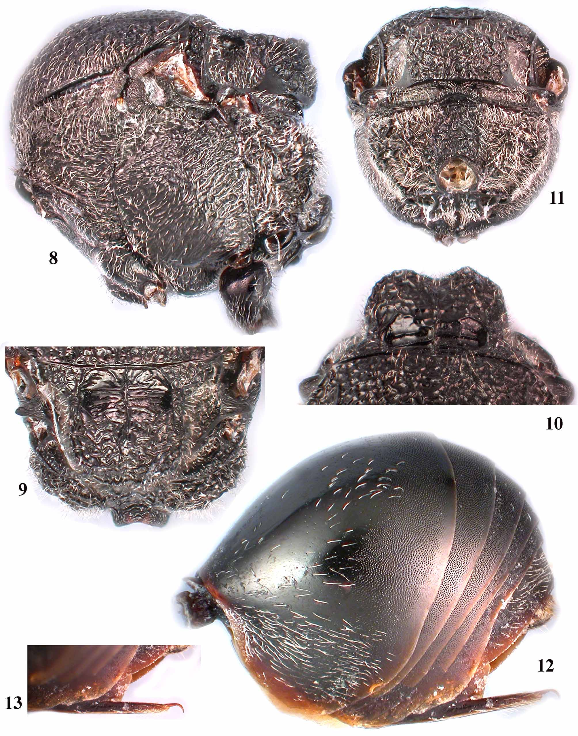
Figs 1–17 View FIGURES 1 – 7 View FIGURES 8 – 13 View FIGURES 14 – 20. 14 – 17
Type material. HOLOTYPE. Female: MEXICO, Monte Escobedo, Zacatecas, Q. eduardi , ( 31.V.2010) ext. 31.V.10. leg. J. Pujade-Villar (deposited in UB).
Etymology. The species is named after the Mexican state, Zacatecas, where the galls were collected.
Diagnosis. The newly described species is characterized by the posterior emargination of the mesoscutellum (posteromedian scutellar depression) ( Fig. 10 View FIGURES 8 – 13 ), and most closely resembles A. fusus and A. nassa . The new species differs from A. nassa by the absense of a heavy dark stripe along the anterior margin of the forewing, which is interrupted by a clear unpigmented cross band in the cell delimited by R1+Sc and Rs+M ( Fig. 14 View FIGURES 14 – 20. 14 – 17 ); furthermore, A. zacatecaensis is reared from Q. eduardi , while A. nassa is reared from Q. castanea (= Q. serrulata ) and Q. mexicana . Amphibolips zacatecaensis and A. fusus are both reared from Q. eduardi and both species have a similar pattern of the forewing pigmentation, but the two species can be differentiated by the shape of the head (quadrangular in A. zacatecaensis and rounded in A. fusus , Figs 1 View FIGURES 1 – 7 , 47 View FIGURES 42 – 48 ), in the tentorial pits (smaller in A. zacatecaensis , Figs 1 View FIGURES 1 – 7 , 47 View FIGURES 42 – 48 ), in the shape of scutellar foveae ( Figs 9 View FIGURES 8 – 13 , 48 View FIGURES 42 – 48 ), and in the color of the metasoma (black in A. zacatecaensis and dark brown in A. fusus ). A. nassa and A. fusus galls have a hard and lignified parenchima while they are soft, with spongious parenchima in A. zacatecaensis . Moreover, galls of A. fusus are more elongate ( Fig. 44–45 View FIGURES 42 – 48 ) than in A. zacatecaensis ( Figs 17–18 View FIGURES 14 – 20. 14 – 17 ) and galls of A. nassa are more elongate than A. zacatecaensis ( Figs 17–18 View FIGURES 14 – 20. 14 – 17 ), spindleshaped ( Figs 42–43 View FIGURES 42 – 48 ).
Description. FEMALE ( holotype) ( Figs 1–14 View FIGURES 1 – 7 View FIGURES 8 – 13 View FIGURES 14 – 20. 14 – 17 ). Head black, except chestnut brown maxillary and labial palpi; antenna, mesosoma, legs and metasoma black.
Head ( Figs 1–4 View FIGURES 1 – 7 ) quadrangular in anterior view, dull rugose, with sparse short white setae, denser on lower face and gena, 1.9 times as broad as long from above, 1.3 times as broad as high in anterior view and narrower than mesosoma. Gena dull rugose, strongly broadened behind eye, visible in anterior view behind eye, as broad as cross diameter of eye; malar space rugose, without striae; height of eye 1.4 times as long as length of malar space. POL nearly equal OOL; length of lateral ocellus 1.3 times as long as LOL; ocelli elongate. Transfacial distance 1.6 times as broad as height of eye; diameter of antennal torulus 2.2 times as long as distance between toruli, distance between torulus and inner margin of eye 0.9 times as long as diameter of torulus; lower face dull rugose, with striae radiating from clypeus and extending to antennal sockets, with denser white setae and narrow elevated rugose median area. Clypeus rounded ventrally, coriaceous, with strongly elevated small, rounded central area, ventrally emarginate, without median incision; anterior tentorial pits deep, epistomal sulcus conspicuously impressed, clypeo-pleurostomal line indistinct. Frons, vertex, interocellar area and occiput uniformly dull rugose. Occiput with strong carina dorsally, postocciput and postgena striate, impressed around occipital foramen, with dense white setae; posterior tentorial pits large, deep, area around them strongly impressed; height of occipital foramen at least 3.0 times as long as height of postgenal bridge; hypostomal carina emarginate, not going around oral foramen, continuing into postgenal sulcus. Labial palpus 3-segmented, maxillary palpus 5-segmented. Antenna ( Fig. 5 View FIGURES 1 – 7 ) with 11 flagellomeres; slightly longer than mesosoma; scape 3.0 times as long as pedicel; pedicel subglobose, slightly broader than long; F1 slightly longer than scape+pedicel and 1.5 times as long as F2; F2 1.3 times as long as F3; F3 nearly equal in length to F4, subsequent flagellomeres shorter, F11 2.1 times as long as F10; whitish placodeal sensilla visible on F5–F11, absent on F1–F4.
Mesosoma ( Figs 7–11 View FIGURES 1 – 7 View FIGURES 8 – 13 ) only slightly longer than high. Pronotum coriaceous dorsally, with numerous strong irregular rugae laterally; propleuron black, coriaceous, concave in mediocentral part. Mesoscutum uniformly dull rugose, subequal, nearly as long as broad in dorsal view (largest width measured across mesoscutum on the level of tegulae base). Notauli indistinct in dull rugose sculpture; anterior parallel lines extending to half length of mesoscutum, slightly elevated, mesoscutum impressed along both sides of lines; parapsidal lines distinct, originating away from posterior margin and extending to nearly half length of mesoscutum; median mesoscutal line absent; parascutal carina short, extending to level of tegula only. Mesoscutellum 0.7 times as long as mesoscutum, uniformly dull rugose, quadrangular, only slightly longer than broad, slightly overhanging metanotum; scutellar foveae large, deep, with parallel transverse strong striae on shiny bottom, with distinct elevated narrow median carina dividing the base of mesoscutellum into two halves; lateral sides of foveae with strong carinae, separating them from dorsoaxillar area. Mesoscutellum with moderately deep posteromedian depression. Mesopleuron, including speculum, uniformly dull rugose, ventral rugae orientated into transverse subparallel striae. Mesopleural triangle rugose; dorsal axillar area delicately rugose; lateral axillar area and axillula coriaceous, with few short, white setae; subaxillular bar smooth, shiny, with parallel sides, its height less than height of metanotal trough, most posterior part extending to half height of mesoscutellum; postalar process long, with parallel striae; metapleural sulcus hidden in dull rugose sculpture. Metascutellum uniformly coriaceous, metanotal trough coriaceous, with dense white setae; ventral impressed area smooth, slightly shorter than height of metascutellum; central propodeal area smooth, shiny, narrow, with numerous strong irregular rugae, mostly orientated transversely; lateral propodeal carinae strong, high, subparallel, slightly curved outwards medially; lateral propodeal area with irregular strong wrinkles and dense white setae; nucha short, surrounded by irregular wrinkles. All legs uniformly black, with dense short white setae; tarsal claws with acute basal lobe ( Fig. 6 View FIGURES 1 – 7 ).
Forewing ( Fig. 14 View FIGURES 14 – 20. 14 – 17 ) longer than body, infuscate, with short dense cilia on margin, heavy dark stripe on anterior margin of forewing continuous, going across radial cell, cells delimited by R1+Sc and Rs+M and by R+Sc, M+Cu1 and M and absent in basal cell; radial cell narrow, long, opened on margin, 3.4 times longer than broad; R1 and Rs nearly reaching wing margin; areolet small, triangular, closed and distinct; Rs+M reaching basalis (M) at its half height.
Metasoma ( Fig. 12 View FIGURES 8 – 13 ) longer than head+mesosoma, slightly longer than high in lateral view; 2nd metasomal tergite occupying nearly half length of metasoma, smooth, shiny, with short sparse setae dorsolaterally and with larger patch of dense setae ventrolaterally; posterior half conspicuously punctate dorsally and laterally and only very narrow posterior band smooth, without punctures; all subsequent tergites dorsally and laterally uniformly and entirely micropunctate, with a narrow smooth band posteriorly on each tergite. Ventral spine of hypopygium ( Fig. 13 View FIGURES 8 – 13 ) robust, long, needle-like, prominent part 6.5 times as long as broad, with two rows of white setae each side, extending beyond apex of spine. Body length 5.5 mm.
Gall ( Figs 15–17 View FIGURES 14 – 20. 14 – 17 ). A rather large, subglobose, slightly spindle-shaped oak bud gall, with a nipple at the top. The body of the gall is quite globose, with greatest diameter near middle of the gall, up to 5.0 cm and 6.8 cm in length; from the middle the gall gradually tapering to a point (nipple) at the top. The gall is very thin-walled, light brown when mature, with smooth and naked surface; spongious internally, with radiating filaments and a central ovate, hard-walled larval chamber, with largest length of 5.0– 6.5 mm.
Biology. Only the female is known, inducing galls on Quercus eduardi Trel. (Section Lobatae of Quercus , red oaks) which is found only in Mexico ( Govaerts & Frodin 1998). The mature gall was collected in May and the dead adult wasp was cut out from the gall.
Distribution. Currently known from the type locality only: Mexico, Zacatecas state, Monte Escobedo.


No known copyright restrictions apply. See Agosti, D., Egloff, W., 2009. Taxonomic information exchange and copyright: the Plazi approach. BMC Research Notes 2009, 2:53 for further explanation.
|
Kingdom |
|
|
Phylum |
|
|
Class |
|
|
Order |
|
|
Family |
|
|
Genus |