
Ophiopsila xmasilluminans, Okanishi & Oba & Fujita, 2019
|
publication ID |
https://doi.org/10.26107/RBZ-2019-0034 |
|
publication LSID |
lsid:zoobank.org:pub:4537754B-0BBE-4388-84E1-5A51DCD507AC |
|
persistent identifier |
https://treatment.plazi.org/id/AA621784-C18B-4A54-9F5F-942EA5AC6605 |
|
taxon LSID |
lsid:zoobank.org:act:AA621784-C18B-4A54-9F5F-942EA5AC6605 |
|
treatment provided by |
Carolina |
|
scientific name |
Ophiopsila xmasilluminans |
| status |
sp. nov. |
Ophiopsila xmasilluminans new species
( Figs. 2–10 View Fig View Fig View Fig View Fig View Fig View Fig View Fig View Fig View Fig )
Ophiopsila pantherina non Koehler, 1898: ― Tan et al., 2014: 413, fig. 19.
Type material. Holotype: ( WAM Z99800 View Materials ) submarine cave (known as “ Thunderdome Cave ”) at Christmas Island, northwestern Australia, depth approximately 10 m, SCUBA diving, coll. Y. Fujita, 27 March 2011 . Paratypes: 1 specimen ( ZRC.ECH.1332), same data as holotype ; 12 specimens (RUMF-ZE-00148, RUMF-ZE-00153, RUMF-ZE-02086, RUMF-ZE-02087, ZRC.ECH.1333, ZRC.ECH.1334, ZRC. ECH.1335, ZRC.ECH.1336, WAM Z99801 View Materials , WAM Z99802, WAM Z99803, WAM Z99804), same data as holotype ; 1 specimen (RUMF-ZE-00152), same locality as holotype , 30 March 2011.
Diagnosis. Disc surface entirely covered by thick skin with embedded small and delicate scales and granules; 3 oral papillae, basically as scales, but sometimes spiniform for one on innermost or middle position; diamond-shaped oral shield with rounded edges, as long as wide; long and flat arm spines, 6 in maximum number; inner tentacle scale narrow, flat and long; arms approximately 18 times longer than disc diameter.
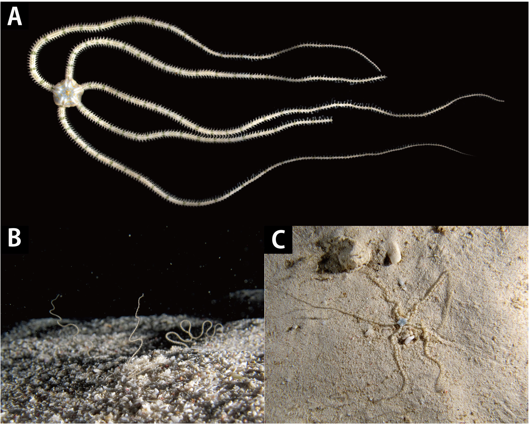
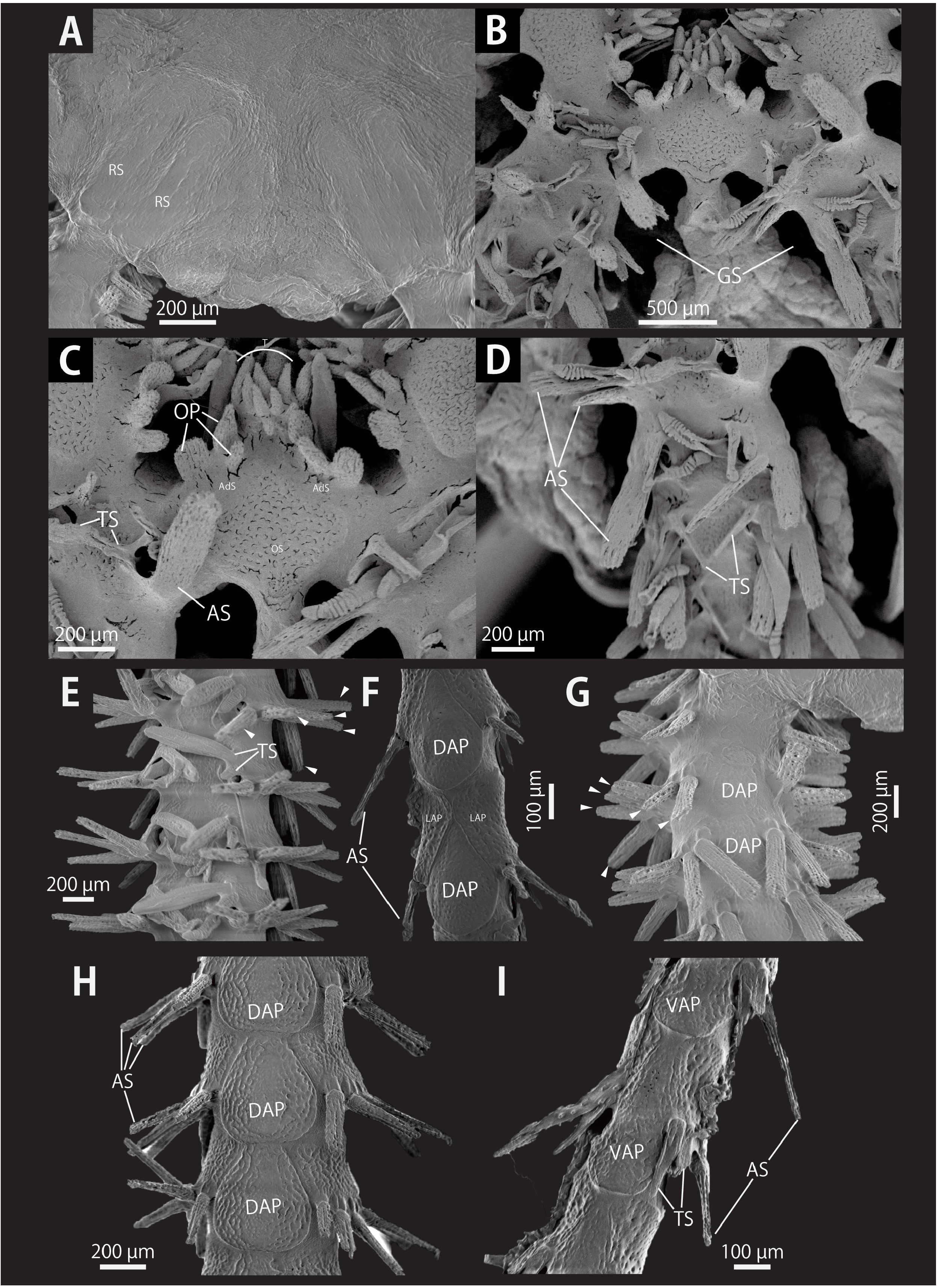
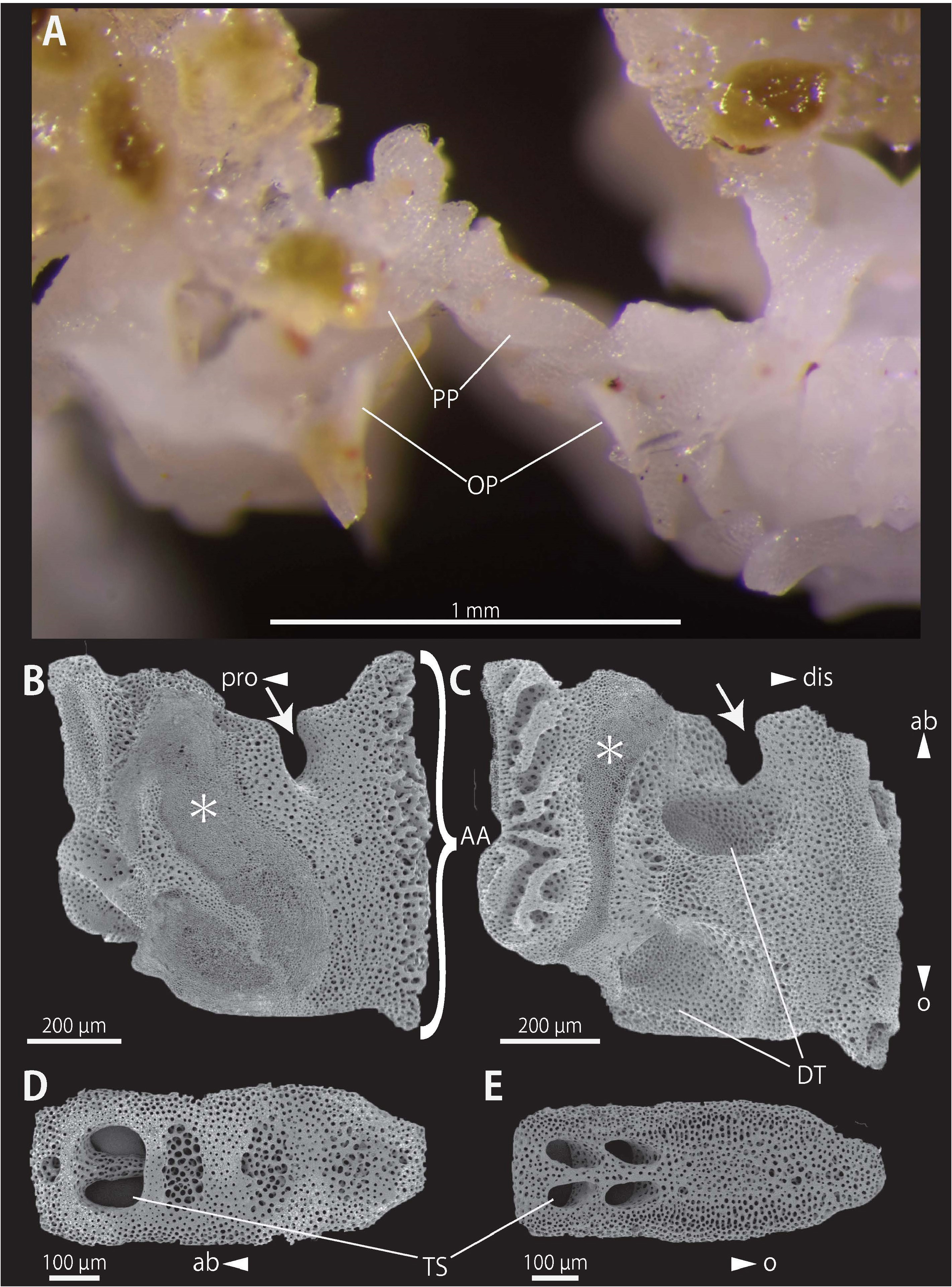
Description of external morphology ( holotype WAM Z99800 View Materials and paratype RUMF-ZE-02087). Disc. Pentagonal, 6.3 mm in diameter ( Fig. 2A View Fig ), aboral surface covered by small round scales, approximately 30 μm in length on central disc and by scales around radial shields, approximately 50–100 μm ( Fig. 3A View Fig ). These ossicles embedded in thick skin ( Figs. 3A View Fig , 5A View Fig ). Radial shields bar-like, 5 to 6 times longer than wide, two-thirds or half the length of disc radius ( Fig. 3B View Fig , 5A View Fig ). On oral surface, adoral shields bar-like, wider than long ( Figs. 3E View Fig , 5C View Fig ), approximately 500 μm in length, 150 μm in width, in contact with the first ventral arm plates ( Fig. 3E View Fig ). Oral plates short, invisible from outside, approximately 300 μm in length, 80 μm in width, contacting each other. Oral shields pentagonal, approximately as long as wide ( Figs. 3E View Fig , 5C View Fig ). All oral shields similar in size and shape: oral shield serving as madreporite unrecognisable in external view ( Fig. 3D, E View Fig ). Interradial oral disc covered only by skin ( Figs. 3F View Fig , 5B View Fig ). Genital slits long, almost extending to disc edge ( Figs. 3F View Fig , 5B View Fig ), approximately 0.2 mm ( Fig. 3F View Fig ). Abradial genital plates visible on side of genital slit, near disc edge, bar-like, approximately 1 mm in length and 0.1 mm wide ( Fig. 3F View Fig ). Near the oral shield, small scales lie immediately outside of the genital slits, approximately 0.1 mm wide and long ( Fig. 3F View Fig ). Two flat, subequal trapezoidal oral papillae and an spiniform oral papilla just below the inner trapezoidal papilla at each opening for the second tentacle of adoral shield ( Fig. 3E View Fig ), sometimes the middle papilla spiniform ( Fig. 5C View Fig ; see also “Variation” section below). Teeth triangular, pointed, forming tooth papillae consisted of 7 to 9 papillae on oral half side of dental plate, and at least two flat, block-like papillae forming vertical row on aboral half of plate ( Figs. 3E View Fig , 5C View Fig ). Second tentacle pore completely inside the mouth slit.
Arms. Five, three arms lacking distal one-fourth to onethird, and two arms remaining almost entire length, 85 mm long. Arms approximately 1.3 mm wide and 1.0 mm high in proximal portion, square in cross section. Arms tapering gradually distally ( Fig. 2 View Fig ).
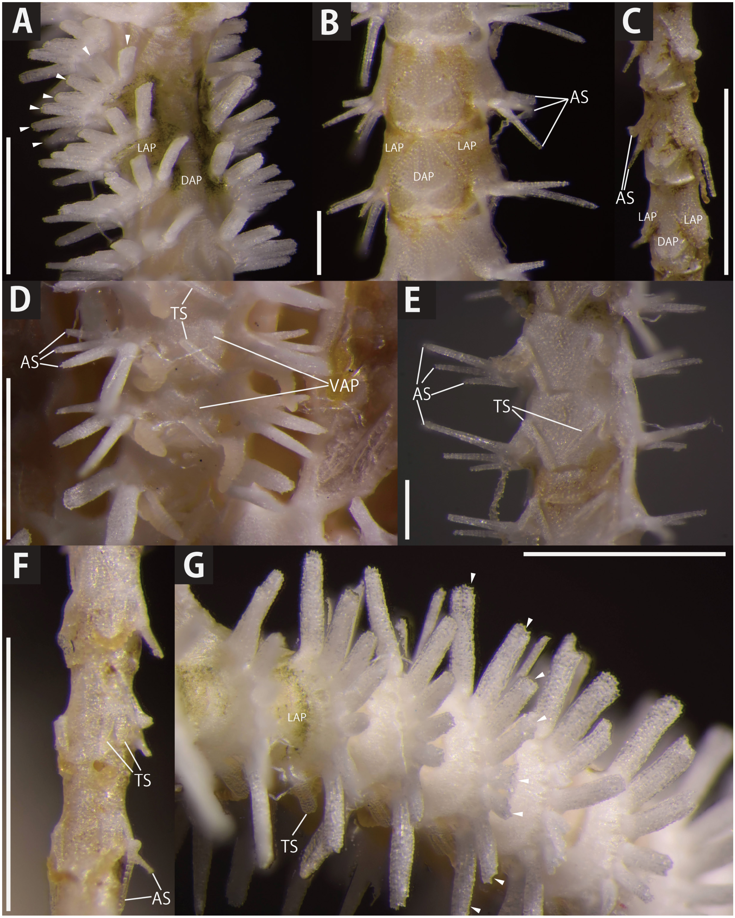
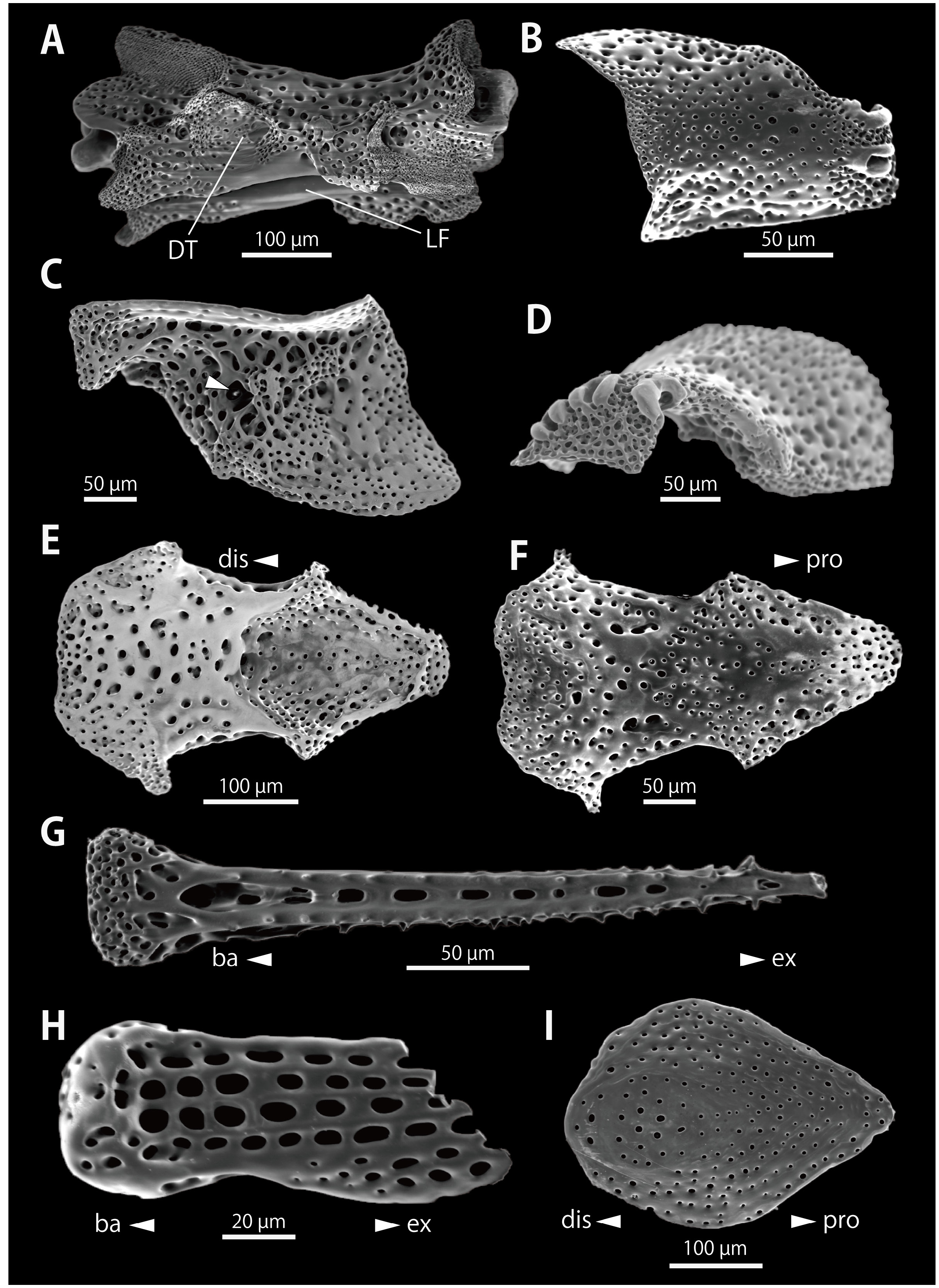
On proximal to middle portion of the arms, ventral arm plates oblong, slightly longer than wide, distal and lateral sides concave, contiguous ( Figs. 4D View Fig , 7D View Fig ). Dorsal arm plate oblong, slightly longer than wide, distal edge slightly rounded, contiguous ( Figs. 4A, B View Fig , 5G, H View Fig ). Lateral arm plates slightly protruding from arm, separated by dorsal and ventral arm plates ( Figs. 4A, B, D, E View Fig , 5G, H View Fig ). On distal portion of the arms, ventral arm plates long, pointed distally, concave on lateral side, and having small points on proximal side ( Figs. 4F View Fig , 5I View Fig , 8E, F View Fig ). Dorsal arm plates also longer than wide, oval, and pointed distally ( Figs. 5I View Fig , 8I View Fig ). Lateral arm plates bearing arm spines, on proximal portion, 7 or 8 long and flat spines, aboral- and oral-most ones the longest, approximately 1.5 to 2 times longer than corresponding arm segment, and others as long as the corresponding arm segment ( Figs. 4A, G View Fig , 5E, G View Fig ). On middle portion, 5 or 6 spines, long and thin, oral-most one the longest, approximately twice as long as corresponding arm segment, and others threefourth length of corresponding arm segment ( Figs. 4B, E View Fig , 5H View Fig ). On distal portion of arms, 2 spiniform arm spines, oral (inner) one longer, approximately two-thirds length of corresponding arm segment and the other one-fourth length of the corresponding arm segment ( Fig. 4C, F View Fig ). Tentacle scales 2, flat, spiniform, inner one longer, two-thirds to the same length of the corresponding arm segment, and outer one half to one-third the length of the corresponding arm segment ( Figs. 4D View Fig , 5 View Fig C–E) on proximal to middle portion of arm. On distal portion of arm, scales more flattened, inner spine half the length of corresponding arm segment, and the other one-fourth length of the corresponding arm segment ( Figs. 4C, F View Fig , 5I View Fig ). Detailed descriptions of ossicles are provided below.
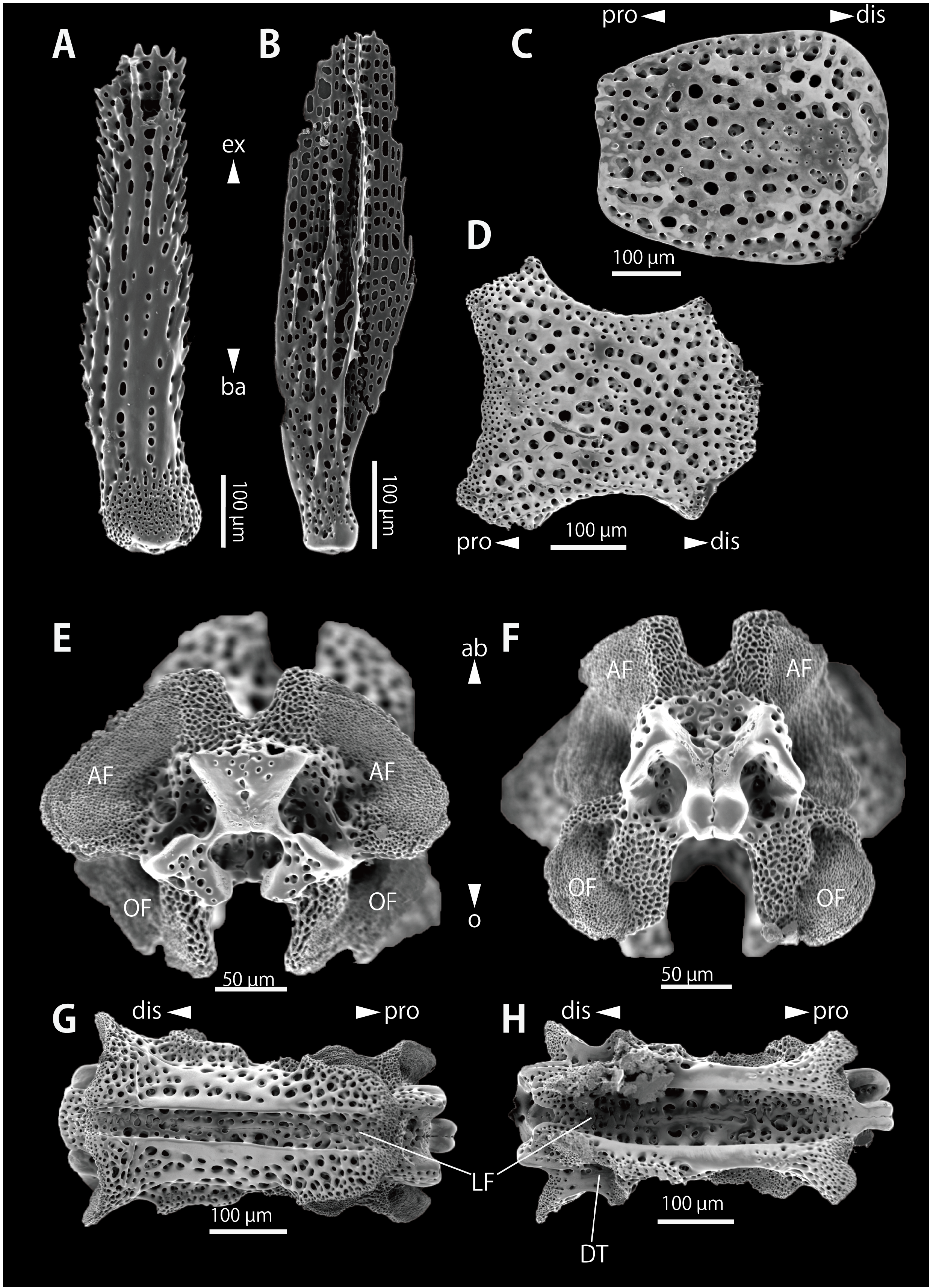
Description of ossicle morphology ( holotype, WAM Z99800 View Materials ). Vertebrae with zygospondylus articulation, large aboral and oral muscle flanges on both distal and proximal sides ( Fig. 6C, D View Fig ). Aboral muscle flanges wider than oral muscle flanges. Longitudinal deep furrows on both aboral and oral sides ( Figs. 6A, B View Fig , 7G, H View Fig , 8A View Fig ). Transverse furrow on proximal portion of the arm ( Fig. 6B View Fig ). Radial water and nerve holes unrecognisable on oral furrow ( Figs. 6A View Fig , 7H View Fig ). Depression for tentacles located on lateral distal side of the vertebra ( Figs. 6A View Fig , 7H View Fig , 8A View Fig ).
Proximal dorsal arm plates trapezoid, as long as wide, distal edge becoming rounder, wider than proximal width ( Fig. 7C View Fig ). On distal portion of arm, dorsal arm plate rhomboid with rounded edge, longer than wide, pointing to proximal edge, in form of a water drop ( Fig. 8I View Fig ). Proximal lateral arm plates strongly arched, sickle-shaped, with pointed oral-proximal and aboral-proximal protrusion with strong constriction, and strongly pointed on oral-distal side ( Fig. 6E, F View Fig ). On inner surface, irregular series of perforations forming a vertical line on the center ( Fig. 6E View Fig ) and two well defined, confluent knobs on oral side ( Fig. 6E View Fig ). Seven or eight equal-sized spine articulations on distal edge ( Fig. 6G View Fig ), composed of parallel, horizontal and equal-sized dorsal and ventral lobes, with muscle and nerve openings inside of lobes ( Fig. 6G View Fig ). A short ridge in proximal space between a pair of lobe ( Fig. 6F View Fig ). Distal lateral arm plates elongated parallelograms with slightly pointed oral-proximal and aboral-proximal protrusions ( Fig. 8B View Fig ). On inner surface, a perforation recognisable centrally, no conspicuous ridge defined ( Fig. 8C View Fig ). Up to three equal-sized spine articulations on distal edge ( Fig. 8D View Fig ), composed of parallel, horizontal and equal-sized dorsal and ventral lobes, muscle and nerve openings unrecognisable ( Fig. 8D View Fig ).
Arm spines long, cylindrical on proximal portion of the arm ( Fig. 7A View Fig ) and on distal portion, spiniform ( Fig. 8G View Fig ) with serrate minute spinelets on lateral surfaces, more conspicuous and numerous than those of proximal portion of arm.
Inner tentacle scales long, flat, thinner on basal side and wider on external side on proximal portion of arm ( Fig. 7B View Fig ), distal tentacle scales shorter ( Fig. 8H View Fig ). Ventral arm plates on proximal portion of arm octagonal, spearhead-shaped, longer than wide, with strongly concave lateral and proximal edges, proximally wider than distally ( Fig. 7D View Fig ), becoming spear shaped distally, pointing to proximal side, concave lateral edges ( Fig. 8E, F View Fig ).
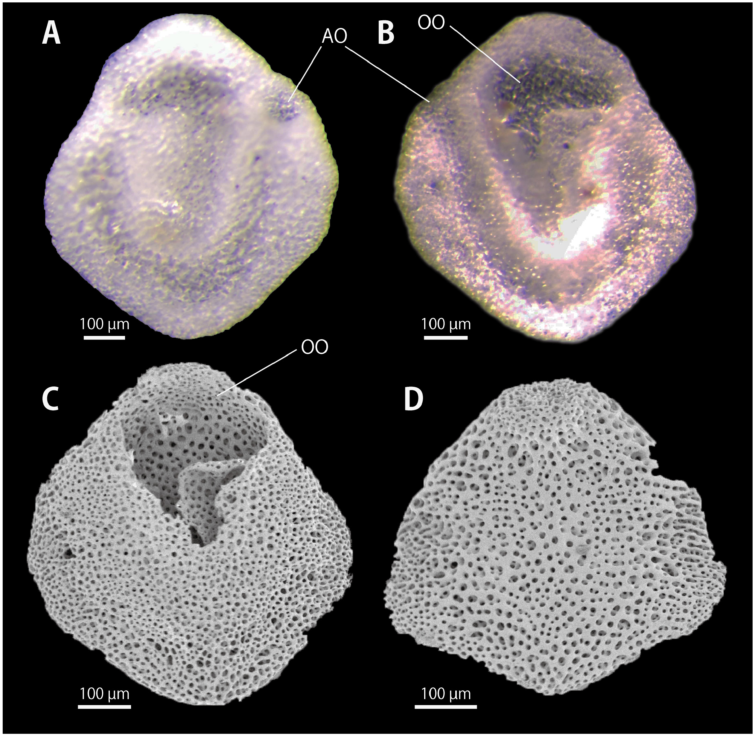
Description of interbrachial frame and oral shields morphology (a paratype, RUMF-ZE-02086). Morphology of interbrachial frame showed the typical morphology of “Type B” as defined by Wilkie & Brogger (2018): two oblong peristomial plates adjoining at their abradial edges on aboral side of oral plates ( Fig. 9A View Fig ); oral plates with deep neural grooves on aboral-distal area ( Fig. 9B, C View Fig ); abradial muscle attachment area of oral plate large and located on a human ear-shaped fringe that projects beyond the lateral profile of the whole plate, the attachment area with radiating interruptions of microstructually differentiated stereom ( Fig. 9B View Fig ); adradial muscle attachment area spoon-shaped and aboral portion expanding like the ‘bowl’ and oral portion narrowing like ‘handle’, and two depressions for tentacles presenting distal side of the muscle attachment area ( Fig. 9C View Fig ); dental plate entire, not divided into several plates, one or two aboral tooth sockets in the form of wide perforations that are divided into two lateral halves by a complete vertical bar ( Fig. 9D, E View Fig ). One of five oral shields have two opening pores, smaller one on oral side ( Fig. 10A View Fig ) and larger one on aboral side ( Fig. 10B, C View Fig ). These pores connecting a spiral canal inside of the shield, which recognised under stereomicroscopic observations ( Fig. 10 View Fig A–C). No canal and pores recognised in the normal oral shield ( Fig. 10D View Fig ).
Colour in life. Aboral periphery of disc and radial shields generally creamy white, with concentric non-continuous yellowish bands on aboral periphery and yellowish spots scattered on aboral center of disc. Green bands present on every 4 to 6 arm segments. Oral disc also creamy white, yellowish spot on oral interradial disc ( Fig. 2 View Fig ).
Variation. A morphological variation was observed in 12 of 15 examined specimens of Ophiopsila xmasilluminans new species. The innermost oral papillae of holotype and 6 paratypes ( WAM Z99800 View Materials , d.d. = 6.3 mm; ZRC.ECH.1333, d.d. = 5.3 mm; ZRC.ECH.1334, d.d. = 5.5 mm; ZRC. ECH.1335, d.d. = 5.7 mm; WAM Z99801 View Materials , d.d. = 5.3 mm; WAM Z99803, d.d. = 5.6 mm; WAM Z99804, d.d. = 5.3 mm) is situated below the other papillae ( Fig. 3E View Fig ) whereas those of 5 paratypes ( ZRC.ECH.1332, d.d. = 7.0 mm; RUMF-ZE-00153, d.d. = 5.0 mm; RUMF-ZE-02087, d.d. = 5.0 mm; ZRC.ECH.1336, d.d. = 5.1 mm; WAM Z99802, d.d. = 5.0 mm) form a horizontal raw on oral edge of adoral shields ( Fig. 5C View Fig ) .
Etymology. The specific name is an adjective in apposition formed as a compound of the island name “ xmas ” and the Latin participle, illuminans, meaning “lighting”, referring to its sampling locality name ( Christmas Island) and luminescence.
Common Japanese name. Dohkutsu-hikari-kumohitode.
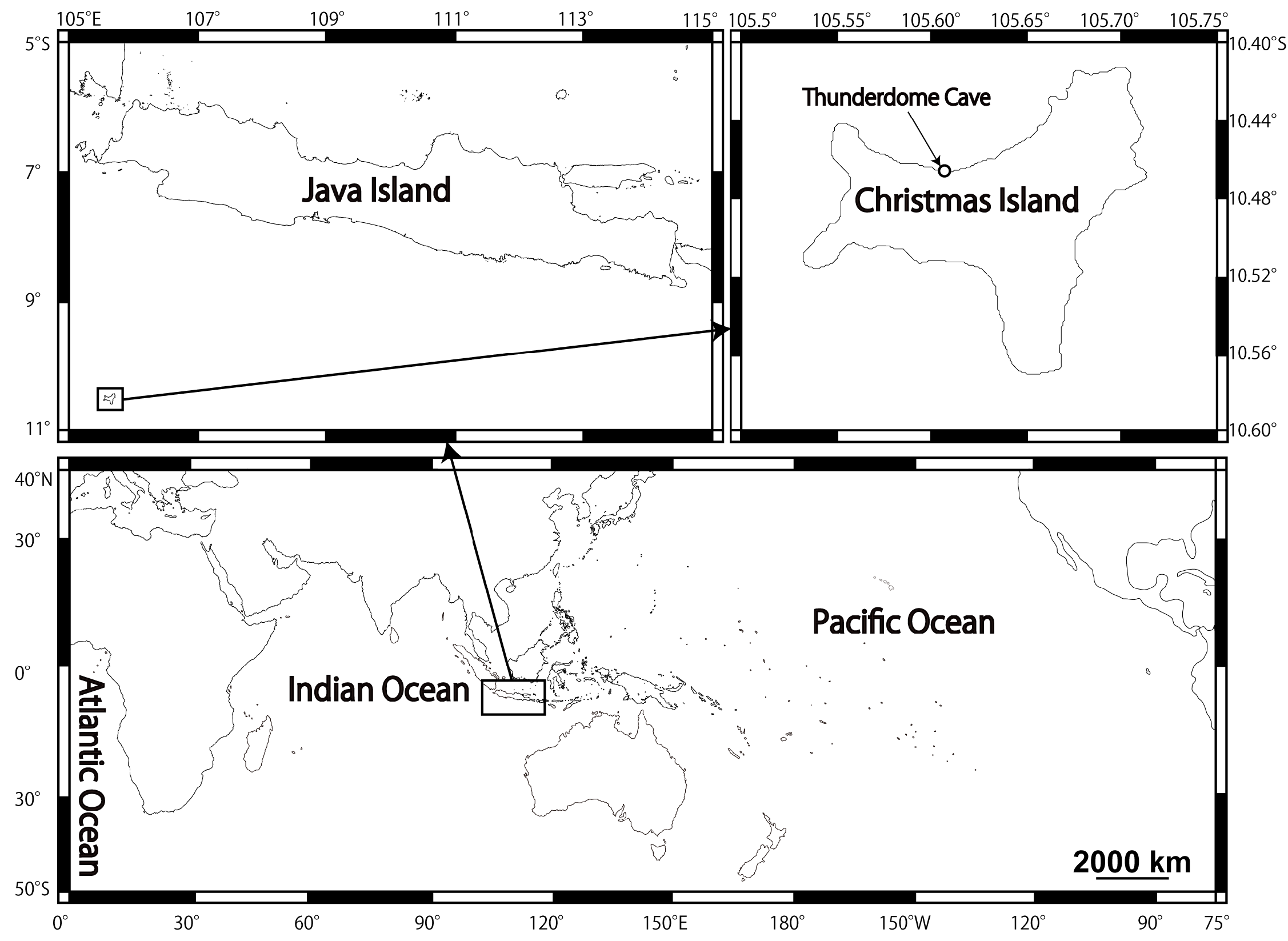
Distribution. Known only from the type locality, a submarine cave called as “Thunderdome Cave”, northern coast of Christmas Island, northwestern Australia, approximately 10 m depth ( type locality, Fig. 1 View Fig ). Tan et al (2014) noted that Ophiopsila pantherina [= this new species] was also found at “Thundercliff Cave”, which is located near Thunderdome Cave. However, specimens of this species were only collected from Thunderdome Cave by the last author (Yoshihisa Fujita).
Habitat. All type specimens of Ophiopsila xmasilluminans new species were collected in sandy bottoms in the submarine cave, with buried disc and arms extended into the water ( Fig. 2B View Fig ).
Remarks. This new species falls within the genus Ophiopsila by virtue of having: extremely long tentacle scales that cross on the mid-line of the oral side of arm; arm spine articulations of lateral arm plates with two smooth, parallel, straight lobes; in the proximal space between them a short ridge; inner side of the lateral arm plates with two merged knobs ( Guille & Jangoux, 1978; O’Hara et al., 2018).
Among the 28 species of Ophiopsila , the present new species is similar to O. pantherina Koehler, 1898 , originally described from King’s Island, Mergui Archipelago, Myanmar and O. timida Koehler, 1930 from Kei Island, Philippines ( Koehler, 1898; Koehler, 1930), in sharing combination of following characteristics: small round scales on disc; contiguous radial shields; 2 or 3 oral papillae on each adoral shield; and narrow and/or long inner tentacle scales on proximal portion of the arm. This new species can be distinguished from these similar species by the covering of the disc, number and shape of oral papillae, shape of oral shield, shape of arm spines, shape of inner tentacle scales and length of arm ( Koehler, 1898, 1930; H. L. Clark, 1918; A. H. Clark, 1921; Murakami, 1963; Baker, 1974; A. M. Clark, 1974; Liao & Clark, 1995). These characters are compared among the three species as follows:
(1) Disc surfaces: Disc of this new species is covered by thick skin with embedded small round scales on aboral side, and only by skins on oral side. Both aboral and oral disc surface of O. pantherina and O. timida are also covered by small round scales ( Koehler, 1898; 1930).
(2) Oral papillae: Three oral papillae on each side of the jaw in this new species; two in O. pantherina and O. timida . Oral papillae are flat, broad scales or inner papillae or middle one sometimes spiniform; all broad scales in O. pantherina and spiniform in O. timida .
(3) Oral shield: Oral shields diamond shaped with rounded edges, as long as wide in the new species and O. pantherina ; elliptical, 2.5 times longer than wide in O. timida .
(4) Arm spines: Shapes of arm spines on proximal portion of arms long and flat in this new species, spiniform in O. pantherina , and cylindrical with thorny tips in O. timida .
(5) Tentacle scales: Inner tentacle scales of this new species and O. timida are narrow, cylindrical and long, and scale long and flat in O. pantherina .
(6) Length of arm: relative length of an arm with maximal length of this new species exceeds 18 times disc diameter; whereas those of O. pantherina and O. timida are 8.5 and 8 times, respectively. The arms of the new species are the longest in Ophiopsila . Other than this new species, the arms are 14 times longer than the disc diameter in O. brevisquama Koehler, 1930 and O. riisei Lütken, 1859 ( Lütken, 1859; Koehler, 1930).
Ecological note. Tan et al. (2014) noted that this new species is bioluminescent, with flashing arms. Our in situ video observations reveal that this species partly flashes its arms where touched (Supplementary file 1). An autotomised and separated arm further flashed and showed active wiggling, suggesting a sacrificial-lure function, which has also been reported for other luminous brittle stars ( Mallefet et al., 2004; Supplementary file 2). We did not measure the spectrum, but the bioluminescence colour observed in the field was green. Although two species of brittle stars are known to show blue emission ( Amphiura filiformis and Amphiura arcystata : e.g., Emson & Herring, 1985; Jones & Mallefet, 2013), the luminescence of other known brittle stars ( Widder et al., 1983), including a congener of the new species, is green ( O. californica ; Brehm & Morin, 1977; Shimomura, 1986). No bioluminescence has previously been observed for cave-dwelling brittle stars, but considering that other coastal congeners also show the same bioluminescence ( O. aranea , O. californica , O. pantherina and O. riseii ; see Grober, 1988; Vanderlinden & Mallefet, 2004; Woolsey et al., 2013), this flashing behaviour is not restricted to cave dwellers. The bioluminescence of ophiuroids has also been considered to be an aposematic signal ( Grober, 1988), an effective defense against predation, most likely for determent ( Deheyn et al., 2000; Jones & Mallefet, 2013; Woolsey et al., 2013), or acting as a “burglar alarm”, for enemies of the predator at night ( Mallefet, 2009). It is difficult to say which hypothesis is likely given that we did not carry out quantitative experiments in this study. The flashing of O. xmasilluminans new species might have a similar function to a “burglar alarm” because its habitat is the interior part of the cave, where it is almost dark, without any daylight. On the other hand, the “lure” hypothesis cannot be rejected because light is also emitted from the autotomised arm (Supplementary file 1, 2). Indeed, potential predators having developed eyes, such as decapods and fishes, live sympatrically in this cave ( Tan et al., 2014). To our knowledge, no bioluminescent species have previously been discovered in submarine anchialine caves ( Bell, 1887; Gibson-Hill, 1947; Marsh, 2000), although it is difficult to say whether O. xmasilluminans new species is a cave-endemic, since adequate inventory surveys around the cave have not yet been done. Our present study suggests that further extensive surveys in these submarine caves and their surrounding environment might reveal the presence of new locally-endemic bioluminescent species which have adapted to distinctive dark or anchialine environments.
In Ophiopsila , sand burying behaviour is known for O. pantherina ( Woolsey et al., 2013) , but it is different from O. xmasilluminans new species: O. pantherina extend almost their entire arms ( Woolsey et al., 2013) into the water, but O. xmasilluminans new species buries the proximal portions of its arms in the sand and extends only the distal portions into the water ( Fig. 2B View Fig ). As suggested in previous studies of ophiuroids ( Bribiesca-Contreras et al., 2013), extraordinarily long arms of species could be an adaptation to cave life. Long arms also possess more tentacles than other species and it may help ophiuroids to find food resources in cave environments, with less predators. More detailed morphological examination of the arms is required to understand the peculiar behaviour of this new species.


No known copyright restrictions apply. See Agosti, D., Egloff, W., 2009. Taxonomic information exchange and copyright: the Plazi approach. BMC Research Notes 2009, 2:53 for further explanation.
|
Kingdom |
|
|
Phylum |
|
|
Class |
|
|
Order |
|
|
Family |
|
|
Genus |
Ophiopsila xmasilluminans
| Okanishi, Masanori, Oba, Yuichi & Fujita, Yoshihisa 2019 |
Ophiopsila pantherina
| Tan HH & Naruse T & Fujita Y & Kiat TS 2014: 413 |