
Tmetonyx cicada (Fabricius, 1780)
|
publication ID |
https://doi.org/10.1080/00222933.2021.1906458 |
|
DOI |
https://doi.org/10.5281/zenodo.5497317 |
|
persistent identifier |
https://treatment.plazi.org/id/5372F948-3013-FFF1-FF10-2B9B964FFD23 |
|
treatment provided by |
Plazi |
|
scientific name |
Tmetonyx cicada |
| status |
|
Mouthparts: Tmetonyx cicada View in CoL
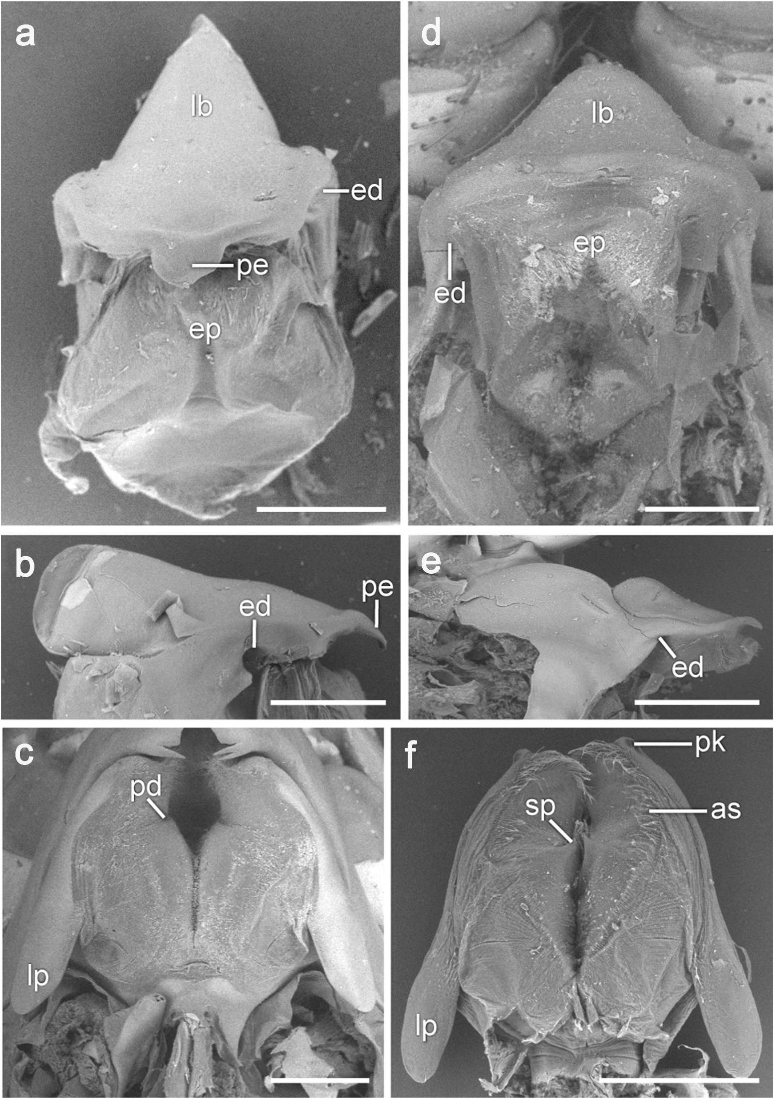
Epistome and upper lip
The epistome is produced, widely conical with a rounded apex and lacks any spines or setae ( Figure 1 View Figure 1 (d)). The epistome and the upper lip are separate. Posterolaterally on the oral surface of the epistome is a pair of small depressions ( ed, Figure 1 View Figure 1 (d)) that receive the articulating knobs of the mandibles.
Mandibles
The hinge line of the mandible is only slightly diagonal (almost transverse) and is located at the posterior end of the mandible ( hl, Figure 2 View Figure 2 (f)). In the resting position, the palps are held anteriorly below the antennae, with article 3 directed laterally ( Figure 5 View Figure 5 (g)). Palpal article 2 is slender and about 4 times as long as article 1 ( Figure 2 View Figure 2 (g)). The mandibles are asymmetrical in the morphology of the incisor, setal row and lacinia mobilis. The large, triangular molar processes ( mo, Figure 2 View Figure 2 (f)) are oriented obliquely to the mandibular body in the posteromedial direction and have narrow, slightly convex triturating surfaces ( ts, Figure 2 View Figure 2 (g)) with ca. 50 transverse ridges carrying densely packed columnar teeth ( Figure 2 View Figure 2 (k)). The triturating surface also bears three crater-like structures ( arrowheads, Figure 2 View Figure 2 (k)) arranged equidistant along the midline of the surface. The setal row ( sr, Figure 2 View Figure 2 (g)) extends from the incisor to the molar process along the medial margin of the mandible and then continues along the medial side of the molar process and the aboral side of the triturating surface ( Figure 2 View Figure 2 (k)). The three apical setae ( ar, Figure 2 View Figure 2 (h)) on each mandible are very large and spine-like, with curved, hooklike tips. In mandibular adduction, these setae on one mandible fit into the gaps between those on the other mandible. The remaining setae of the setal row are thin and hairlike, they have about the same size throughout the length of the row and are especially densely spaced along the distal side of the molar, where they form a thick brushlike coat. In the resting position, the triturating surfaces of the molar processes and the spine rows of the opposite mandibles are tightly pressed against each other ( Figure 2 View Figure 2 (f)). A low ridge ( mmr, Figure 2 View Figure 2 (g)) carrying a row of simple setae extends along the middle of the oral side of the molar process from the edge of the triturating surface to the base of the molar. The whole oral surface of the molar process is pubescent, with denser pubescence on the proximal side of the molar ridge. The left incisor ( Figure 2 View Figure 2 (f,j)) has a wide and straight cutting edge and bears a single small tooth at its ventral end. The cutting edge of the right incisor ( in, Figure 2 View Figure 2 (g,h)) is shorter, curved, and in some specimens is armed ventrally with two small teeth ( Figure 2 View Figure 2 (h)). The cutting edges of the incisors are oriented in a diagonal plane; in the adducted position, the left incisor overlaps the right incisor anteriorly. Midway between the incisor and the palp is a small, rounded articulating knob that fits into the corresponding depression on the oral side of the upper lip (see above). The lacinia mobilis is present only on the left mandible and has been reduced to a small, curved peg ( arrowhead, Figure 2 View Figure 2 (i)).
Paragnaths
A pair of paragnaths ( Figure 1 View Figure 1 (f)) bound the mouth posteriorly; in the resting position, the mandibular bodies are closely pressed against their lateral sides and the paragnaths cover completely their molar processes. The left paragnath is slightly larger than the right and the distal tip of the right paragnath lies below the tip of the left paragnath. The paragnaths are slightly longer than wide and are fused only at their bases. Proximolateral surfaces on the oral side of each paragnath are finely pubescent. These fields of pubescence are bordered medially by an arch-shaped longitudinal row of longer simple setae ( as, Figure 1 View Figure 1 (f)). Lateral to this setal row at the distal end of each paragnaths is a small knoll ( pk, Figure 1 View Figure 1 (f)) that fits into the depression on the mandible posterior to the incisor. A small, conical spine ( sp, Figure 1 View Figure 1 (f)) with a pointed tip projects from the medial face of each paragnath, about one-third of their length from the distal tips. The posterior face of each paragnath forms a posteriorly directed lateral lappet with a rounded tip ( lp, Figure 1 View Figure 1 (f)).
Maxillulae
The maxillulae lie in a horizontal plane ( ml, Figure 5 View Figure 5 (g)) and are closely pressed against the paragnaths and mandibles. A short, triangular coxa is attached to the cephalothorax with a wide articulating membrane ( mmp, Figure 5 View Figure 5 (g)). The inner plate ( ml ip, Figure 3 View Figure 3 (e,f)) is small, cylindrical, about twice as short as the outer plate. Its blunt apical tip bears two large flexible pappose setae with curved tips ( pi, Figure 3 View Figure 3 (e,f)). The basipod extends widely from the coxa and has an additional medial joint with the inner plate. The outer plate ( ml op, Figure 3 View Figure 3 (e,f)) is wide and leaf-shaped. In the distal portion of the plate, there are two rows of large slender spine teeth with a 7/4 crown arrangement diagnostic for the Uristidae ( Figure 3 View Figure 3 (e,h)): 7 teeth (st1–7) in an apical row (st1 1–2-cuspidate, st2 1–3-cuspidate, st3 2-cuspidate, st4 3-cuspidate, st5 3–4-cuspidate, st6 5-cuspidate, st7 4–6-cuspidate) and 4 teeth (stA-D) in a subapical row (stA 2-cuspidate, stB 3–4-cuspidate, stC and stD 3-cuspidate). The distal portion of the outer plate also has two longitudinal bands of simple setae ( bo, Figure 3 View Figure 3 (e,f,h)) located along the medial margin and on the oral surface of the plate. The palp ( ml pl, Figure 3 View Figure 3 (e–g)) is 2-articulate and lacks setation; article 1 is triangular, about as long as wide; article 2 is long and curved towards the midline. Wide distal ends of both palps are armed with eight short spine-like setae ( smp, Figure 3 View Figure 3 (g)) and with two flexible sensory setae ( arrows, Figure 3 View Figure 3 (g)) located between spines 6 and 7 and dorsally to the last eighth spine (counting spines ventral to dorsal). In the resting position, the opposite palps meet at the midline behind the outer plates of the maxillipeds, with the left palm slightly overlapping the right one anteriorly ( Figure 3 View Figure 3 (e,g)).
Maxillae
The left and right maxillae are roughly equal in size and shape ( Figure 4 View Figure 4 (e)). Small, triangular coxae ( mx cx, Figure 4 View Figure 4 (e)) arise from a common extension of the sternum. The surface of the coxa lacks setation. The basipod and both inner and outer plates extend from the coxa anteriorly in a horizontal plane ( mx, Figure 5 View Figure 5 (g)) partly concealing the larger maxillulae from below. The anteromedially oriented basipod is basally wide ( Figure 4 View Figure 4 (e)), then tapers apically and curves to form an anteriorly oriented inner plate ( mx ip, Figure 4 View Figure 4 (e,f)). The inner plate carries three closely spaced parallel rows of flexible setae along the medial margin ( Figure 4 View Figure 4 (f,h)). The setae in the outer ( ri3, Figure 4 View Figure 4 (h)) and inner ( ri1, Figure 4 View Figure 4 (h)) rows are about twice as wide and long as those in the middle row ( ri2, Figure 4 View Figure 4 (h)). The setae in the outer row are unilaterally serrated in the distal half ( Figure 4 View Figure 4 (h)); the remaining setae are simple. On the aboral surface of the inner plate near the setal row is a small field of sparsely spaced simple setae ( fi, Figure 4 View Figure 4 (f)). The outer plate ( mx op, Figure 4 View Figure 4 (e,f)) is lancet-shaped and carries distally small simple setae on the oral surface and two rows of flexible setae along the medial margin ( Figure 4 View Figure 4 (g)): the setae in the outer row ( ro2, Figure 4 View Figure 4 (g)) are wide and unilaterally serrated in the distal half, those in the inner row ( ro1, Figure 4 View Figure 4 (g)) are thin and simple.
Maxillipeds
Maxillipeds consist of a fused coxa ( mp cx, Figure 5 View Figure 5 (g,h)), basipods ( mp bs, Figure 5 View Figure 5 (g,h)) with endites (inner plates), ischia ( mp is, Figure 5 View Figure 5 (g–i)) with endites (outer plates) and palps. The coxa is oriented in the vertical plane ( mp cx, Figure 5 View Figure 5 (g)), but near its distal tip it curves forward so that the remaining maxilliped articles lie in the horizontal plane covering completely the other mouthparts. The basipods touch across the midline but do not fuse ( Figure 5 View Figure 5 (h)). An array of long, simple setae extends along the medial and distal margins on the aboral surface of basipods and there are tufts of simple setae at the distolateral corners of the basipods and ischia ( Figure 5 View Figure 5 (h)). The inner plates ( mp ip, Figure 5 View Figure 5 (i,j)) gradually widen from the base to the apex; their anterior margins are obliquely bevelled. In the resting position, the opposite plates meet each other in the middle ( Figure 5 View Figure 5 (i,j)). The medial portion of the anterior margin of each inner plate is extended into a triangular process that bears three nodular spines ( ns, Figure 5 View Figure 5 (k)). The anterior and medial margins on the oral surface of the plates carry a row of large setae; the medial of these setae are pappose ( psi, Figure 5 View Figure 5 (j)) and those at the anterior margin lack setules ( ssi, Figure 5 View Figure 5 (j)). The rest of the oral surface of the plates is evenly covered with thin simple setae. The outer plates ( mp op, Figure 5 View Figure 5 (g–i)) are about twice as large as the inner plates and have straight medial and arcuate lateral margins. The medial and distal margins of the outer plates are armed with small subquadrate teeth and covered with a row of simple setae on their aboral surfaces ( Figure 5 View Figure 5 (l)); the remaining surface of the plates lacks setation. Numerous small irregularly spaced pores ( Figure 5 View Figure 5 (l)) are located on the oral surfaces of the outer plates along their medial margins. The palps are 4-articulate ( Figure 5 View Figure 5 (g–i)); their articles 1 and 2 are flattened and wide (but less so than in A. nugax ) and cover the maxillae laterally. The medial margins of all palpal articles bear a row of long simple setae. In the resting position, the palps do not cross or meet at the midline, but the right palp lies slightly anterior to the left palp ( Figure 5 View Figure 5 (h)).


No known copyright restrictions apply. See Agosti, D., Egloff, W., 2009. Taxonomic information exchange and copyright: the Plazi approach. BMC Research Notes 2009, 2:53 for further explanation.
|
Kingdom |
|
|
Phylum |
|
|
Class |
|
|
Order |
|
|
SuperFamily |
Lysianassoidea |
|
Family |
|
|
Genus |