
Anonyx nugax (Phipps, 1774)
|
publication ID |
https://doi.org/10.1080/00222933.2021.1906458 |
|
DOI |
https://doi.org/10.5281/zenodo.5497315 |
|
persistent identifier |
https://treatment.plazi.org/id/5372F948-3019-FFF4-FF10-2E879528FB19 |
|
treatment provided by |
Plazi |
|
scientific name |
Anonyx nugax |
| status |
|
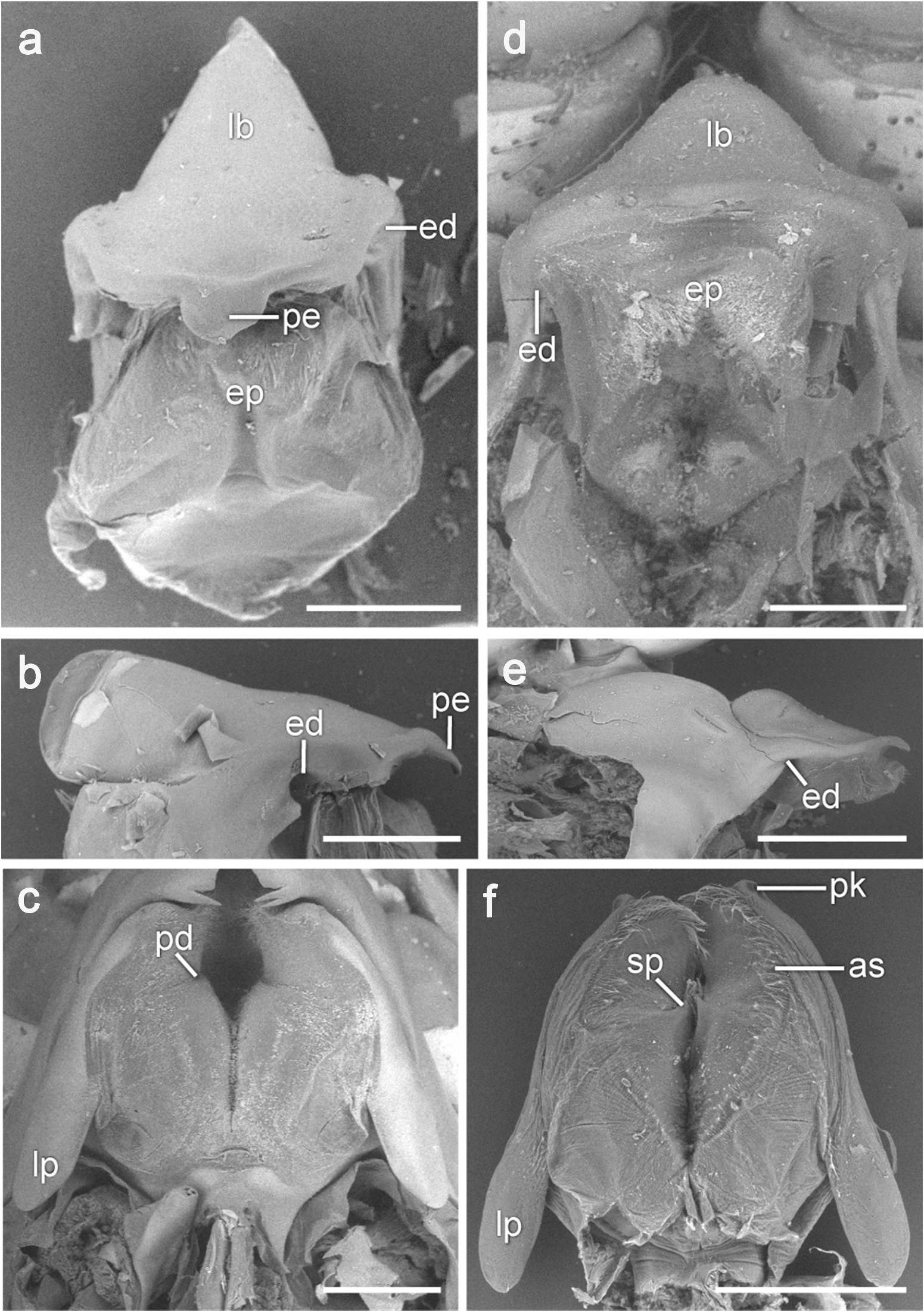
Mouthparts: Anonyx nugax View in CoL
Epistome and upper lip
The epistome ( Figure 1 View Figure 1 (a,b)) is produced, conical, subacute and lacks any spines or setae on the aboral surface, but is evenly covered with simple setae on the oral surface. The ventral margin of the epistome bears a rounded medial projection ( pe, Figure 1 View Figure 1 (a,b)) that in the resting position fits between the mandibular incisors. The epistome and the upper lip are separate. Posterolaterally on the oral surface of the epistome is a pair of deep depressions ( ed, Figure 1 View Figure 1 (a,b)) that receive the articulating knobs of the mandibles.
Mandibles
The hinge line of the mandible is slightly oblique and is located at the posterior end of the mandible ( hl, Figure 2 View Figure 2 (a)). In the resting position, the palps are held upward so that they lie between the antennae ( md pl, Figure 5 View Figure 5 (a)), with article 3 directed anteriorly. The mandibles are asymmetrical in the morphology of the incisor, setal row and lacinia mobilis. The molar process ( mo, Figure 2 View Figure 2 (a–d)) is large, roughly rectangular and is oriented obliquely to the mandibular body in the posteromedial direction. In the resting position, the tips of the opposite molar processes do not meet at the midline lying a short distance apart ( Figure 2 View Figure 2 (a)). The tip of each molar process is slightly rounded; it is densely pubescent and lacks a triturating surface ( Figure 2 View Figure 2 (d)). The setal row is located on the oral side of the mandible near its medial margin and is oriented perpendicular to the blade of the incisor. It starts about 100–150 µm from the incisor ( Figure 2 View Figure 2 (b,c)) and reaches the basal portion of the molar process ( sr, Figure 2 View Figure 2 (d)). The three apical setae (near the incisor) on each mandible are very large and spine-like ( ar, Figure 2 View Figure 2 (a–c)); the remaining setae are much thinner and hairlike. Since the setal row is located some distance from the medial margin of the mandible, in the resting position the setae of the opposite mandibles are kept widely apart ( Figure 2 View Figure 2 (a)), but it is clear that in mandibular adduction the three apical setae of one mandible interdigitate with the corresponding setae on the other mandible. The blades of the incisors are oriented in a nearly vertical plane; in the adducted position the left incisor overlaps the right incisor anteriorly ( Figure 2 View Figure 2 (a)). Both incisors have a wide and straight cutting edge ( Figure 2 View Figure 2 (a–c,e)), but the edge of the left incisor is slightly longer ( Figure 2 View Figure 2 (a)). The medial ends of both incisors bear long conical teeth ( Figure 2 View Figure 2 (a)), with two teeth located on the left ( ti, Figure 2 View Figure 2 (b)) and one on the right mandible ( ti, Figure 2 View Figure 2 (c)). The teeth are arranged such that in mandibular adduction the right tooth fits into the gap between the left teeth. Midway between the incisor and the palp is a large, conical articulating knob ( mk, Figure 2 View Figure 2 (b)) that fits into the corresponding depression on the oral side of the upper lip (see above). The lacinia mobilis ( arrow, Figure 2 View Figure 2 (b); lm, 2(e)) is present only on the left mandible and has been reduced to a small, curved peg ( Figure 2 View Figure 2 (e)). The palp is 3-articulate and located opposite the molar; its article 2 is slender and about 3 times as long as article 1.
Paragnaths
A pair of paragnaths ( Figure 1 View Figure 1 (c)) bound the mouth posteriorly; in the resting position the mandibular bodies are closely pressed against their lateral sides and the paragnaths cover completely their molar processes. The paragnaths are symmetrical, only slightly longer than wide and are fused only at their bases. Both oral and aboral surfaces of the paragnaths are densely pubescent, except their lateral margins, which lack setae. On the oral surface of each paragnath there is a depression ( pd, Figure 1 View Figure 1 (c)) into which the molar processes of the mandibles fit. The aboral face of each paragnath forms a posteriorly directed lateral lappet with a pointed tip ( lp, Figure 1 View Figure 1 (c)).
Maxillulae
The maxillulae lie in a horizontal plane ( ml, Figure 5 View Figure 5 (a)) and are closely pressed against the paragnaths and mandibles. A short triangular coxa is attached to the cephalothorax with a wide articulating membrane ( mmp, Figure 5 View Figure 5 (a)). The inner plate ( ml ip, Figure 3 View Figure 3 (a,b)) is about twice as short as the outer plate; it has an elongated narrow basis and an expanded club-shaped distal portion. The inner plate carries apically two large pappose setae with long needle-like setules ( pi, Figure 3 View Figure 3 (a,b,d)). The basipod extends narrowly from the coxa and is articulated medially with the inner plate by a cuticular membrane and a triangular knob. The outer plate ( ml op, Figure 3 View Figure 3 (a,b,d)) is wide, almost cylindrical in shape. In the distal portion of the plate, there are two rows of large slender spine teeth with a 7/4 crown arrangement diagnostic for the Uristidae ( Figure 3 View Figure 3 (a–c)): 7 teeth (st1–7) in an apical row (st1–4 one-cuspidate, st5–6 two-cuspidate, st7 three-cuspidate) and 4 teeth (stA-D) in a subapical row (stA one-cuspidate, stB three-cuspidate, stC one-cuspidate, stD threecuspidate). The outer plate also bears a U-shaped band of simple setae ( bo, Figure 3 View Figure 3 (b–d)); the two arms of this band extend along the medial margin and oral surface of the plate and are linked just proximal to st7. The palp ( ml pl, Figures 3 View Figure 3 (a–c), 4(a) and 5(a)) is 2-articulate; article 1 is triangular, about as long as wide, article 2 is long and curved towards the midline. The surface of the palp lacks setae, except a small setal field on the oral side. Wide distal ends of both palps are armed with 8 short spine-like setae ( smp, Figure 3 View Figure 3 (c)). In the resting position, the opposite palps meet at the midline behind the outer plates of the maxillipeds, with the left palp overlapping the right one anteriorly ( Figure 3 View Figure 3 (a)).
Maxillae
The left and right maxillae are roughly equal in size and shape ( Figure 4 View Figure 4 (a)). Small, triangular coxae ( mx cx, Figure 4 View Figure 4 (a)) arise from a common extension of the sternum. The surface of the coxa lacks setation. The basipod and both inner and outer plates extend from the coxa anteriorly in a horizontal plane ( mx, Figure 5 View Figure 5 (a)) partly concealing the large maxillulae from below ( Figure 4 View Figure 4 (a)). The anteromedially oriented basipod is basally wide ( Figure 4 View Figure 4 (a)), then tapers apically and curves to form an anteriorly oriented lancet-shaped inner plate ( mx ip, Figure 4 View Figure 4 (a,b)). The inner plate carries three closely parallel rows of simple setae along the medial margin ( Figure 4 View Figure 4 (b,d)): the setae in the inner ( ri1, Figure 4 View Figure 4 (d)) and outer ( ri3, Figure 4 View Figure 4 (d)) rows are spine-like and much wider and longer than those in the middle row ( ri2, Figure 4 View Figure 4 (d)). The setae progressively increase in size towards the apex, but the most proximal seta in the inner row ( arrow, Figure 4 View Figure 4 (b,d)) is markedly larger than the others. A U-shaped field of thin simple setae encloses the three setal rows on either side and proximally ( bi, Figure 4 View Figure 4 (b,d)). The outer plate ( mx op, Figure 4 View Figure 4 (a,b)) is lancet-shaped and carries distally a small field of simple setae on the oral surface and two rows of simple setae along the medial margin ( Figure 4 View Figure 4 (c)): the setae in the outer row ( ro2, Figure 4 View Figure 4 (c)) are much wider and almost twice as long as those in the inner row ( ro1, Figure 4 View Figure 4 (c)).
Maxillipeds
Maxillipeds consist of a fused coxa ( mp cx, Figure 5 View Figure 5 (a–c)), basipods ( mp bs, Figure 5 View Figure 5 (a–c)) with endites (inner plates), ischia ( mp is, Figure 5 View Figure 5 (a–c)) with endites (outer plates) and palps. The coxa is oriented in the vertical plane ( mp cx, Figure 5 View Figure 5 (a)), but near its distal tip it curves forward so that the remaining maxilliped articles lie in the horizontal plane covering completely the other mouthparts. The basipods touch across the midline but do not fuse ( Figure 5 View Figure 5 (b)). A few long simple setae are located along the distal and medial margins on the aboral surface of basipods and the distolateral corners of the basipods and ischia; a row of simple setae extends across the aboral surface of the ischia and continues along the medial margin of all palpal articles ( Figure 5 View Figure 5 (b,c)). The inner plates ( Figure 5 View Figure 5 (e)) are very small, subrectangular; in the resting position, the opposite plates do not touch across the midline. Each plate bears apically three large pappose setae ( ppi, Figure 5 View Figure 5 (e)) that project from a deep longitudinal notch on the oral surface of the plate. Lateral to these setae on the apical tip of the plate is a pair of larger and another pair of smaller cuspidate setae ( cpi, Figure 5 View Figure 5 (e,f)). The outer plates ( mp op, Figure 5 View Figure 5 (a–d)) are large, lancet-shaped, about twice as long as the inner plates, finely serrated along the medial and anterior margins ( Figure 5 View Figure 5 (d)) and lack setation except for a row of small reduced setae along the medial margin ( Figure 5 View Figure 5 (d)). The palps are 4-articulate ( Figure 5 View Figure 5 (a–c)); their articles 1 and 2 are wide, flattened and cover the maxillae laterally. In the resting position, the palps cross each other at the midline ( Figure 5 View Figure 5 (b)).


No known copyright restrictions apply. See Agosti, D., Egloff, W., 2009. Taxonomic information exchange and copyright: the Plazi approach. BMC Research Notes 2009, 2:53 for further explanation.
|
Kingdom |
|
|
Phylum |
|
|
Class |
|
|
Order |
|
|
SuperFamily |
Lysianassoidea |
|
Family |
|
|
Genus |