
Dunkleosteus amblyodoratus, Carr & Hlavin, 2010
|
publication ID |
https://doi.org/10.1111/j.1096-3642.2009.00578.x |
|
DOI |
https://doi.org/10.5281/zenodo.10545517 |
|
persistent identifier |
https://treatment.plazi.org/id/5415C76B-FF9C-FFA8-9BE8-D55AFD2351D8 |
|
treatment provided by |
Valdenar |
|
scientific name |
Dunkleosteus amblyodoratus |
| status |
sp. nov. |
DUNKLEOSTEUS AMBLYODORATUS SP. NOV.
( FIGS 6 View Figure 6 , 7 View Figure 7 )
Diagnosis: A Dunkleosteus species possessing a tapered prehypophysial region of the parasphenoid, with anterolateral contact faces for posterior processes of the anterior superognathals.
Holotype: UM 101105 ( Figs 6 View Figure 6 , 7 View Figure 7 ), an isolated nuchal plate, with the transverse thickened portion of the left paranuchal plate and an incomplete parasphenoid plate.
Etymology: From amblys meaning blunt and doratos meaning spear (Greek); referring to the form of the prehypophysial region of the parasphenoid plate.
Occurrence and stratigraphy: Collected from Ontario, Canada ( Fig. 2A View Figure 2 ). The exact locality data are not available, but the specimen was recovered from the Kettle Point Formation (Upper Devonian, but the exact stratigraphic level was not recorded; Fig. 2B View Figure 2 ). The specimen was found within a carbonate concretion that is typical for the Kettle Point Formation.
SKULL ROOF AND PARASPHENOID
Dunkleosteus amblyodoratus sp. nov. is known only from fragmented parasphenoid, nuchal, and paranuchal plates. The external surface of the skull roof is worn. Tubercles are visible only in one area of the paranuchal plate where an outer layer is missing, revealing tubercles on a deeper surface (representing an earlier generation of tubercles). The punctate tubercles are evenly spaced.
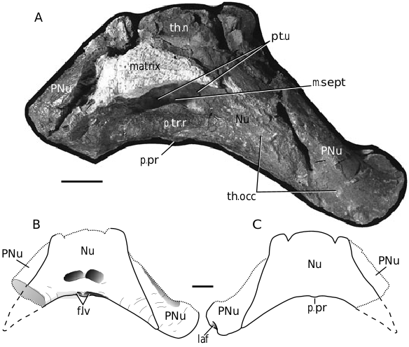
The nuchal plate (Nu; Fig. 6 View Figure 6 ) outline is trapezoidal with a transverse anterior margin, with reduced indentations forming a shallow W-shape (suggesting an interdigitation with the central plates). The exact nature of this shape is unclear because of some weathering. The internal surface possesses a transverse occipital thickening (th.occ; Fig. 6A View Figure 6 ) that is continuous with that of the paranuchal plate. Double pits (pt.u; Fig. 6A View Figure 6 ) are present and bounded anteriorly by the nuchal thickening (th.n; Fig. 6A View Figure 6 ), and posteriorly by a well-developed transverse ridge (p.tr.r; Fig. 6A View Figure 6 ). Individual pits are separated by a median septum (m.sept; Fig. 6A View Figure 6 ) that does not cross the posterior transverse ridge. A posterior process (p.pr; Fig. 6A, C View Figure 6 ) is present, and is bounded laterally by shallow paired fossae (f.lv; Fig. 6B View Figure 6 ).
Only the transverse occipital thickening of the paranuchal plate is preserved (PNu; Fig. 6 View Figure 6 ). The thickening is massive and continues to the lateral articular fossa (laf; Fig. 6C View Figure 6 ). Only a portion of the left fossa remains, but it suggests the presence of a large and well-developed ginglymoid articulation between the head and thoracic armour.
The parasphenoid (Psp; Fig. 7 View Figure 7 ) is incomplete, with only its anterior half preserved. The prehypophysial region (pre.reg; Fig. 7 View Figure 7 ) is tapered, and is as long as it is wide. The lateral edges of the prehypophysial region are thickened, providing possible contact facets for the anterior superognathal plates (cf.ASG; Fig. 7 View Figure 7 ).


| UM |
University of Marburg |
No known copyright restrictions apply. See Agosti, D., Egloff, W., 2009. Taxonomic information exchange and copyright: the Plazi approach. BMC Research Notes 2009, 2:53 for further explanation.