
Amerodectes sicalis, Mironov & González-Acuña, 2011
|
publication ID |
https://doi.org/10.11646/zootaxa.3057.1.1 |
|
DOI |
https://doi.org/10.5281/zenodo.4623074 |
|
persistent identifier |
https://treatment.plazi.org/id/546E87CE-030E-FF85-FF11-E436FA7790E0 |
|
treatment provided by |
Plazi |
|
scientific name |
Amerodectes sicalis |
| status |
sp. nov. |
Amerodectes sicalis sp. n.
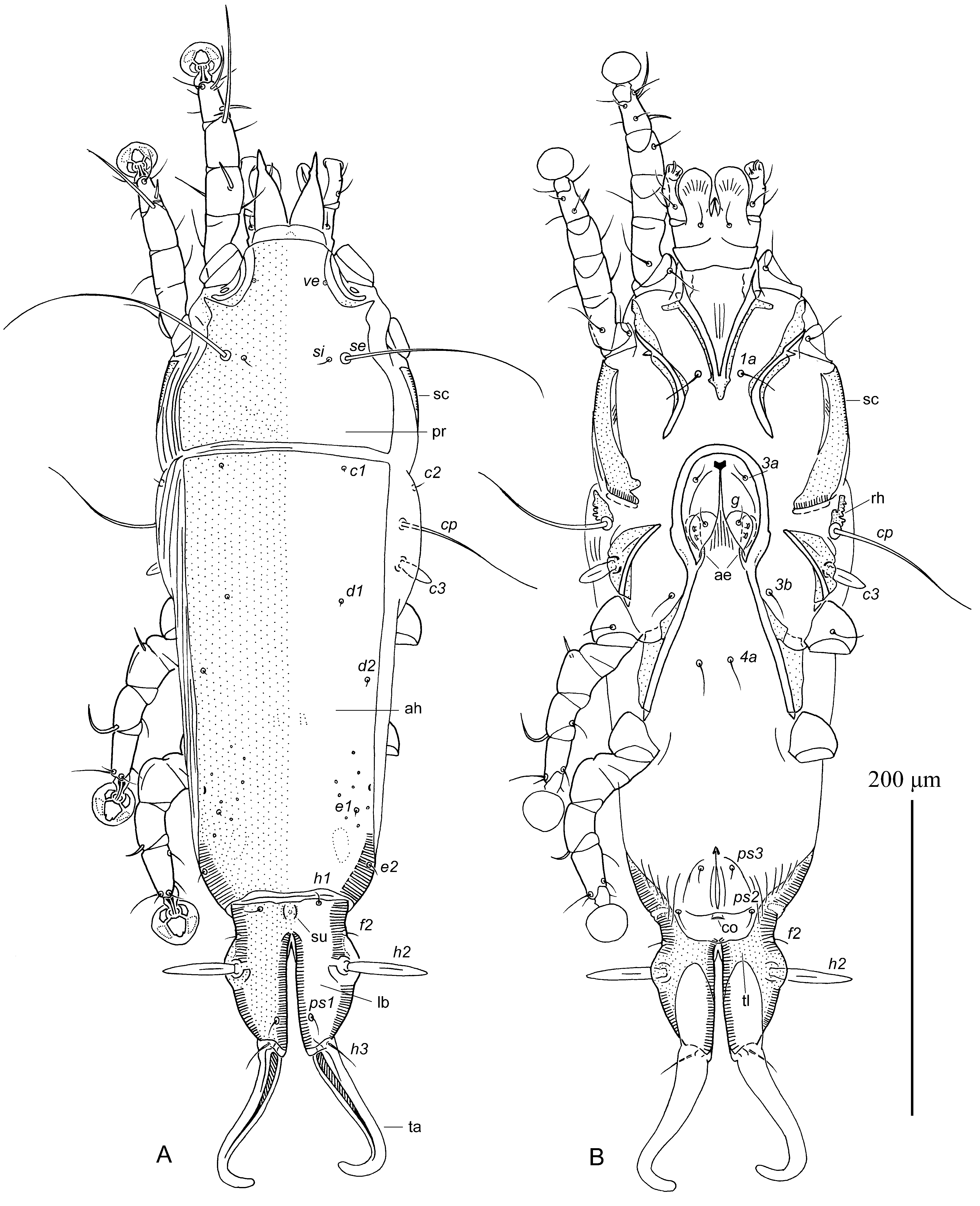
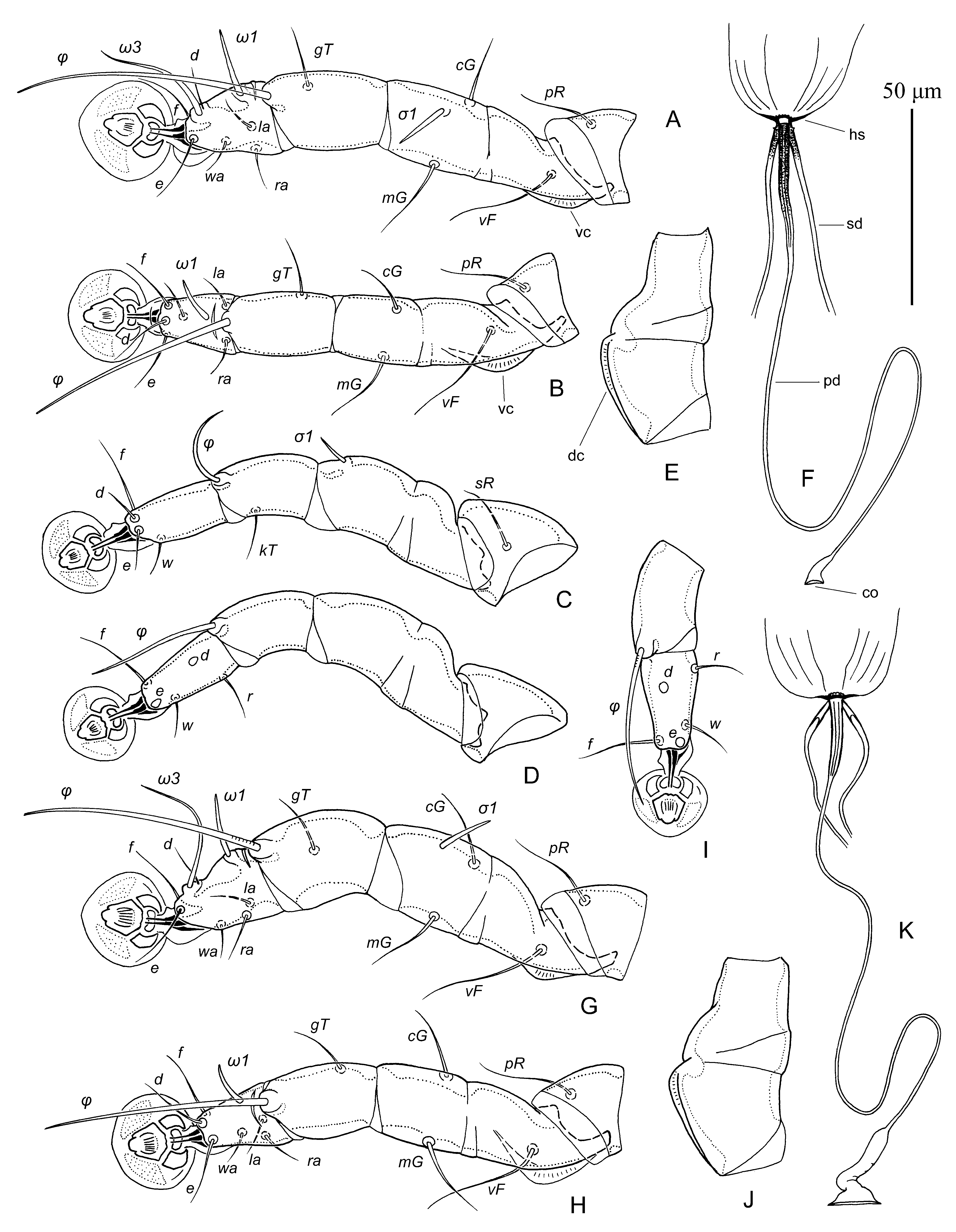
( Figs. 3 View FIGURE 3 G–K, 4, 5)
Type material. Male holotype ( ZISP 4595 View Materials ), 1 male and 2 female paratypes from the Grassland Yellow Finch Sicalis luteola (Sparman) ( Emberizidae ), CHILE: Bío Bío Region, Concepción Province, Concepción , 36°48'53"S 73°1'45"W, 2 November 2007, coll. D.A. González-Acuña. GoogleMaps
Type depository. Holotype and all paratypes—ZISP.
Description. MALE ( holotype, measurements for 1 paratype in parentheses). Idiosoma, length × width, 365 (370) × 145 (160), length of hysterosoma 235 (245). Prodorsal shield: 118 (115) × 125 (126), lateral margins entire, posterior margin almost straight, antero-lateral extensions elongate and acute, median area with small pore-like lacunae; scapular setae se separated by 68 (64) ( Fig. 4 View FIGURE 4 A). Setae ve present. Humeral shields rudimentary, situated ventrally. Setae cp and c2 situated on soft tegument. Subhumeral setae c3 lanceolate, 22 (18) × 7 (6). Hysteronotal shield: greatest length 240 (250), width in anterior part 122 (118), anterior margin straight, posterior part with small pore-like lacunae. Distance between prodorsal and hysteronotal shields 10 (7). Opisthosomal lobes approximately as long as wide at base; posterior ends of lobes roughly rounded, with blunt extensions at bases of setae h2 and h3. Terminal cleft shaped as an inverted U with divergent branches, 24 (29) long. Supranal concavity present, semicircular. Setae f2 situated anterior to bases of setae ps2. Setae h1 situated at level of anterior end of terminal cleft. Setae h3 whip-like, 75 (65) long; setae ps2 75 (68) long; setae ps1 minute, filiform, about 10 long, situated on margin of terminal cleft approximately at level of setae ps2. Distance between bases of dorsal setae: c2:d2 97 (100), d2:e2 84 (86), e2:h3 59 (51), d1:d2 38 (36), e1:e2 20 (28), h1:ps2 11 (15), h2:h2 48 (55), h3:h3 37 (39), ps2:ps2 64 (70).
Epimerites I fused into a narrow V, fused part with tridentate posterior end ( Fig. 4 View FIGURE 4 B). Coxal fields I, II without extensive sclerotized areas. Rudimentary sclerites rEpIIa absent. Coxal fields II, III open. Coxal fields IV without sclerotized areas. Epimerites IVa absent. Genital arch of moderate size, 27 (25) × 40 (38); basal sclerite of genital apparatus with semicircular posterior margin; aedeagus sword-shaped, 106 (104) long, extending to anterior end of terminal cleft; genital papillae not connected by bases. Genital and adanal shields absent. Anal suckers 11 (12) in diameter, corolla smooth. Opisthoventral shields occupying posterior half of lobes and narrow area at level of anal suckers, inner margins without extension, setae ps3 approximately at midlevel of anal suckers. Distance between ventral setae: 3b:3a 9 (10), 3a:4a 37 (38), 4a:g 42 (46), g:ps3 57 (56), ps3:ps3 71 (73), ps3:h3 31 (33).
Femora I, II with ventral crests, other segments of legs I, II without processes. Solenidion σ 1 of genu I 13 (14) long, situated at midlevel of segment; genual setae cG I, II and mG I, II setiform, noticeably thickened in basal part ( Figs. 3 View FIGURE 3 G, H). Seta d of tarsus II subequal to corresponding setae f, seta d of tarsi III shorter than corresponding seta f. Tarsus IV 27 (29) long, without apical process; seta d situated in basal half of segment; solenidion φ of tibia IV extending to midlevel of ambulacral disc ( Fig. 3 View FIGURE 3 I).
FEMALE ( 2 paratypes). Idiosoma, length × width, 510–535 × 155–175, length of hysterosoma 365–380. Prodorsal shield: general form as in male, minute lacunae in anterior part present or absent, 133–137 × 137–144, setae se separated by 80–82 ( Fig. 5 View FIGURE 5 A). Setae ve present, rudimentary. Humeral shields represented by small rudimentary sclerites situated anterior to bases of setae cp. Setae c2 and cp situated on soft tegument. Setae c3 lanceolate, 18–22 × 7–8. Distance between prodorsal and hysteronotal shields 8–10. Anterior and lobar parts of hysteronotal shield separated dorsally by narrow transverse band of soft tegument, but remain connected ventro-laterally by thin sclerotized bands ( Fig. 5 View FIGURE 5 B). Anterior hysteronotal shields almost rectangular, anterior margin straight, greatest length 280–305, width at anterior margin 135–145, surface without lacunae. Length of lobar region 98–100, greatest width 86–95, lobar shield with incision on posterior margin, therefore lateral parts of this shield connected each other by narrow transverse band. Supranal concavity present, poorly outlined. Terminal cleft as a very narrow V, extending beyond level of setae h2, 65–77 long, width at level of lobar apices 14–16. Setae h1 on anterior margin of lobar shield; setae h1 and f 2 in trapezoid arrangement. Setae h2 spindle-like, 43–46 × 7–8. Setae ps1 near inner margins of opisthosomal lobes. Setae h3 16–22 long, about 1/5 of terminal appendages. Distance between dorsal setae: c2:d2 113–120, d2:e2 130–142, e2:h2 62–73, h2:h3 46–50, d1:d2 40–44, e1:e2 30–40, h1:h2 35–40, h1:h1 32–42, h2:h2 68–77.
Epimerites I fused into a Y with very short sternum; fused part smooth, without lateral extensions. Lateral parts of coxal fields I, II without large sclerotized areas ( Fig. 5 View FIGURE 5 B). Epimerites IVa absent. Translobar apodemes of opisthosomal lobes present, wide, not fused to each other anterior to terminal cleft. Epigynum with small lateral extensions at level of epimerites III, greatest width 58–65; apodemes of oviporus fused with epimerites IIIa. Pseudanal setae filiform, setae ps2 approximately at level of posterior end of anal opening, distance between setae: ps2:ps2 47–50, ps3:ps3 15–20, ps2:ps3 28–31. Primary spermaduct with short enlargements (15-20 long) in proximal part near head of spermatheca and also in distal part forming bursa copulatrix; secondary spermaducts approximately twice as long as proximal enlargement of primary spermduct ( Fig. 3 View FIGURE 3 K).
Femur I, II with ventral crest, other segments of these legs as in male. Solenidion σ 1 of genu I short, 12–13 long, situated at midlevel of segment. Genual setae cG I, II and mG I, II setiform, slightly enlarged basally. Setae d of tarsus II subequal to corresponding setae f; setae d of tarsi III–IV shorter than corresponding setae f. Genu IV dorsally inflated, with wide longitudinal dorsal crest ( Fig. 3 View FIGURE 3 J), genu III without noticeable dorsal crest.
Differential diagnosis. Amerodectes sicalis sp. n. is most similar to A. phrygilus described above by the loss of the humeral shields on the dorsal side of hysterosoma in both sexes and by having long filiform setae h3 and tridentate sternum in males. Amerodectes sicalis differs from A. phrygilus by the following features: in males, the humeral shields are represented by minute rudiments situated anterior to the bases of setae cp, and the aedeagus extends to the anterior end of terminal cleft; in females, the lateral parts of lobar shield are connected each other only by narrow transverse band bearing the supranal concavity. In males of A. phrygilus , any remnants of the humeral shields are completely absent, and the aedeagus extends to the midlevel of anal opening; in females, the lobar shield is entire, without any area of soft tegument posterior to the supranal concavity ( Fig. 2 View FIGURE 2 A).
Etymology. The specific epithet is taken from the generic name of the type host and is a noun in apposition.
No known copyright restrictions apply. See Agosti, D., Egloff, W., 2009. Taxonomic information exchange and copyright: the Plazi approach. BMC Research Notes 2009, 2:53 for further explanation.
|
Kingdom |
|
|
Phylum |
|
|
Class |
|
|
Order |
|
|
Family |
|
|
Genus |