
Pseudechiniscus ehrenbergi, Roszkowska & Grobys & Bartylak & Gawlak & Kmita & Kepel & Kepel & Parnikoza & Kaczmarek, 2020
|
publication ID |
https://doi.org/10.11646/zootaxa.4763.4.1 |
|
publication LSID |
urn:lsid:zoobank.org:pub:0DE45665-F3A9-474B-B438-1022FABB6BD1 |
|
DOI |
https://doi.org/10.5281/zenodo.3804847 |
|
persistent identifier |
https://treatment.plazi.org/id/554F87D9-0772-F746-00C1-FE7D552DCCF1 |
|
treatment provided by |
Carolina |
|
scientific name |
Pseudechiniscus ehrenbergi |
| status |
sp. nov. |
1. Pseudechiniscus ehrenbergi View in CoL sp. nov. Roszkowska, Grobys, Bartylak & Kaczmarek
( Tables 1–2 View TABLE 1 View TABLE 2 , Figs 1–4 View FIGURE 1 View FIGURE 2 View FIGURE 3 View FIGURE 4 )
Pseudechiniscus View in CoL sp. 1 ( Grobys et al. 2020)
Pseudechiniscus aff. suillus View in CoL [Ca6] MONGOLIA C 2595-V06 ( Cesari et al. 2020)
Material examined: 35 animals ( holotype (female) and 34 paratypes ( 31 females, 3 males)) mounted on microscope slides in Hoyer’s medium, 30 animals prepared for SEM and 5 prepared for barcoding.
Description
Animals (measurements and statistics in Tables 1 View TABLE 1 and 2 View TABLE 2 )
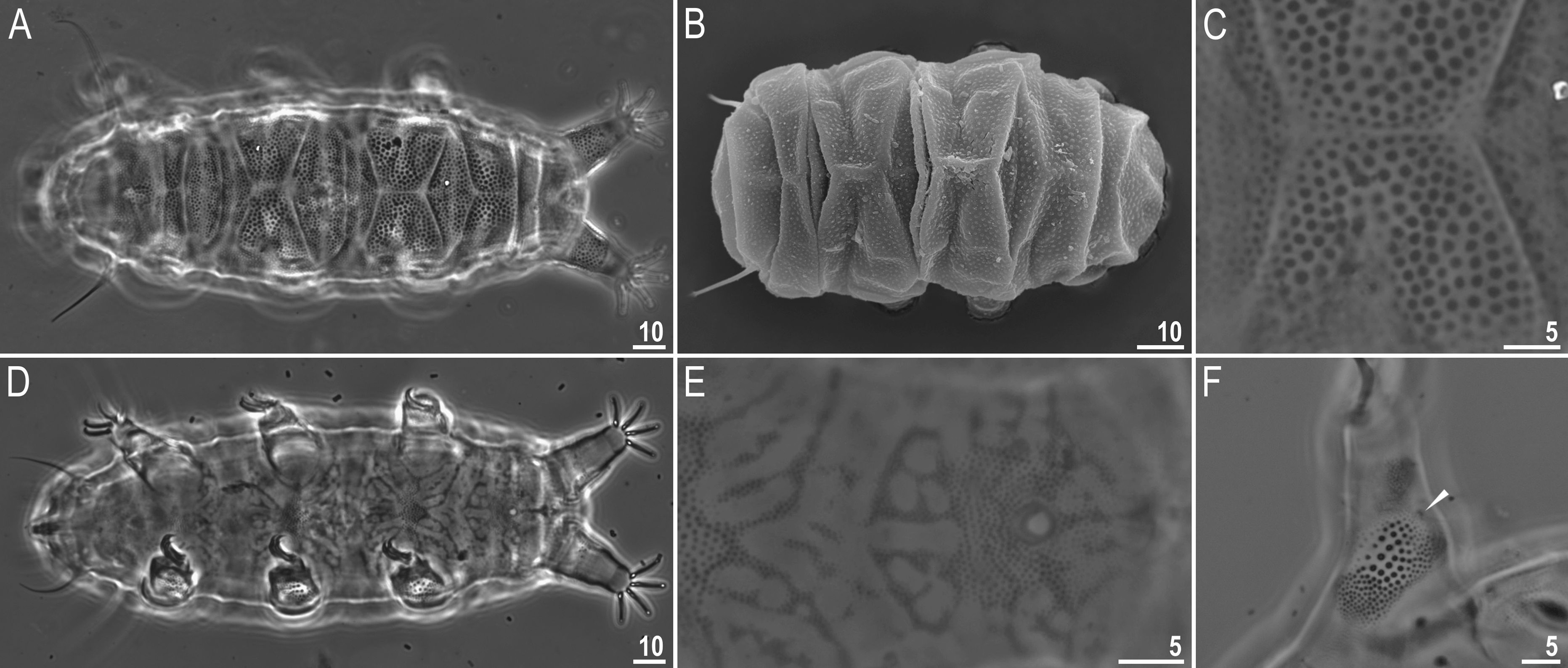
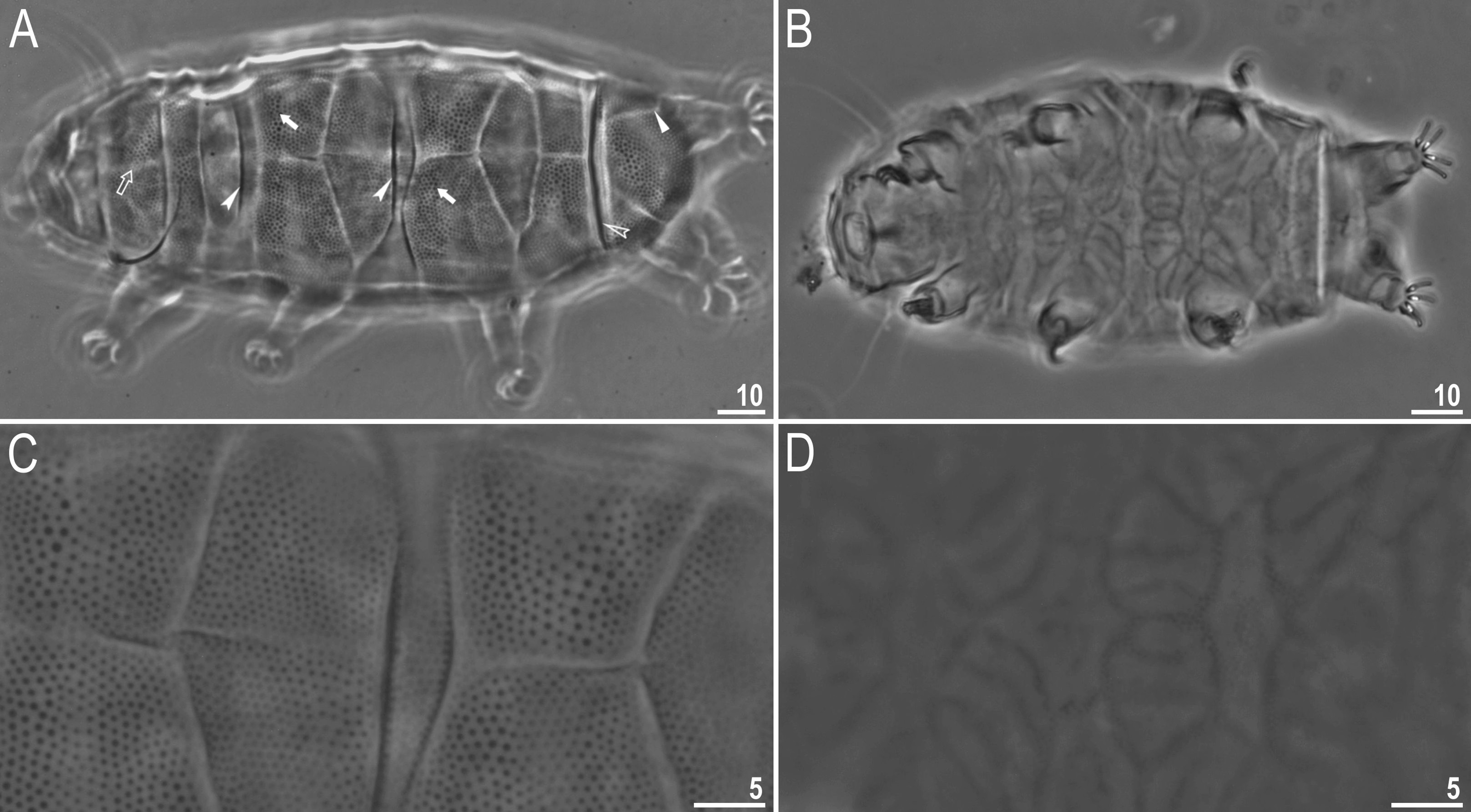
Females. Body ( Figs 1 View FIGURE 1 , 2 View FIGURE 2 ) yellow-orange in living specimens (transparent after mounting on slides), black eyes present after mounting on slides. Apart from the head appendages (cirri interni and externi and spherical cephalic papillae [secondary clavae]), only lateral cirrus A present (with finger-like [primary] clavae near the base) ( Fig. 2B View FIGURE 2 ). Cephalic papillae smaller than clavae.
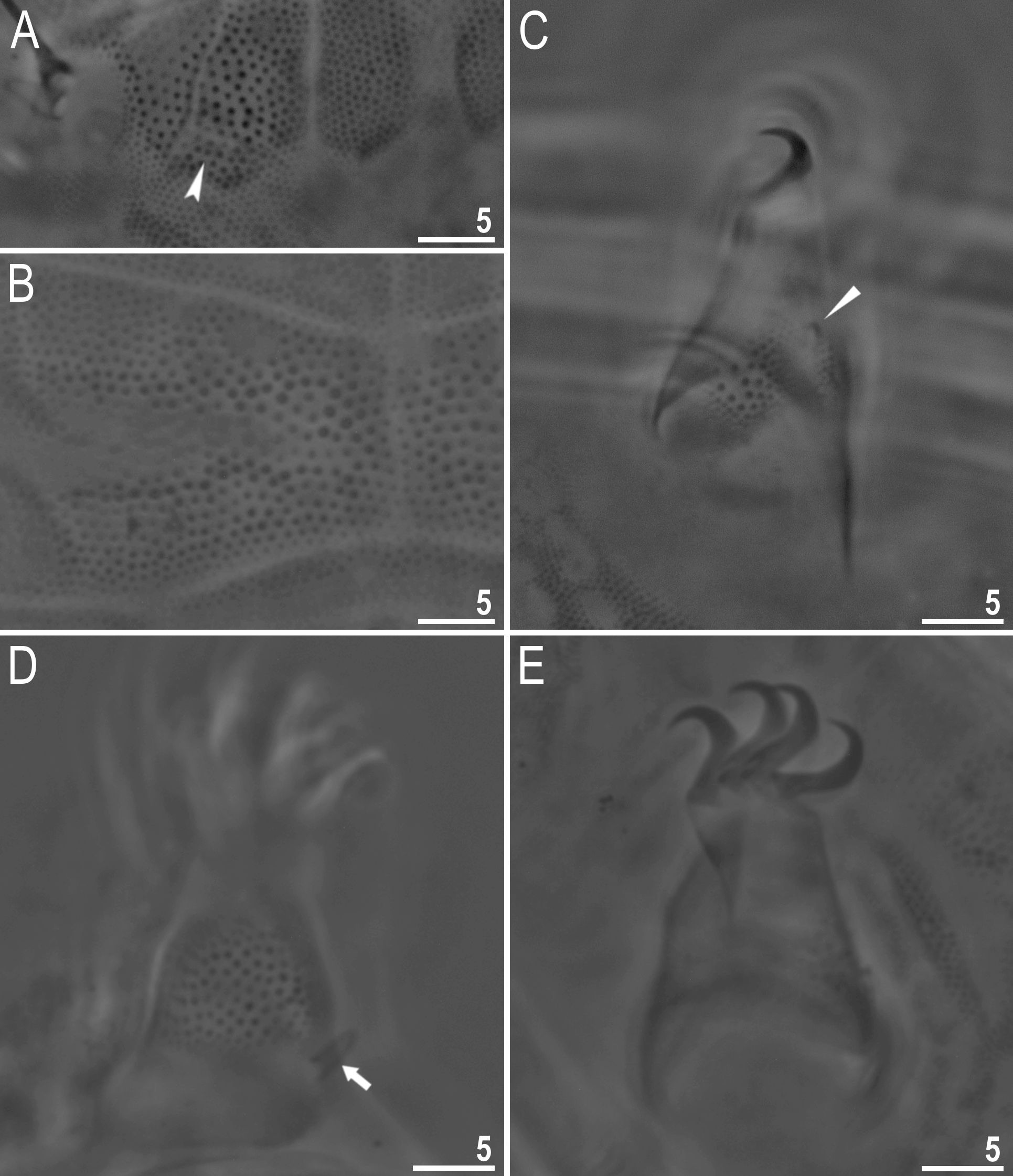
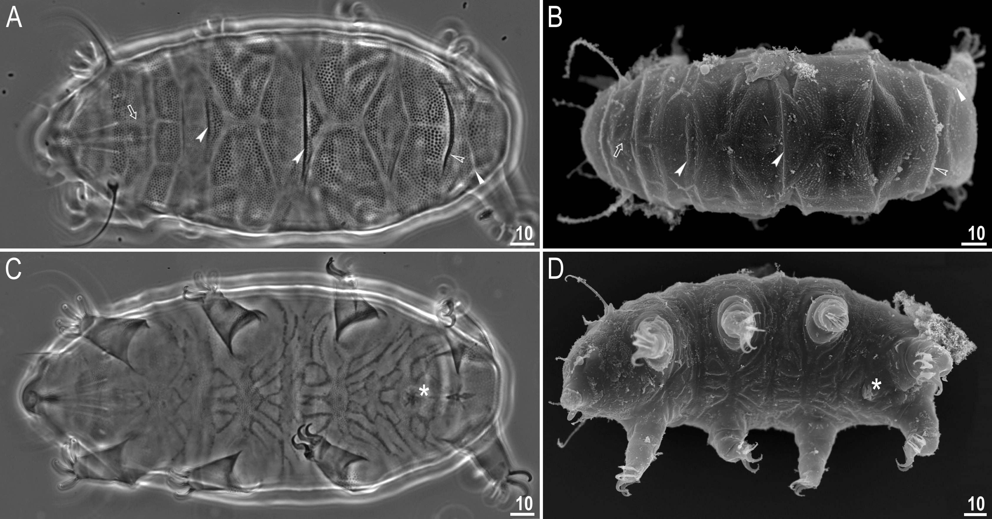
Dorsal plates with small hemispherical granules/upper ends of cuticular pillars (dots in LM) 0.3–0.8 μm in di- ameter, densely (spaces between granules 0.2–1.1 μm) and uniformly distributed and not joined by striae ( Figs 2A View FIGURE 2 , 3 View FIGURE 3 A–B). Granules/upper ends of cuticular pillars slightly larger in the centre of the plates.
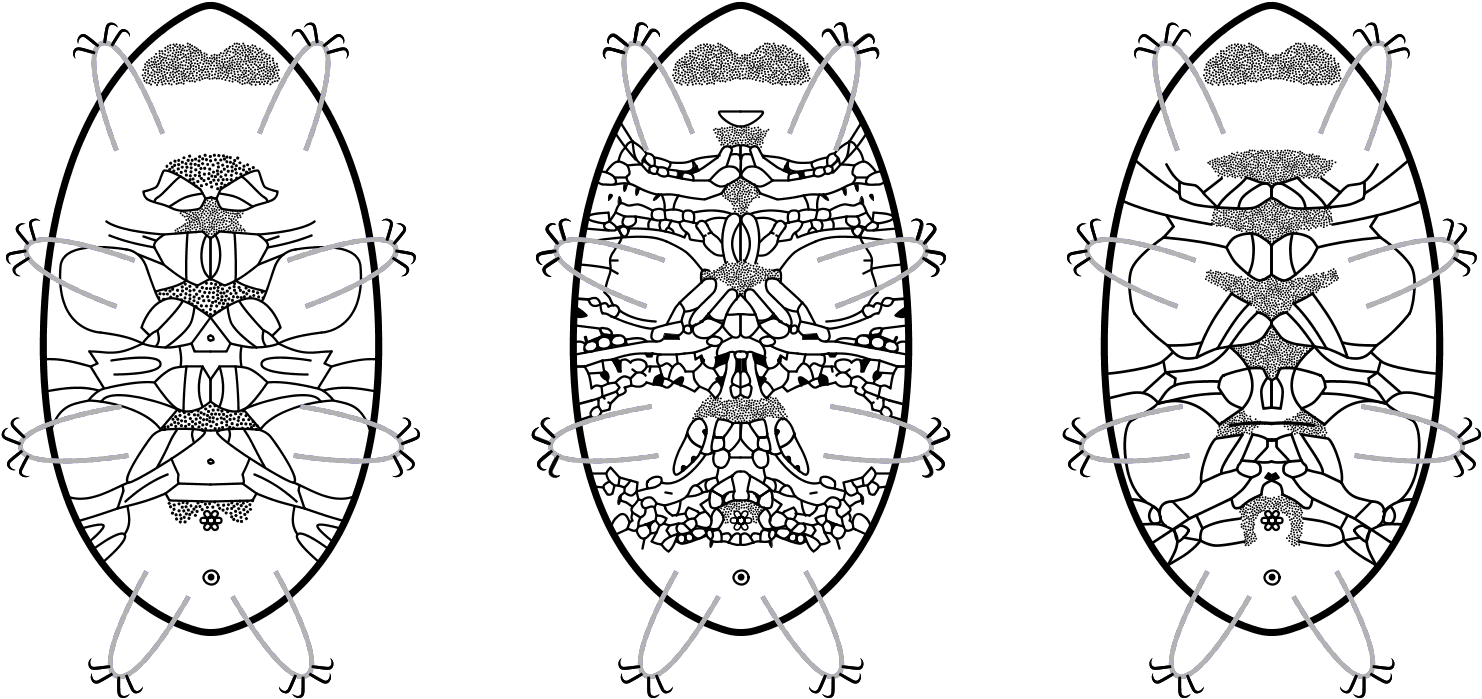
Dorsal plates typical for the genus Pseudechiniscus (cephalic plate (cp), neck plate (np) scapular plate (scp), median plates (m1, m2, m3), paired segmental plates I and II (s1, s2), pseudosegmental plate (psp), and the caudal plate (cap), see also Dorsal and ventral plates and sculpture in Grobys et al. (2020)) well developed. The cp with W-shaped pattern divided into five parts. The scp divided by a transversal fold, forming a long narrow stripe in posterior part of the plate. This narrow stripe often divided by three longitudinal folds resulting in four parts/subplates ( Fig. 2A View FIGURE 2 ). Entire scp divided by median longitudinal fold into two parts ( Fig. 2 View FIGURE 2 A–B, empty arrow). Lateral portions of the scp appear detached from the dorsal plate, forming small plate-like structures separated from the scp by a thin bright stripe ( Fig. 3A View FIGURE 3 , indented arrowhead). Plates m1 and m2 divided in two portions by transverse fold, plate m3 undivided ( Fig. 2 View FIGURE 2 A–B, filled indented arrowheads). Laterally to the median plates, lateral intersegmental plates (lip) present. On the plates s1 and s2 darker stripes (folds in SEM) visible ( Fig. 2 View FIGURE 2 A–B, filled arrows). The psp divided by a longitudinal fold. Posterior margin of psp straight, i.e. without projections, teeth or spines ( Fig. 2 View FIGURE 2 A–B, empty indented arrowheads). The cap concave with two Y-shaped bifurcated ridges ( Fig. 2B View FIGURE 2 , filled arrowhead). Ventral cuticle with tiny granulation (formed by dense granules/upper ends of cuticular pillars, 0.1–0.4 μm) forming a unique pattern ( Figs 1 View FIGURE 1 , 2 View FIGURE 2 C–D). Ventral patches of granulation present (granulation 0.5–0.6 μm in diameter, spaces between granules 0.5–1.0 μm) with configuration PG:I-II-III-IV-VI-VIII g ( Figs 1 View FIGURE 1 , 2 View FIGURE 2 C–D). The female gonopore with the typical six-petal rosette. ( Fig. 2 View FIGURE 2 C–D, asterisks).
The outer cuticle on legs I–III with round patches of granulation (with larger granules but more sparse in the centre and smaller and denser in peripheral parts), on legs IV with uniform wide stripes of granulation (slightly larger in the centre of these stripes) ( Fig. 3 View FIGURE 3 C–D). Triangular spine on leg I absent, instead a small papilla-like structure present, but very hardly visible under LM ( Figs 2D View FIGURE 2 , 3C View FIGURE 3 filled arrowheads). Dentate collar on leg IV absent. A finger-like papillae on leg IV present ( Fig. 3D View FIGURE 3 , filled arrow). External claws of all legs smooth, internal with spurs directed downwards, identical on legs I–IV ( Fig. 3E View FIGURE 3 ).
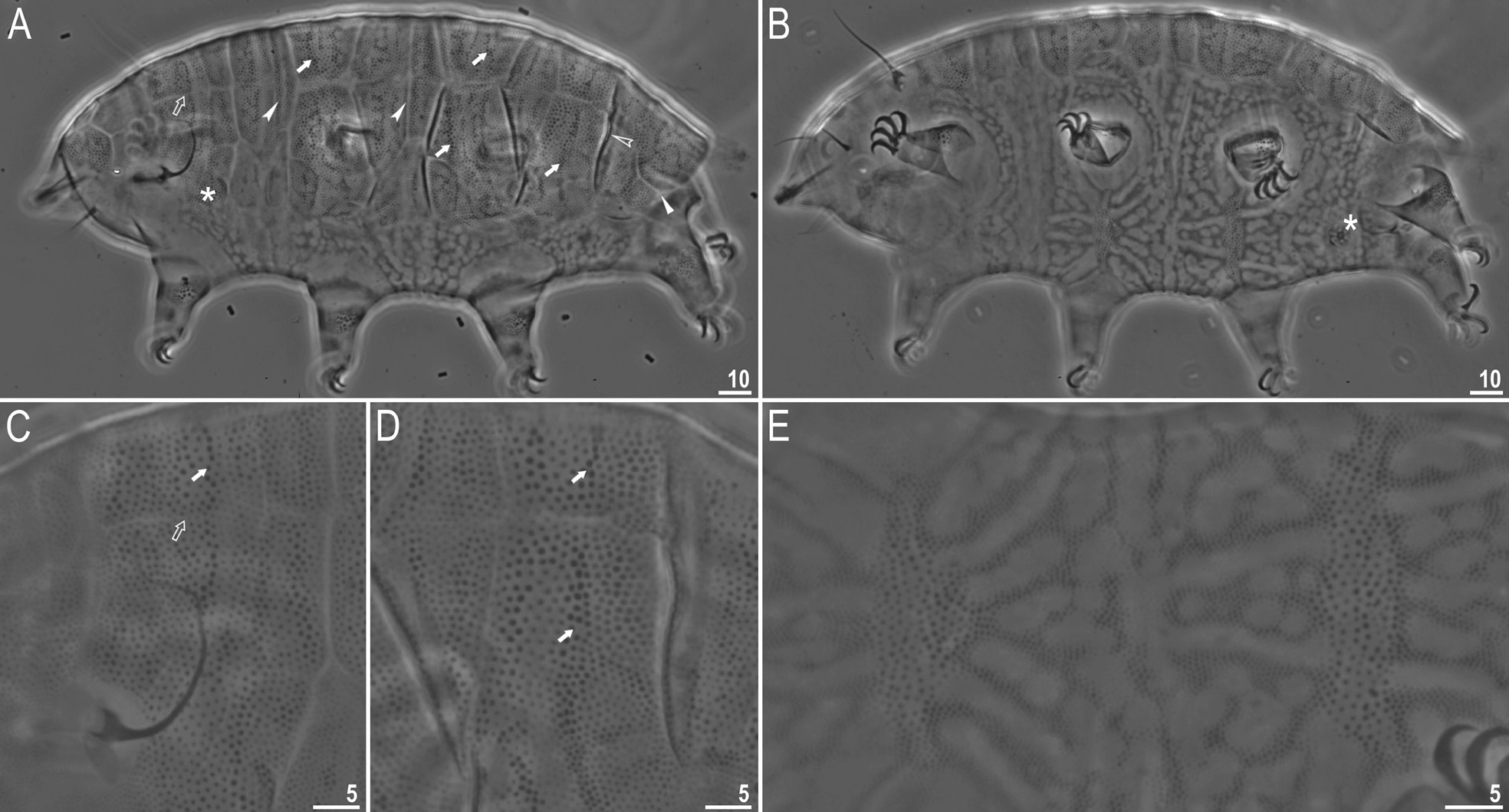
Males ( Figs 1 View FIGURE 1 , 4 View FIGURE 4 ). Body slenderer than in females. Dorsal plates and body appendages arranged identically like in females. Head appendages clearly longer than in females (compare Tables 1 View TABLE 1 and 2 View TABLE 2 ). Dorsal granules/upper ends of cuticular pillars larger than in females (0.4–1.3 μm in diameter), densely (spaces between granules 0.5–1.3 μm) and uniformly distributed, not joined by striae ( Fig. 4 View FIGURE 4 A–C). Ventral pattern (granules/upper ends of cuticular pil- lars, 0.1–0.4 μm) of different shape than in females ( Figs 1 View FIGURE 1 , 4 View FIGURE 4 D–E). Patches of granulation with configuration PG: I-II-III-IV-VI-VIII a (granules/upper ends of cuticular pillars, 0.4–0.7 μm) ( Figs 1 View FIGURE 1 , 4 View FIGURE 4 D–E). The gonopore round and without six-petal rosette and with more distinct PG in anterior and lateral parts ( Fig. 4E View FIGURE 4 ). The leg sculpture similar to females but with larger granulation ( Fig. 4F View FIGURE 4 ). Triangular spine on leg I absent, instead a small papilla-like structure present, but very hardly visible under LM ( Fig. 4F View FIGURE 4 , filled arrowhead).
Juveniles. Unknown.
Larvae. Unknown.
PG
PG
PG
PG
PG
PG
PG
PG
. suillus ( Ehrenberg, 1853) Pse. ehrenbergi sp. nov. Pse. ehrenbergi sp. nov. Pse. dastychi sp. nov. female female male female Pse. angelusalas sp. nov. Pse. lacyformis sp. nov. Pse. indistinctus sp. nov. female female female
DNA sequences
We obtained good quality sequences for the applied molecular markers:
- COI sequence (GenBank: MN528470 View Materials ), 686 bp long;
- ITS-2 sequence (GenBank: MN537866 View Materials ), 405 bp long.
Etymology.
We dedicate this species to the distinguished German zoologist Christian Gottfried Ehrenberg, who described Pse. suillus , the first species in the genus Pseudechiniscus .
Type locality.
45°53’10’’N, 10°50’11’’E, 140 m asl: Italy, Trentino Province, Riva del Garda, on the way to the Bastion, moss from dead wood, 07.07.2016, coll. Daria Grobys and Hanna Kmita.
Type depositories.
Holotype: slide IT40/7 and 25 paratypes (slides: IT40/*, where the asterisk can be substituted by any of the following numbers: 3, 7, 9, 3/S, 4/S, 5/S, 6/S, 7/S) are deposited at the Department of Animal Taxonomy and Ecology, Institute of Environmental Biology, Adam Mickiewicz University in Poznań, Uniwersytetu Poznańskiego 6, 61-614 Poznań, Poland; 16 paratypes (slide IT40/10) are deposited at the collection of Binda and Pilato, Museum of the Department of Animal Biology ‘Marcello La Greca’, University of Catania, Italy.
Morphological differential diagnosis*
*only measurements of adult females are used in differential diagnosis
Pseudechiniscus ehrenbergi sp. nov. differs specifically from:
1. Pse. angelusalas sp. nov., known only from Madagascar (the present study), by: a small papilla-like structure on leg I present, different ventral pattern ( Figs 1 View FIGURE 1 , 2 View FIGURE 2 C–D for Pse. ehrenbergi sp. nov. vs Figs 1 View FIGURE 1 , 7B View FIGURE 7 for Pse. angelusalas sp. nov.), different position of PG VIII (PG VIII placed around gonopore in Pse. ehrenbergi sp. nov. vs PG VIII placed only above gonopore in Pse. angelusalas sp. nov.), lower sp of cephalic papillae ( 12.8–17.1 in Pse. ehrenbergi sp. nov. vs 18.3–20.8 in Pse. angelusalas sp. nov.), lower sp of clavae ( 17.7–20.6 in Pse. ehrenbergi sp. nov. vs 22.4–25.7 in Pse. angelusalas sp. nov.), lower sp of cirrus A ( 95.1–112.6 in Pse. ehrenbergi sp. nov. vs 129.2–152.2 in Pse. angelusalas sp. nov.), lower cirrus A /body length ratio (14–17% in Pse. suillus vs 19–22% in Pse. angelusalas sp. nov.), higher cirri interni / externi length ratio (72–85% in Pse. ehrenbergi sp. nov. vs 66–71% in Pse. angelusalas sp. nov.), longer papillae on leg IV (2.8–3.3 μm in Pse. ehrenbergi sp. nov. vs 2.0–2.4 μm in Pse. angelusalas sp. nov.) and longer claws (see Tables 1 View TABLE 1 and 4).
2. Pse. beasleyi, known only from China ( Li et al. 2007), by: scp not divided in anterior part (scp divided into four parts in Pse. beasleyi), smaller granules of dorsal sculpture (0.3–0.8 μm in Pse. ehrenbergi sp. nov. vs up to 1.6 μm in Pse. beasleyi), shorter cirri interni and externi (7.1–8.6 and 9.0–11.7 μm respectively in Pse. ehrenbergi sp. nov. vs 10.4–15.7 and 13.1–18.3 μm respectively in Pse. beasleyi), different claw lengths arrangement (shortest claws II and III, and longest claws IV in Pse. ehrenbergi sp. nov. vs shortest claws I and II, and longest claws III and IV in Pse. beasleyi) and shorter claws (see Table 1 View TABLE 1 in this paper and Table 2 View TABLE 2 in Li et al. 2007).
3. Pse. chengi, known only from China ( Xue et al. 2017), by: a small papilla-like structure on leg I present, plates m1 and m2 divided in two portions by transverse fold (unndivided in Pse. chengi), shorter claws I–III and lower sp of claws I–II (see Table 1 View TABLE 1 herein and Table 2 View TABLE 2 in Xue et al. 2017).
4. Pse. clavatus , known only from Spain ( Mihelčič 1955), by: different shape of clavae (finger-like in Pse. ehrenbergi sp. nov. vs club-shaped in Pse. clavatus ) and normally developed cephalic papillae (reduced in Pse. clavatus ).
5. Pse. dastychi sp. nov., known only from Antarctica ( Dastych 1984 and present study), by: a small papilla-like structure on leg I present, different ventral pattern ( Figs 1 View FIGURE 1 , 2 View FIGURE 2 C–D for Pse. ehrenbergi sp. nov. vs Figs 1 View FIGURE 1 , 5 View FIGURE 5 C–D for Pse. dastychi sp. nov.), different ventral PG configuration (PG:I-II-III-IV-VI-VIII g in Pse. ehrenbergi sp. nov. vs PG:I-II-III-IV-V-VI-VII-VIII a in Pse. dastychi sp. nov.), dorsal granules not joined by striae, narrower scp (20.3–26.6 μm in Pse. ehrenbergi sp. nov. vs 27.5–33.0 μm in Pse. dastychi sp. nov.), shorter cirri interni (7.1–8.6 μm in Pse. ehrenbergi sp. nov. vs 10.4–12.7 μm in Pse. dastychi sp. nov.), shorter cirri externi (9.0–11.7 μm [ sp=39.6–49.6] in Pse. ehrenbergi sp. nov. vs 15.9–19.1 μm [ sp=54.7–61.4] in Pse. dastychi sp. nov.), shorter cirri A (21.6–26.8 μm [ sp=95.1–112.6] in Pse. ehrenbergi sp. nov. vs 40.0–45.0 μm [ sp=135.2–149.1] in Pse. dastychi sp. nov.), lower cirrus A /body length ratio (14–17% in Pse. ehrenbergi sp. nov. vs 22–26% in Pse. dastychi sp. nov.), higher cirri interni/externi length ratio (72–85% in Pse. ehrenbergi sp. nov. vs 62–69% in Pse. dastychi sp. nov.) and shorter claws (see Tables 1 View TABLE 1 and 3).
6. Pse. facettalis , known from distant localities throughout the world ( McInnes 1994). Based on present study, an inaccurate description of this species makes it impossible to differentiate this taxon from Pse. ehrenbergi sp. nov.. See also Morphological differential diagnosis of Pse. suillus and Discussion in the paper Grobys et al. (2020).
7. Pse. indistinctus sp. nov., known only from Norway (present study), by: a small papilla-like structure on leg I present, different ventral pattern ( Figs 1 View FIGURE 1 , 2 View FIGURE 2 C–D for Pse. ehrenbergi sp. nov. vs Figs 1 View FIGURE 1 , 13 View FIGURE 13 C–D for Pse. indistinctus sp. nov.), different ventral PG configuration (PG:I-II-III-IV-VI-VIII g in Pse. ehrenbergi sp. nov. vs PG: I-II-III-IV-V-VI-VIII a in Pse. indistinctus sp. nov.), granules on cap similar in size to other dorsal plates (granules visibly larger on cap in comparison with other dorsal plates in Pse. indistinctus sp. nov.), lower sp of cirri externi ( 39.6–49.6 in Pse. ehrenbergi sp. nov. vs 54.3–59.3 in Pse. indistinctus sp. nov.), shorter cirri A (21.6–26.8 μm [ sp=95.1–112.6] in Pse. ehrenbergi sp. nov. vs 27.6–34.2 μm [ sp=123.0–144.9] in Pse. indistinctus sp. nov.), lower cirrus A /body length ratio (14–17% in Pse. ehrenbergi sp. nov. vs 18–22% in Pse. indistinctus sp. nov.), higher cirri interni/externi length ratio (72–85% in Pse. ehrenbergi sp. nov. vs 62–70% in Pse. indistinctus sp. nov.) and lower spur/branch length ratio of all claws (17–24% in Pse. ehrenbergi sp. nov. vs 28–34% in Pse. indistinctus sp. nov.).
8. Pse. juanitae , known from Austria, Brazil ( type locality), Italy and Galapagos Islands ( McInnes 1994). Based on present study, an inaccurate description of this species makes it impossible to differentiate this taxon from Pse. ehrenbergi sp. nov.. See also Morphological differential diagnosis of Pse. suillus and Discussion in the paper Grobys et al. (2020).
9. Pse. lacyformis sp. nov., known only from Norway (the present study), by: a small papilla-like structure on leg I present, different ventral pattern ( Figs 1 View FIGURE 1 , 2 View FIGURE 2 C–D for Pse. ehrenbergi sp. nov. vs Figs 1 View FIGURE 1 , 10B View FIGURE 10 for Pse. lacyformis sp. nov.), different position of PG VIII (PG VIII placed around gonopore in Pse. ehrenbergi sp. nov. vs PG VIII placed above gonopore in Pse. lacyformis sp. nov.), shorter cirri interni (7.1–8.6 μm [ sp=32.3–37.4] in Pse. suillus vs 10.6–14.0 μm [ sp=48.4–53.9] in Pse. lacyformis sp. nov.), shorter cirri externi (9.0–11.7 μm [ sp=39.6–49.6] in Pse. suillus vs 14.1–19.4 μm [ sp=66.5–77.8] in Pse. lacyformis sp. nov.), shorter clavae (4.0–4.7 μm [ sp=17.7– 20.6] in Pse. ehrenbergi sp. nov. vs 5.1–6.4 μm [ sp=23.4–26.8] in Pse. lacyformis sp. nov.), lower sp of cirrus A ( 95.1–112.6 in Pse. ehrenbergi sp. nov. vs 114.1–142.5 in Pse. lacyformis sp. nov.) and lower sp of papillae on leg IV ( 11.3–13.7 in Pse. ehrenbergi sp. nov. vs 14.7–17.2 in Pse. lacyformis sp. nov.).
10. Pse. megacephalus , known only from Austria ( type locality) and Turkey ( McInnes 1994), by: different shape of cephalic papillae (spherical in Pse. ehrenbergi sp. nov. vs mushroom-like in Pse. megacephalus ), absence of papilliform projection between external buccal cirri and cirri A.
11. Pse. suillus , known only from Italy and Portugal ( Grobys et al. 2020), by: different ventral pattern ( Figs 1 View FIGURE 1 , 2 View FIGURE 2 C–D for Pse. ehrenbergi sp. nov. vs Figs 1 View FIGURE 1 herein and 4C–D in Grobys et al. (2020) for Pse. suillus ), well developed ventral patches of granulation, shorter cirri interni (7.1–8.6 μm [ sp=32.3–37.4] in Pse. ehrenbergi sp. nov. vs 8.7–11.1 μm [ sp=44.0–49.6] in Pse. suillus ), lower sp of cephalic papillae ( 12.8–17.1 in Pse. ehrenbergi sp. nov. vs 19.1–24.3 in Pse. suillus ), shorter cirri externi (9.0–11.7 μm [ sp=39.6–49.6] in Pse. ehrenbergi sp. nov. vs 12.0–16.8 μm [ sp= 62.1–75.0] in Pse. suillus ) lower sp of clavae ( 17.7–20.6 in Pse. ehrenbergi sp. nov. vs 20.9–26.8 in Pse. suillus ), shorter cirrus A (21.6–26.8 μm [ sp=95.1–112.6] in Pse. ehrenbergi sp. nov. vs 28.4–34.4 μm [ sp=134.9–156.9] in Pse. suillus ) and lower sp of papillae on leg IV ( 11.3–13.7 in Pse. ehrenbergi sp. nov. vs 14.7–18.4 in Pse. suillus ).
12. Pse. xiai , known only from China ( Wang et al. 2018), by: different ventral pattern ( Figs 1 View FIGURE 1 , 2 View FIGURE 2 C–D for Pse. ehrenbergi sp. nov. vs Figs 1B, F View FIGURE 1 and 2E View FIGURE 2 in Wang et al. 2018 for Pse. xiai ) and higher cirrus A /body length ratio (17–21% in Pse. ehrenbergi sp. nov. vs 13–16% in Pse. xiai ).
Genotypic differential diagnosis
The ranges of genetic distances between Pse. ehrenbergi sp. nov. and species of the genus Pseudechiniscus , for which DNA sequences are available in GenBank, are as follows:
COI: 0.4–29.3% (22.4% on average), with the most similar being Pse. aff. suillus ( MK804906 View Materials , Cesari et al. 2020) and the least similar being Pseudechiniscus sp. ( KJ857008 View Materials , Velasco-Castrillón et. al. 2015 (described in Gen- Bank as Echiniscus sp.); for more details see Discussion section in Grobys et al. 2020).
ITS-2: 7.7–35.5% (25.1% on average), with the most similar being Pse. dastychi sp. nov. ( MN537865 View Materials , present study) and the least similar being Pse. lacyformis sp. nov. ( MN537868 View Materials , present study).


No known copyright restrictions apply. See Agosti, D., Egloff, W., 2009. Taxonomic information exchange and copyright: the Plazi approach. BMC Research Notes 2009, 2:53 for further explanation.
|
Kingdom |
|
|
Phylum |
|
|
Class |
|
|
Order |
|
|
Family |
|
|
Genus |
Pseudechiniscus ehrenbergi
| Roszkowska, Milena, Grobys, Daria, Bartylak, Tomasz, Gawlak, Magdalena, Kmita, Han- Na, Kepel, Andrzej, Kepel, Marta, Parnikoza, Ivan & Kaczmarek, Łukasz 2020 |
Pseudechiniscus
| Grobys et al. 2020 |
Pseudechiniscus aff. suillus
| Cesari et al. 2020 |