
Nipponnemertes Friedrich, 1968
|
publication ID |
https://doi.org/ 10.1080/00222930600833800 |
|
DOI |
https://doi.org/10.5281/zenodo.4672302 |
|
persistent identifier |
https://treatment.plazi.org/id/573F4468-FFC0-FFCD-FE44-F433252DFAA4 |
|
treatment provided by |
Carolina |
|
scientific name |
Nipponnemertes Friedrich, 1968 |
| status |
|
Genus Nipponnemertes Friedrich, 1968
Species
Nipponnemertes bimaculata ( Coe 1901)
Specimen
USNM 1072180; 10 slides of stained serial section, including transverse sections of the anterior portion of the body (4) and tail (4) and longitudinal sections of the midgut (2). Another specimen was not sectioned (it dried out after it was preserved).
Locality
Obstruction Pass , between Obstruction Island and Orcas Island, Washington (Lat. 48 ° 36 9 W, Long 122 ° 49 9 W), and Satellite Channel, Vancouver Island, British Columbia, Canada, August 22, 1964 .
Description
External features
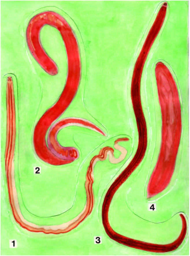
The specimen collected in Obstruction Pass was only 8 mm long and 0.5 mm wide, but the one from Satellite Channel was 18 cm long and 6 mm wide ( Figure 1.3 View Figure 1 ). When contracted, it was 10 cm long and 1 mm wide.
The body of the first specimen was slender and cylindrical in the posterior portion ( Figure 13a View Figure 13 ). The head was somewhat triangular in shape and had two dark brown markings on an otherwise whitish background ( Figure 13a View Figure 13 ). The rest of the body was brown on both the dorsal and the ventral sides, except at the posterior end, where it was whitish ( Figure 13b View Figure 13 ). Behind the dorsal markings of the head were two oblique cephalic grooves. A longitudinal cephalic groove was visible as a long line running in the colourless midline of the anterior region of the body and continuing into the post-cephalic region. Five right and three left ocelli were arranged on the lateral margins of the head outside the dark brown markings. Two pairs of ocelli were located just behind the transverse cephalic grooves; one member of each pair was larger than the other ( Figure 13a View Figure 13 ).
The head of the specimen collected in Satellite Channel had a rounded outline when viewed from above ( Figure 13c View Figure 13 ). The color of the body was vermilion and the posterior end, with a slight protrusion, lacked the whitish coloration seen in the small specimen from Obstruction Pass ( Figure 13c, d View Figure 13 ). The head had a pair of brown patches. Two groups of 13 ocelli were arranged close to the lateral margins of the head and two groups of eight were arranged near the posterior edges of the brown markings ( Figure 13c View Figure 13 ). Two pairs of large ocelli were located behind the oblique cephalic grooves. There was a longitudinal cephalic groove in the midline of the anterior portion of the body. The ventral portions of the oblique cephalic grooves ran forward along the lateral margin of the head and met at the anterior tip of the head, where the proboscis pore was visible as a short line ( Figure 13e View Figure 13 ).
Body aeall, musculature and parenchyma
The epidermis contains elliptical unicellular glands sparsely arranged in a row ( Figure 14a View Figure 14 ). The thickness of the dermis averages 40 mm. The body wall musculature is well-developed and has a layer of fasciated diagonal muscles between the outer circular and inner longitudinal layers ( Figure 14a View Figure 14 ). The outer circular muscle layer is only 7 mm thick, while the inner longitudinal muscle layer is three or four times this thickness, averaging 23 mm. The longitudinal layer is not divided anteriorly by connective tissue and there are no cephalic retractor muscles. Dorsoventral muscles are well-developed and parenchyma is moderate in extent.
Rhynchodeum, rhynchocoel and proboscis
The proboscis pore is situated at the anterior end of the head immediately behind the opening of the frontal organ ( Figure 14b, c View Figure 14 ). The proboscis sheath reaches nearly to the posterior end of the body ( Figure 14d View Figure 14 ). The rhynchocoel has a wickerwork of interwoven circular and longitudinal muscles ( Figure 14a View Figure 14 ). The rhynchodeum has a circular muscle sphincter ( Figure 14e View Figure 14 ). The pre-cerebral septum is of the closed type and is composed of radial muscles derived from the longitudinal muscle layer of the body wall ( Figure 14f View Figure 14 ).
The proboscis diaphragm has the central stylet apparatus consisting of a conical basis about 40 mm wide; the content of this, in the section, was extruded ( Figure 14g View Figure 14 ). The basis rests on a large bolster consisting of interwoven circular and longitudinal muscles ( Figure 14g View Figure 14 ). Posterior to the bolster, the stylet bulb consists of a plate-like muscular wall, a thick layer of glandular cells in its anterior wall, thick connective tissue, and interlacing circular muscles similar to those of Nipponnemertes fernaldi ( Iwata 2001) and Satellitenemertes satellitensis of the present report ( Figure 14h View Figure 14 ). There are two accessory stylet pouches, each containing eight stylets. The proboscis has 16 nerves.
Alimentary canal
Near the anterior end of the head, the oesophagus opens into the rhynchodeum ( Figure 15a View Figure 15 ). The oesophagus does not have a caecum; according to Coe (1905); however, a rather extensive oesophageal caecum extends beneath the stomach of this species. The oesophagus and the stomach have no circular muscles. The stomach has two deep folds and is about the length of the brain. The pylorus is long and two times the length of the stomach. The intestinal caecum has a pair of anterior pouches and three pairs of lateral diverticula. The anterior pouches are short and have a length of about 110 mm. They are situated a long distance behind the posterior end of the brain, and are about as long as the brain. The intestinal diverticula are unbranched.
The anus opens on the ventral side of the posterior end of the body ( Figure 15b View Figure 15 ). The rectum, 80 mm long, has a sphincter of circular muscle fibres ( Figure 15c View Figure 15 ). The lateral nerves are connected to each other by a commissure on the dorsal side of the rectum ( Figure 15d View Figure 15 ). A short distance (20 mm) anterior to the commissure, the blood vessels anastomose into a slender vessel ( Figure 15e View Figure 15 ). The rhynchocoel ends a little anterior to this anastomosis ( Figure 14d View Figure 14 ).
Blood υascular system
The blood vascular system has three longitudinal vessels. The dorsal vessel, originating from the right cephalic vessel, enters the rhynchocoel wall and forms a median vascular plug ( Figure 15f View Figure 15 ). The lateral vessel makes a large lacuna-like enlargement immediately behind the cerebral sensory organ ( Figure 15g View Figure 15 ). The cephalic vessels enter the brain ring without giving off cerebral vessels. There are no pseudometameric anastomoses of blood vessels.
Nerυous system
The brain and lateral nerves are covered by a thin layer of fibrous connective tissue. The nervous system has no neurochord cells or accessory lateral nerves. The mid-dorsal nerve extends anteriorly beyond the brain ( Figure 14a View Figure 14 ). The lateral nerve cords have myofibrillae in a connective tissue band on the medial side of the fibre core and peripheral nerves ( Figure 15g View Figure 15 ).
The ventral ganglia are separated from the dorsal ganglia posteriorly. The dorsal ganglia have no bifurcated fibre core. There is a pair of foregut (splanchnic) nerves originating from the ventral ganglia immediately behind the ventral commissure.
Special sensory organs and frontal organ
The cephalic grooves have secondary transverse grooves. There is a lengthwise row of six ocelli on the left side of the anterior portion of the head, a row of four ocelli on the right side and two ocelli on each side behind the cephalic grooves. The ocelli behind the cephalic grooves are remarkably large, being 55 mm in diameter ( Figures 15h View Figure 15 and 16a View Figure 16 ). The frontal organ and frontal glands are well-developed: the former forms a short duct, opening at the anterior end of the head ( Figure 14b View Figure 14 ); the latter reaches posteriorly to the anterior end of the brain. The cephalic glands are moderately developed along the lateral sides of the head and reach posteriorly to the anterior end of the brain. Sub-muscular glands are small and situated in the ventral side of the head.
The cerebral organ is large, extends behind the cerebral ganglia ( Figure 16b, c View Figure 16 ), and is covered by a neurilemma. The cerebral organ canal branches into a medial sensory canal and a lateral sac ( Figure 16 View Figure 16 d–f); the former has a cyanophilic glandular mass and the latter an eosinophilic glandular mass ( Figure 16c, g View Figure 16 ). The cerebral organ opens laterally into a cephalic groove of the head ( Figure 16h View Figure 16 ). The sensory canal is evident as a small number of cells surrounding the inner lumen of the cerebral organ canal ( Figures 15h View Figure 15 and 16a View Figure 16 ).
Excretory and reproductiυe systems
The excretory tubules extend from the level immediately behind the cerebral organ to the level of the posterior portion of the pylorus. The two efferent ducts, originating in the posterior portion of the nephridial region, are short and run lateral to the lateral nerve cords, opening on the ventral side of the body. The male gonads are immature.


No known copyright restrictions apply. See Agosti, D., Egloff, W., 2009. Taxonomic information exchange and copyright: the Plazi approach. BMC Research Notes 2009, 2:53 for further explanation.
|
Kingdom |
|
|
Phylum |
|
|
Class |
|
|
Order |
|
|
Family |