
Sanjuannemertes aeilloaesi, Iwata, 2006
|
publication ID |
https://doi.org/ 10.1080/00222930600833800 |
|
DOI |
https://doi.org/10.5281/zenodo.4672308 |
|
persistent identifier |
https://treatment.plazi.org/id/573F4468-FFF8-FFF4-FF58-F590258FFA57 |
|
treatment provided by |
Carolina |
|
scientific name |
Sanjuannemertes aeilloaesi |
| status |
|
Sanjuannemertes aeilloaesi gen., sp. nov.
Type specimen
Holotype: USNM 1072181 About USNM ; 22 slides of stained serial section, including transverse sections of the anterior portion of the body (9) and tail (5) and horizontal sections of the midgut (8).
Type locality San Juan Channel, Washington, USA (48 ° 34.3 9 N, 123 ° 1.7 9 W), depth 62 fathoms,
October 21, 1964.
Description
External features
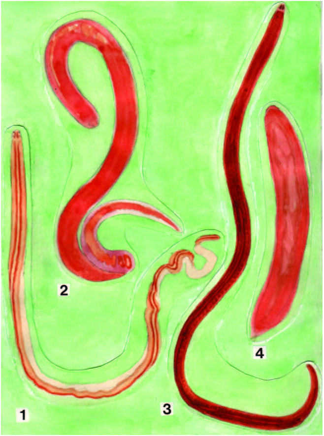
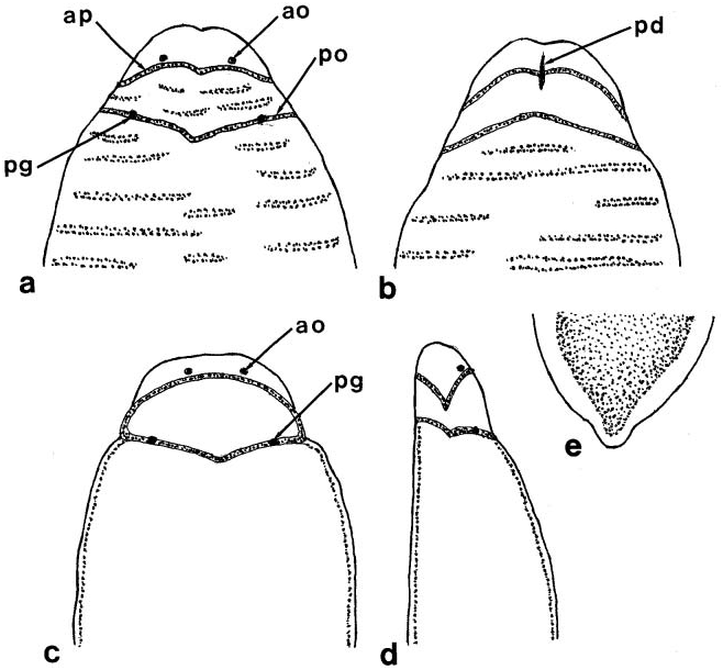
The body was small and cylindrical, 15 mm long and 1.5 mm wide ( Figure 1.4 View Figure 1 ). When contracted, it was 10 mm long and 2 mm wide. The anterior edge of the head was rounded ( Figure 17a, c View Figure 17 ) or slightly indented ( Figure 17b View Figure 17 ). The posterior end of the body had a papilla-like protrusion ( Figure 17e View Figure 17 ). There were two oblique cephalic grooves on both the dorsal and the ventral sides of the head ( Figure 17 View Figure 17 a–d). On the ventral side, the mouth and proboscis pore are in a slit-like depression that crosses the anterior cephalic groove ( Figure 17b View Figure 17 ). The color of the body was orange or light vermilion. On both the dorsal and the ventral surfaces of the anterior portion of the body there were numerous narrow, pale orange or whitish transverse lines. The whitish proboscis apparatus could be seen through the body wall; the basis of the central stylet was evident as a milky white patch. The four ocelli, visible on the dorsal side, were arranged in the form of a trapezoid. The two anterior ocelli were closer than those of the posterior pair and were situated just in front of the anterior cephalic groove, whereas the other pair was situated underneath the posterior one ( Figure 17a, c, d View Figure 17 ).
Body aeall, musculature and parenchyma
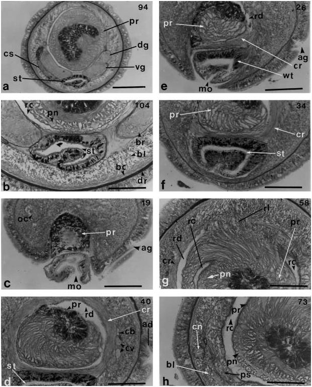
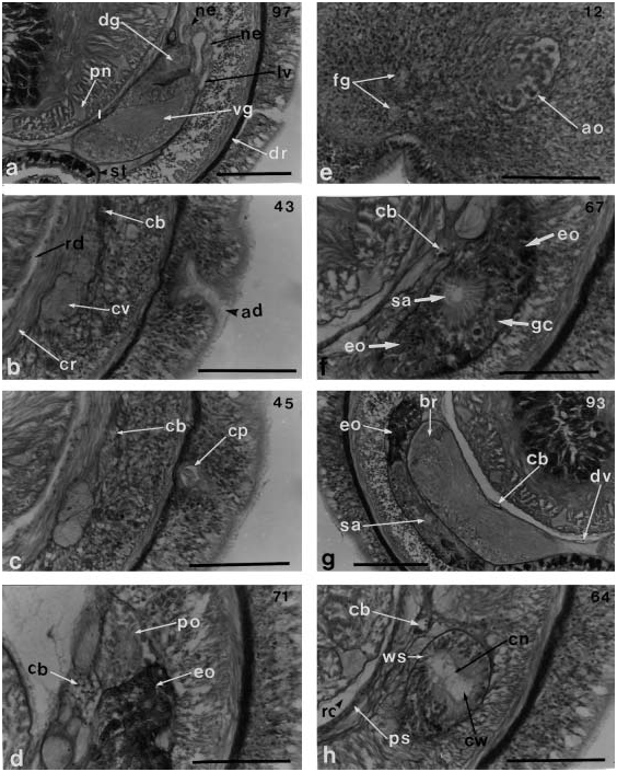
The body in transverse section is circular in both the cephalic and midgut regions ( Figure 18a View Figure 18 ). The epidermis is of uniform thickness (about 60 mm). Large unicellular glands are distributed over the entire body and are especially well developed in the midgut region. Cyanophilic glands are not present between them. In the cephalic region, the epidermis and longitudinal muscle layer of the body wall are nearly the same in thickness, and glandular cells are confined to the upper portion. The dermis, 5–10 mm thick, onefourth to one-tenth the thickness of the epidermis ( Figure 18b View Figure 18 ). The body wall musculature consists of a thin outer circular layer (10 mm thick) and an inner longitudinal layer (60 mm thick). There is sparse lattice-type diagonal muscle layer between them. The longitudinal layer is not divided into inner and outer portions anteriorly. There are no head retractor muscles related to the longitudinal muscle layer. Dorsoventral and radial muscle fibres, as well as parenchyma, are absent.
Rhynchodeum, rhynchocoel and proboscis
The proboscis pore, 60 mm long and 110 mm wide, is a mid-ventral furrow. With the mouth it forms a common atrium ( Figures 17b View Figure 17 , 18c View Figure 18 and 19d View Figure 19 ). Its wall is 15 mm thick and lacks cilia and glands. The rhynchodeum is circular in transverse section owing to the proboscis protruded into it and is covered by a thick layer (20–60 mm) of circular muscles ( Figure 18d View Figure 18 ). The rhynchodeum measures 260 mm long and is at first cylindrical, but then broadens to a width of 300–450 mm ( Figure 18 View Figure 18 d–h). At the posterior end of the rhynchodeum, where its circular muscle sphincter disappears, the pre-cerebral septum is formed by radial longitudinal muscles derived from the longitudinal muscle layer in the dorsal side of the body; the proboscis insertion also becomes evident ( Figure 18g View Figure 18 ). The lateral and ventral sides of the body contain no radial longitudinal muscles in the precerebral septum ( Figure 18h View Figure 18 ).
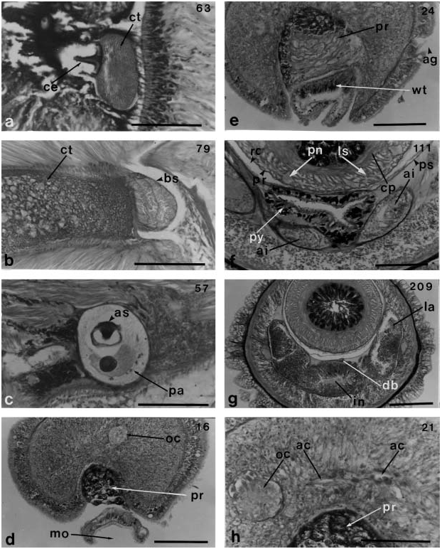
The proboscis diaphragm has a central stylet and a vase-shaped basis 400 mm long, 70 mm wide at its top and 60 mm wide at its middle portion ( Figure 19a, b View Figure 19 ). In sections, the basis shows a strong affinity for eosin and its content is not extruded. The central stylet measures 40 mm long ( Figure 19a View Figure 19 ). The basis of the central stylet rests on a small bolster, is surrounded by a thick layer of circular muscles ( Figure 19b View Figure 19 ) and, in sections, shows no posteriorly inserted accessory stylet. The stylet bulb consists of a thick wall of circular muscles, 300 mm wide. There are two accessory stylet pouches, each containing two stylets ( Figure 19c View Figure 19 ). The proboscis has 10 nerves.
Alimentary canal
The alimentary canal has four major divisions; stomach, pylorus, midgut (with anteriorly directed caecum and lateral diverticula) and hind gut. The mouth and the proboscis pore open into a common atrium on the ventral side of the head; they are not joined together ( Figures 17b View Figure 17 , 18c, e View Figure 18 and 19d, e View Figure 19 ). There is no oesophagus; the mouth leads directly to the stomach (500 mm long). The stomach has a deep lengthwise fold for its entire length and is far longer (160 mm) than the brain ( Figure 18e, f View Figure 18 ). The stomach has no diverticulum.
The pylorus is long, measuring one and one-sixth times the length of the stomach. The intestinal caecum has a pair of short anterior pouches 40 mm long ( Figure 19f View Figure 19 ) and three pairs of lateral diverticula, beginning immediately behind the posterior end of the brain. The intestinal canal has long lateral diverticula ( Figure 19g View Figure 19 ). The hindgut is 100 mm long and ends at the anus.
Blood υascular system
The blood vascular system has three longitudinal vessels. The two cephalic vessels lateral to the rhynchodeum anastomose above the rhynchodeum near the tip of head to form a simple vascular loop 200 mm wide, 10 mm high and 20 mm long. The vascular loop is not a continuous space ( Figure 19h View Figure 19 ). Farther posteriorly, the cephalic vessels do not form large lacunae and do not give off cerebral vessels. They are slender, at first running alongside the rhynchocoel or near the dorsolateral side of the cerebral organ canal, and then above the cerebral organs, but do not enter the brain ring ( Figures 18d, h View Figure 18 and 20 View Figure 20 b–d, f, h). Near the posterior end of the cerebral organ, the cephalic vessels extend down between the brain lobes and the rhynchocoel, and fuse into a dorsal vessel; this does not form a median vascular plug inside the proboscis sheath. A lateral vessel from the cephalic vessels, on the other hand, comes down outside the cerebral ganglia ( Figure 20a View Figure 20 ). There are no pseudometameric anastomoses of blood vessels. The dorsal and lateral vessels anastomose at a level, 40 mm from the posterior end of the body.
Nerυous system
The brain and lateral nerves are covered by a thin layer of fibrous connective tissue. The cerebral ganglia are small; they lie immediately outside the large rhynchocoel and have neither neurochord cells nor an inner neurilemma. The nervous system has no accessory lateral nerves. The dorsal and ventral ganglia are not demarcated externally. The ventral ganglia are not distinctly separated from the dorsal ganglia except posteriorly ( Figures 18a View Figure 18 and 20a, g View Figure 20 ).
The dorsal and ventral fibre cores, measuring 140 mm in length, are not divided anterior to the middle portion of the brain; the dorsal cores (30 mm long), are less voluminous than the ventral cores ( Figure 18a View Figure 18 ). The ventral ganglia behind the posterior end of the dorsal ganglia do not immediately extend laterally to form the lateral nerve cords ( Figure 20a View Figure 20 ). The dorsal ganglia do not have bifurcated fibre cores.
The dorsal commissure, curving dorsally for 650 mm, is 20 mm in dorsoventral thickness and 30 mm in longitudinal thickness. The ventral commissure is straight and shorter, measuring 300 mm long and 50 mm in dorsoventral thickness and 60 mm in longitudinal thickness; it is situated much farther posteriorly than the dorsal commissure, there being 160 mm between the transverse levels at which they lie.
A proboscis nerve trunk arises from the anterior surface of the brain at the root of the dorsal commissure and extends upward, soon branching into 10 nerves. The pair of foregut nerves originate from the ventral ganglia and run anteriorly along the lateral side of the stomach. There are no transverse connectives.
Special sensory organ and frontal organ
Of the two oblique cephalic grooves, the anterior one is narrow and has a ridge bearing long cilia ( Figure 18e View Figure 18 ). The anterior grooves on both the dorsal and ventral sides of the head are united laterally and, at the point of union, form a short canal that extends nearly to the dermis ( Figure 20b, c View Figure 20 ).
The anterior two ocelli are buried in the middle portion of the head, where the longitudinal musculature is dominant ( Figure 20e View Figure 20 ), but the posterior ones are situated above the posterior portion of the cerebral sensory organs ( Figure 20d View Figure 20 ). All four ocelli are the same size, measuring 60 mm wide, 90 mm high and 60 mm long ( Figure 20d, e View Figure 20 ). Each ocellus consists of an outer layer of ocular cells and an internal cytoplasmic portion.
The frontal organ and frontal glands are not well-developed; the former opens on the ventral side of the anterior end of the head, while the latter forms a small mass of glands, 20 mm long ( Figure 20e View Figure 20 ). Cephalic glands and sub-muscular glands are absent.
The cerebral sensory organs are large and reach the lateral sides of the brain lobes. The one on the right side of the head is crescentic in section, 230X 90 mm in diameter in its anterior portion ( Figure 20f View Figure 20 ); whereas on the left side is flattened between the body wall and the brain, being 70 mm wide, 420 mm high and 110 mm long in the middle portion ( Figure 20g View Figure 20 ). It has a sensory canal that is 50X 70 mm in diameter and possesses welldeveloped eosinophilic glands on both the dorsal and ventral sides ( Figure 20f, g View Figure 20 ). The ganglionic mass is found lateral to the sensory canal ( Figure 20f, g View Figure 20 ).
The cerebral organ canal, ciliated and 120 mm long, begins at the mid-lateral side of the anterior cephalic groove of the head, and extends posteriorly inside the longitudinal muscle layer of the body wall ( Figures 17d View Figure 17 and 20h View Figure 20 ). The canal consists of a medial part with a sensory function and a ciliated outer wall that is widened outward forming a sac ( Figure 20h View Figure 20 ). A slender nerve from the lateroventral corner of the dorsal ganglion enters the posterior end of the cerebral organ.
Excretory and reproductiυe system
The excretory system is weakly developed and quite short in the foregut region. No excretory tubules winding around and along the lateral blood vessels have been observed. The single efferent ducts on each side opens dorsolaterally. The gonads are immature.
No known copyright restrictions apply. See Agosti, D., Egloff, W., 2009. Taxonomic information exchange and copyright: the Plazi approach. BMC Research Notes 2009, 2:53 for further explanation.
|
Kingdom |
|
|
Phylum |
|
|
Class |
|
|
Order |
|
|
Family |
|
|
Genus |