
Dinocrocuta Schmidt-Kittler, 1976
Peigné, Stéphane, 2016, Carnivora, Geodiversitas 38 (2), pp. 197-224 : 210-213
|
publication ID |
https://doi.org/10.5252/g2016n2a4 |
|
publication LSID |
urn:lsid:zoobank.org:pub:CDDFC6DE-E4D2-4001-9E8A-9B1CD6815B18 |
|
persistent identifier |
https://treatment.plazi.org/id/591C87F1-FFAC-3325-FC69-EC53F55CF828 |
|
treatment provided by |
Felipe |
|
scientific name |
Dinocrocuta Schmidt-Kittler, 1976 |
| status |
|
Genus Dinocrocuta Schmidt-Kittler, 1976
TYPE SPECIES. — Dinocrocuta algeriensis (Lydekker, 1884) by original designation.
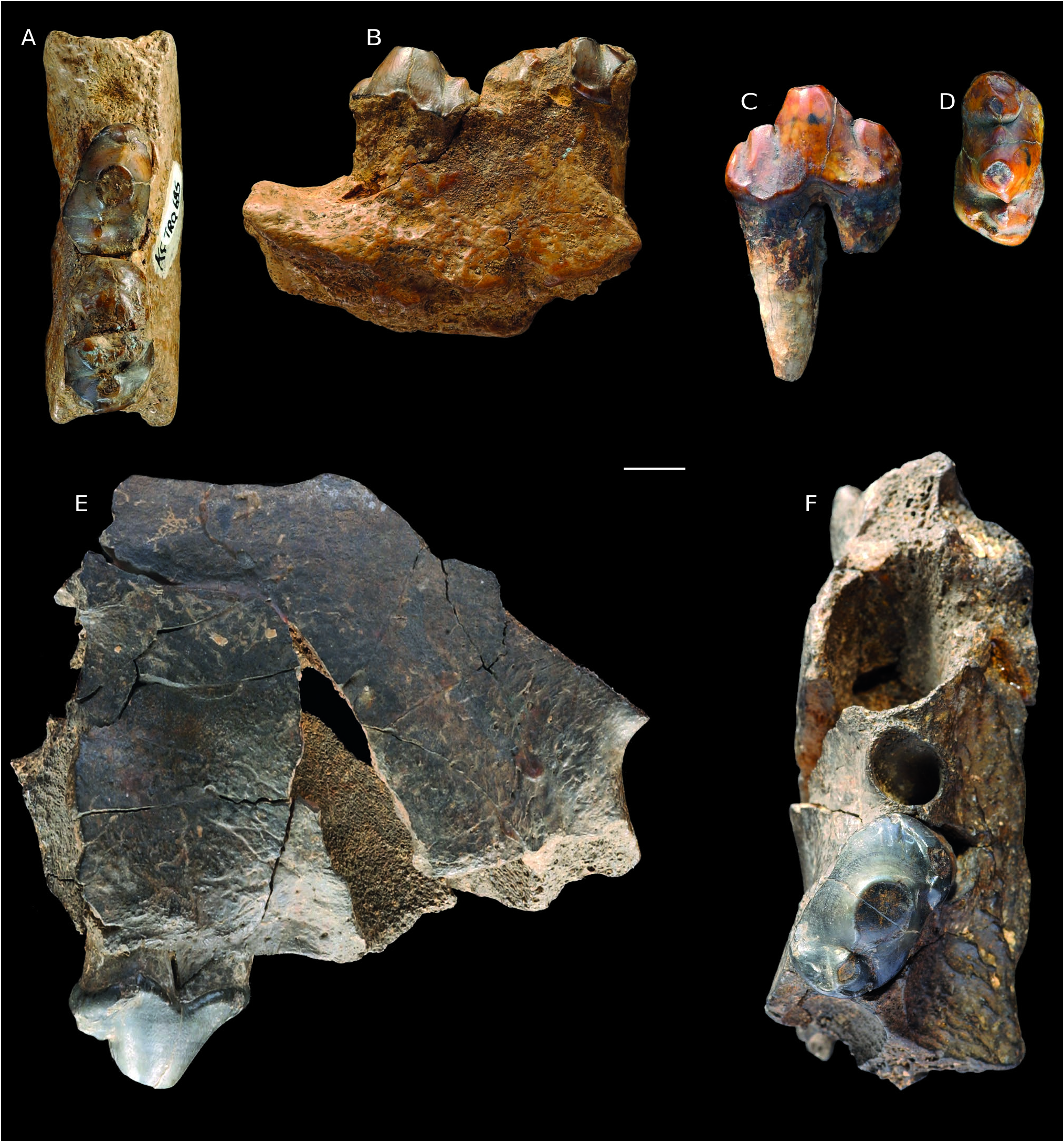
Dinocrocuta senyureki ( Ozansoy, 1957) ( Figs 5 View FIG , 6 View FIG ; Tables 6, 7)
Machairodus aphanistus – Sayar 1953: fig. 1.
REFERRED MATERIAL FROM KÜÇÜKÇEKMECE. — MNHN.F.TRQ685, fragment of left hemimandible with p2-3; KÇ 58, right p4; KÇ 60, fragment of right maxilla with P2; TRQ948, proximal half of left ulna, lacking most of the olecranon.
DESCRIPTION
Mandible ( Fig. 5A, B View FIG ; Table 6)
Only an anterior fragment of the dentary is preserved, but it is thick (TMp3 = 23 mm) and robust. The post-canine diastema is long and there is no alveolus for a p1.
Dentition
Lower dentition ( Fig. 5 View FIG A-D; Table 6). The two premolars preserved in MNHN.F.TRQ685 display a moderate, horizontal wear pattern, the p2 being less heavily worn than the p3. The p2 is set slightly obliquely in the dentary and relative to the p3 (see Fig. 5A View FIG ). A major part of the mesial root is visible well above the dorsal rim of the dentary so that in lateral view the tooth seems to be oriented backwards. It bears a strong, but not particularly bulbous, main cuspid; the crown width increases distad; the mesial accessory cuspid is small, slightly lingual, and displays a small wear facet; the distal accessory cuspid is slightly more prominent than the mesial one, centrally located, and is followed by a short cingulid. The p3 is both worn and damaged so that it is not possible to assess the relative development of the accessory cuspids. However, the tooth does not show the bulbous morphology of the p3 of durophagous species such as Adcrocuta spp. On the contrary, p3 appears to be an enlarged version of the p2. The isolated p4 KÇ 58 is moderately worn. It differs from the p2 and p3 by a more slender main cuspid and more developed accessory cuspids, where the mesial cuspid is larger than the distal cuspid. The distal accessory cuspid is followed by a short cingulid. The distal rim of the tooth is curved distally but rectilinear more mesially along the contact with the carnassial (see Fig. 5D View FIG ).
Upper dentition ( Fig. 5E, F View FIG ; Table 7). The fragmentary right maxilla KÇ 60 preserves the P2. Only the alveoli of the I3, canine, and P1 are preserved. That of the I3 is not complete but indicates a tooth larger than the P1. The canine alveolus is oval, approximately 32 mm in length and 25 mm in width. The single, rounded alveolus of P1 indicates that it was relatively large (alveolus length is 13.9 mm, alveolus width is 12 mm). Very short diastemata separate the P1 from the adjacent teeth. The main cusp and distal accessory cusp of P2 display a moderate, horizontal wear pattern. The tooth is wide relative to its length; it has roughly the same width mesial to the main cusp as distal to that cusp. There is no mesial accessory cusp but a strong cingulum that is particularly prominent mesiolingually. Contrary to the p2, the distal accessory cusp of P2 is lingually located; the distal cingulum is very reduced.
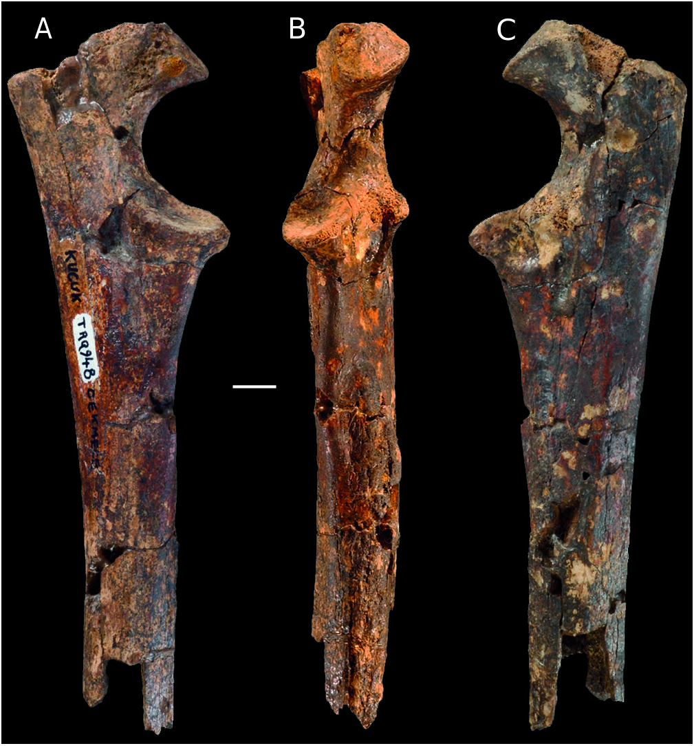
Postcranium ( Fig. 6 View FIG )
A fragmentary ulna is the only postcranial bone from Küçükçekmece that might be assigned to D. senyureki . Unfortunately, the olecranon is not preserved. Overall the shaft is slender and the articular part is narrow compared to that of Indarctos and Machairodus , two genera also present at the site. On the anconeal process, the articular surface for the humerus extends proximally less than in the bears, felids, and hyaenids used in comparison ( Ursus sp. , Indarctos arctoides , Panthera tigris (Linnaeus, 1758) and P. leo (Linnaeus, 1758) , Machairodus aphanistus , Amphimachairodus giganteus (Wagner, 1848) , Crocuta crocuta (Erxleben, 1777) , Hyaena hyaena (Linnaeus, 1758)) , especially on the lateral side. The morphology of the radial notch is typical of that in hyaenids: the lateral extremity of the coronoid process is very short and poorly projected, the articular facet for the articular circumference of the radius forms a very thin surface, and a deep, marked area (presumably for ligament attachment at the proximal radioulnar joint) is present just distal to the radial notch. Overall MNHN.F.TRQ948 looks very like the ulna of roughly contemporaneous hyaenids such as Adcrocuta eximia (e.g., MNHN.F.PIK3247, proximal fragment of right ulna from Pikermi, late Miocene of Greece) and that of extant hyaenas ( Crocuta crocuta , Hyaena hyaena ) though the shaft of the fossil specimen is clearly more robust than in the extant taxa.
COMPARISONS AND DISCUSSION
We follow previous authors by recognizing the family Percrocutidae as distinct from the Hyaenidae Gray, 1821 (see Werdelin 1996, for a historical background). Here I restrict comparisons to the late Miocene middle and large-sized percrocutids, i.e. the species of the genus Dinocrocuta . Sayar (1953: fig. 1) figured KÇ 60 as a maxilla fragment of the sabre-toothed felid Machairodus aphanistus , but the premolar preserved and the size of the alveoli indicate a clear distinction from those of a sabre-toothed cat. This specimen (KÇ 60, Fig. 5E, F View FIG ) is assigned to Dinocrocuta senyureki based on the overall size of the dentition, the morphology of P2, with its distinctive mesial cingulum, and the presence at the site of additional specimens of the same species. Dinocrocuta senyureki was erected by Ozansoy (1957) and described in detail later ( Ozansoy 1961, 1965) based on specimens from Yassiören (MN9, late Miocene, Turkey). The syntype content is not clear in 1957 but is detailed in 1961. Some of the specimens listed by Ozansoy (1961) are stored in the MNHN and comprise: MNHN.F.TRQ1010 (= Yas 62 in Howell & Petter 1985; Ozansoy 1957: pl. 2; Ozansoy 1961: pl. 2, figs 3, 4), a fragment of left hemimandible with p2, fragmentary p3, p4-m1; TRQ1009 (= Yas 59 in Howell & Petter 1985; Ozansoy 1961: figs 4, 5, pl. 2, figs 1, 2; Ozansoy 1965: pl. 3, figs 3, 4), a fragment of right mandible with c-m1;TRQ1008 (= Yas 60 in Howell & Petter 1985; Ozansoy 1961: figs 2, 3, pl. 1, figs 1-3; Ozansoy 1965: pl. 3, figs 1, 2), a fragment of left maxilla with P2-M1; and TRQ1007 (= Yas 61 in Howell & Petter 1985), a fragment of right maxilla with P3-4 belonging to the same individual; a few additional, more fragmentary specimens (see Ozansoy 1961, 1965). In his publications Ozansoy did not clearly select a type specimen, however. Howell & Petter (1985) regarded TRQ1010 (the only specimen figured by Ozansoy in 1957) as the type specimen of Dinocrocuta senyureki . This specimen is a lectotype, by subsequent designation ( Howell & Petter 1985). The material from Küçükçekmece is fragmentary, but the assignment of this material to Dinocrocuta senyureki is well supported by the comparison with specimens from the type locality. I did not find any difference between the lectotype TRQ1010 (Yassiören) and TRQ695 (Küçükçekmece). Both specimens share the absence of p1, the oblique orientation on the dentary and the morphology of p2, the absence of marked bulbous morphology on p3. In the upper dentition, the distinct mesiolingual cingulum around the crown basis of P2 has the same development in TRQ1007 from Yassiören and KÇ 60 from Küçükçekmece. This is also true for the p4 KÇ 58 from the latter site, which shows cuspids and cingulum as developed as in the p4 of the specimens from Yassiören listed above. Additional specimens from the Sinap were collected more recently and described by Viranta & Werdelin (2003). This sample comes from Loc. 108 (10 Ma in age; Kappelman et al. 2003) and Loc. 12 (9.6 Ma in age; Kappelman et al. 2003), both in the MN9 (early Vallesian) biostratigraphic level and from Loc. 37, MN10 (late Vallesian). These localities yielded a few postcranial bones and many dental remains ( Tables 6, 7). Among this new sample from Yassiören the elements that also are known from Küçükçekmece are morphologically very similar. In addition to the type locality, D. senyureki has been described from the Turkish sites of Eşme Akçaköy (MN9, early Vallesian), Kayadibi (MN11, early Turolian) and Inönü (MN10-11, late Vallesian-early Turolian), which yielded dental elements that are not comparable with those from Küçükçekmece (Schmidt-Kittler 1976). Elsewhere, the species is also known at Sahabi (late Miocene, Libya) from right and left hemimandibles of a single individual, an isolated P2 ( Howell 1987: fig. 4), and several postcranial specimens. There are differences in proportions or size of teeth or between teeth ( Table 6), but the intraspecific variability of the sample from Yassiören alone indicates great variability in size of D. senyureki . The P2 from Küçükçekmece is larger than in the other specimens of the species, but this is the sole difference and the known sample is limited. Therefore the material from Küçükçekmece is assigned to the same species.
Other species of Dinocrocuta are known from late Miocene sites in the Old World, especially D. minor ( Ozansoy, 1965) , D. salonicae (Andrews, 1918) , although generic assignment of this species is still debated ( Koufos 1995; Zhang 2005), D. gigantea ( Schlosser, 1903) and D. algeriensis ( Arambourg, 1959) . The morphological distinction between the species of Dinocrocuta , which has been discussed elsewhere (e.g., Howell & Petter 1985; Spassov & Koufos 2002; Zhang 2005), is not easy. The main reason to assign the material from Küçükçekmece to D. senyureki rather than to D. minor is the size, with D. minor being much smaller based on the holotype (by monotypy) and probably single known specimen of this species (but see Viranta & Werdelin 2003: 182),
MNHN.F.TRQ1011 (fragment of left hemimandible with i1-m1 = Yas 58 in Howell & Petter 1985; Ozansoy 1965: pl. 4, fig. 1). The p2 of the holotype of D. minor is narrower distally than in D. senyureki , but it is hard to tell whether this difference is significant considering the limited samples. Except size, one of the diagnostic features of D. senyureki is the presence on P3 of a strong mesial accessory cusp, but this tooth is not preserved in the material from Küçükçekmece. According to Zhang (2005), compared to D. senyureki , D. gigantea is much larger and has proportionally larger p2/P2. The specimens from Küçükçekmece fit this picture and show a more reduced p2 compared to p3 than in most specimens of D. gigantea . Dinocrocuta algeriensis is based on a sample from Oued el Hamman (= Bou Hanifia; late Miocene, Algeria; Arambourg 1959: fig. 8A, 9D, 10C, 11, 12D, pl. 2, pl. 3, fig. 1-4) representing at least three individuals, two (young) adults (the holotype MNHN.F-1951.9-174, the paratype MNHN.F-1951.9-172, MNHN.F-1951.9-29,
MNHN.F-1951.9-75, MNHN.F-1951.9-76) and one juvenile (MNHN.F-1951.9-19 and a couple of isolated, unnumbered teeth). An isolated P3 (MNHN.F.AMA9) from Menacer (= Marceau in Arambourg 1959) is also assigned to this species. Like D. gigantea , Dinocrocuta algeriensis differs from our material by a enlarged p2, especially relative to p3, and a thicker P2.
MNHN.F.TRQ948 is an important specimen because percrocutid postcranial remains are rare ( Howell 1987; Zhang & Xue 1996; Viranta & Werdelin 2003). Nevertheless, it is far too fragmentary to address morphological distinction between the skeleton of hyaenids and percrocutids.
No known copyright restrictions apply. See Agosti, D., Egloff, W., 2009. Taxonomic information exchange and copyright: the Plazi approach. BMC Research Notes 2009, 2:53 for further explanation.