
Microrhagus triangularis (Say, 1823)
|
publication ID |
https://doi.org/10.5281/zenodo.5182118 |
|
publication LSID |
lsid:zoobank.org:pub:1DEC04DB-99DB-466B-838B-2C337251632E |
|
DOI |
https://doi.org/10.5281/zenodo.5191280 |
|
persistent identifier |
https://treatment.plazi.org/id/594DB57A-EE78-BA67-57F2-86DEFD7DFB85 |
|
treatment provided by |
Felipe |
|
scientific name |
Microrhagus triangularis (Say, 1823) |
| status |
|
Microrhagus triangularis (Say, 1823)
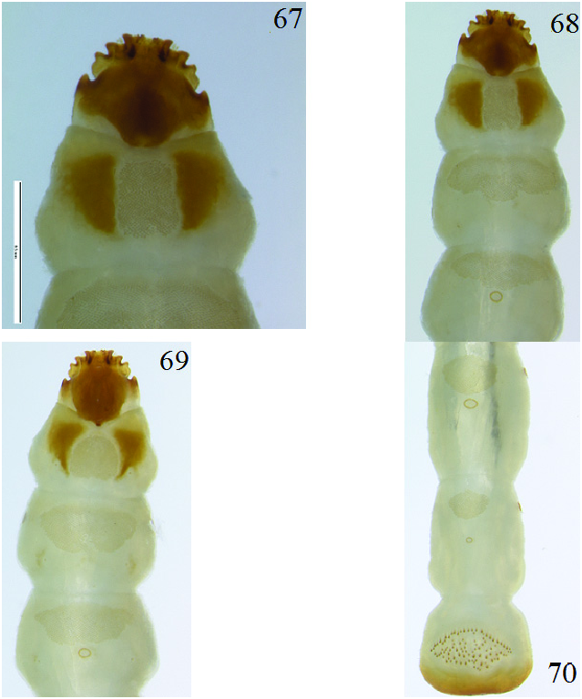
Fifth instar
( Fig. 66–70)
Diagnosis. Rectangular-shaped prothoracic microtrichial patch and oval shaped, somewhat enlarged areoles will distinguish M. triangularis from M. lecontei .
Specimens Examined. Eight larvae collected at USA: WISCONSIN: Dane County, Marshall Park , N43° 05.533’, W-89° 28.975’, 16 September 2013, Robert L. Otto, in rotten log (2 larvae) GoogleMaps ; Oconto County, Rueckert’s private property, T29N R17E sec 16, 10 April 2007, 16 April 2007, Robert L. Otto, in white rotten maple log (2 larvae) ; Oconto County, Quartz Hill Trail , N45° 22.449’, W-88° 37.765’, 16 April 2010, Robert L. Otto, in rotten aspen log (4 larvae). Larvae are deposited in GERP and WIRC GoogleMaps .
Description. Length, 8.0–10.0 mm. Width, 1.0 mm. Orthosomatic, elateriform. Body cylindrical, sides parallel, cream-yellow with head, prothoracic sclerome patches and caudal end of abdominal segment IX dark brown. Setae either indistinct or absent. Legs reduced to simple setae. Dorsal and ventral microtrichial patches slightly darker in color compared to their surrounding areas ( Fig. 66).
Head ( Fig. 67 View Figures 67–70 ): Strongly flattened, prognathous and inserted into prothorax. Dorsal cephalic disc sub-circular. Very weak median carina present on dorsal cephalic disc. Venter unmodified, without furrows or ridges. Base and lateral sides of the head capsule unsclerotized. Anterior portion of head capsule heavily sclerotized. Each lateral side of head capsule with four projections. Basal lateral projections enlarged. Lateral sides between first and second lateral projections strongly sinuate. Second through fourth lateral projections directed laterally. Antennae minute, arising between the third and fourth lateral projections. Scape not visible. Pedicel elongate. Sensorum and flagellum sub-equal in length. Sensory papillae indistinct. Mandibles minute, resting in the mesal acumination of the head capsule. Each mandible heavily sclerotized, oval with two outwardly projecting teeth. Labial and maxillary palpi indistinct. Ligula, mala, lacinia and galea not visible. Hypostomal rods absent.
Prothorax ( Fig. 68–69 View Figures 67–70 ): Sub-equal to subsequent two thoracic segments. Tergum with pair of triangular-shaped scleromes extending from base up three-fourths the length of the segment then diverge towards lateral sides and converge to point of origin; caudal and lateral sides with undefined edges. Rectangular-shaped microtrichial patch present between scleromes. Sternum with pair of internally bent sub–triangular-shaped scleromes present converging towards median of segment. Sub–circularshaped microtrichial patch present between scleromes. Both surfaces without areoles.
Meso- and metathorax: Terga with elliptical-shaped microtrichial patch. Sterna with semicircularshaped microtrichial patch. Mesothorax variable; either with small, circular areole or absent altogether on both surfaces. Metathorax with small circular areole beneath patch, near middle of each segment on both surfaces. Metathorax with reduced spiracles.
Abdomen: Segments I–IX sub-equal in length and width. Terga I–VIII with microtrichial patch that successively change from oval on segment I to a small triangle on segment VIII. Sterna I–VIII with microtrichial patch that successively change from elliptical on segment I to a small oval on segment VIII. Areoles variably sized, largest on segments II–VI on both surfaces, circular to oval-shaped; positioned beneath patch, near middle of each segment. Tergum IX without microtrichial patch and areole; sternum ( Fig. 70 View Figures 67–70 ) heavily sclerotized at caudal half with prominent, semicircular circumanal asperities. Urogomphi absent on segment IX. Spiracles annular-biforous, with rounded spiracular collar.
Distribution. Microrhagus triangularis is known from CANADA: New Brunswick, Nova Scotia, Ontario, Québec; USA: Alabama, Arkansas, Connecticut, District of Columbia, Florida, Georgia, Illinois, Indiana, Iowa, Kansas, Kentucky, Louisiana, Maryland, Massachusetts, Michigan, Minnesota, Mississippi, Missouri, New Hampshire, New Jersey, New York, North Carolina, Ohio, Pennsylvania, South Carolina, Tennessee, Texas, Virginia, West Virginia and Wisconsin ( Muona 2000; Majka 2007; Webster et al. 2012). All specimens used in this study came from Wisconsin.
Biology. Microrhagus triangularis is one of the most common species found in eastern North America and some biological information is now known. Muona (2000) wrote M. triangularis was found on dogwood ( Cornus sp. ; Cornaceae ). Majka (2007) took the species using a car net in Nova Scotia. Webster et al. (2012) collected seven specimens in July and August. One specimen was taken by beating foliage in a mixed forest. The remaining six specimens were taken from Lindgren funnel traps placed in an old red pine forest, a mature hardwood forest with American beech, sugar maple, and ash, an old-growth eastern white cedar forest, and an old red oak forest.
In Wisconsin I found M. triangularis in a variety of forest systems including floodplain forest, northern dry-mesic forest, northern hardwood swamp, northern mesic forest, northern wet-mesic forest, Pine Barrens, southern hardwood swamp, and southern mesic forest. I collected one adult specimen on goldenrod ( Solidago sp. ; Asteraceae ) in 1994. Another adult specimen was swept through understory at Lone Rock in 1994. Many larvae were extracted from white rotten, moist maple and aspen ( Populus sp. ; Salicaceae ) logs from 2007 through 2009. Searching in conifers has yielded no larvae, which may indicate this species is a deciduous specialist. I observed larvae tunneling parallel with the wood grain, leaving no trails behind them. Many larvae were extracted at least 2.5–3.0 cm beneath the surface. Like many other observed species, larvae of M. triangularis construct a pupal chamber near the surface and assume a U-shaped position. I observed no noticeable difference between previous instars and prepupal larval stage. Pupation requires about two to three weeks. Most recently, many adults were collected from purple prism traps during mid-June through the end of August in northeastern Wisconsin. Collectors in Wisconsin found M. triangularis in Malaise traps, sweeping grass and herbaceous understory, in Lindgren funnel traps, and on girdled ash trees.
Seven adults emerged on 5 June, 2010. Adults were placed in a vial with a piece of wood to observe their behavior and attempt to induce breeding. No eggs or first instars were observed following completion of adult observations. Like other eucnemid species, M. triangularis are capable of snapping into the air when placed on their backs. Adults were observed to quiver their extended antennae while in captivity. Adult beetles are short lived, lasting about one to two weeks in captivity before dying. It is likely M. triangularis completes its development in one year under optimal conditions, but may arrest its development under less optimal conditions.
No known copyright restrictions apply. See Agosti, D., Egloff, W., 2009. Taxonomic information exchange and copyright: the Plazi approach. BMC Research Notes 2009, 2:53 for further explanation.
|
Kingdom |
|
|
Phylum |
|
|
Class |
|
|
Order |
|
|
Family |
|
|
Genus |