
Pseudogonatodes furvus Ruthven 1915
|
publication ID |
https://doi.org/10.11646/zootaxa.4915.1.3 |
|
publication LSID |
lsid:zoobank.org:pub:A2B1245F-0665-49DA-B476-89F80FDAA75D |
|
DOI |
https://doi.org/10.5281/zenodo.4457940 |
|
persistent identifier |
https://treatment.plazi.org/id/5F6F87C7-FFA0-FFC4-FF46-85679EDB527C |
|
treatment provided by |
Plazi |
|
scientific name |
Pseudogonatodes furvus Ruthven 1915 |
| status |
|
Pseudogonatodes furvus Ruthven 1915
Pseudogonatodes furvus Ruthven 1915:2
Lepidoblepharis intermedius Boulenger-Ruthven 1922:58
Pseudogonatodes furvus Ruthven : Parker 1926:297, Dunn 1944:86, Vanzolini 1967:2, Vanzolini 1968:92 Mechler 1968:350, Huey & Dixon 1970:538, Peters & Donoso-Barros 1970:242, Ayala et al. 1984:135, Ayala 1986:561, Sánchez-C. et al. 1995:318, Avila-Pires & Hoogmoed 2000:209, Carvajal-Cogollo et al. 2012:810, Rangel-Ch. et al. 2012:337, Rueda-Solano et al. 2014:609, Esqueda et al. 2016:18.
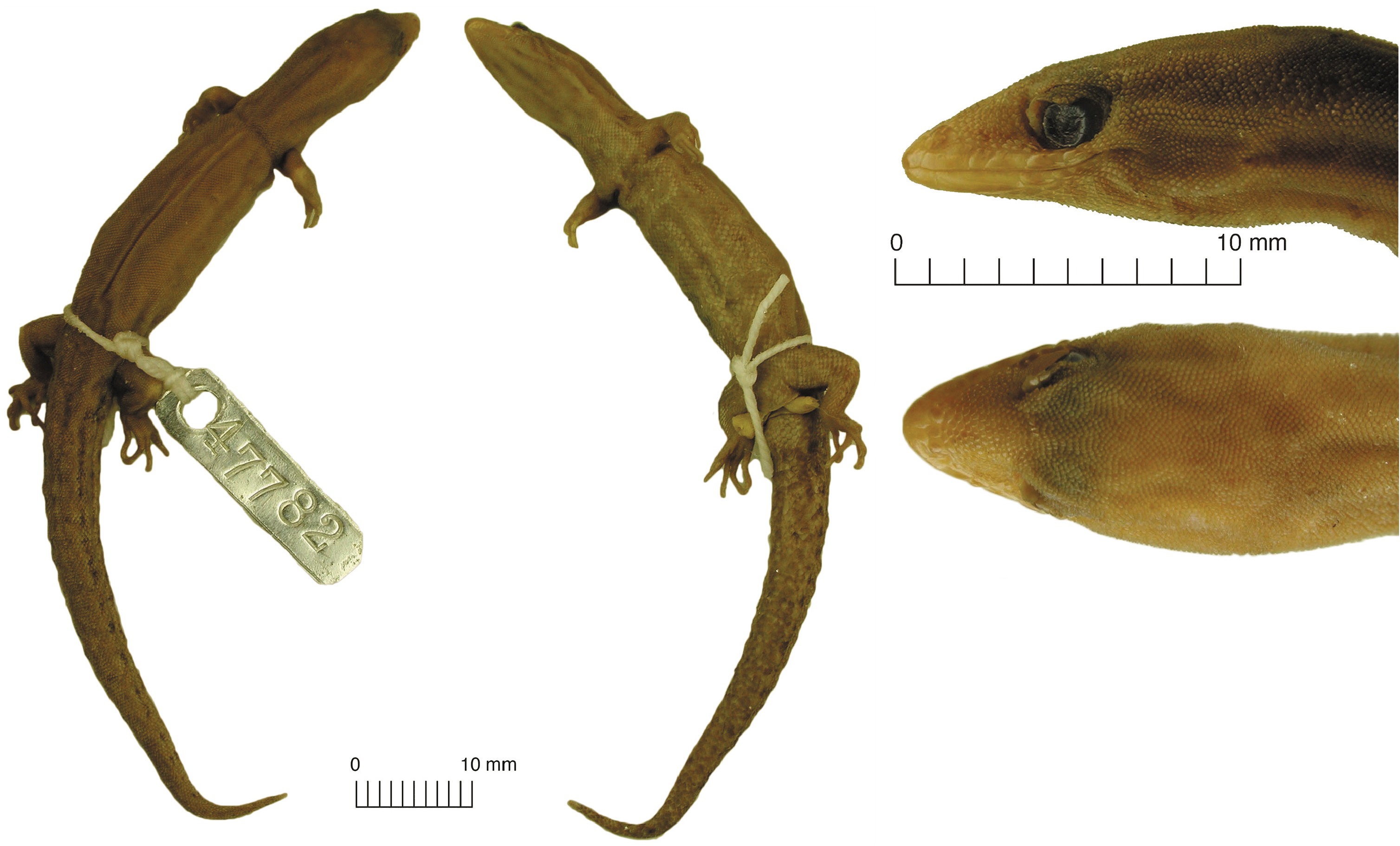
Holotype. UMMZ 47782 View Materials , adult male collected July 20, 1913 by Frederick M. Gaige.
Paratopotype. UMMZ 44783. According to Ruthven (1915), a “much mutilated” specimen found in the stomach contents of the snake Mastigodryas boddaerti ( Sentzen, 1796) .
Type locality. Colombia, Departamento del Magdalena, Distrito de Santa Marta, from “San Lorenzo (altitude of 5000 feet [~ 1500 m asl.]), Santa Marta Mountains”. The Serranía de San Lorenzo is a peripheral relief of the northwestern sector of the SNSM, which reaches 2800 m asl. Nevertheless, the site currently called “San Lorenzo” ( i.e. Estación Experimental San Lorenzo) is located above 2000 m asl. Since the holotype was collected at 1500 m asl., we restrict the type locality to “Vereda Bella Vista, Sierra Nevada de Santa Marta”, corregimiento de Minca, Distrito de Santa Marta , Magdalena , Colombia.
Examined specimens. MCZ: Herp:R-29700, adult male collected 1928 by Philip J. Darlington at Río Frío , municipio de Zona Bananera, Magdalena, elevation unknown. ICN-R 3501–02 , two juvenile specimens collected 1986 by D. M. Harris at San Pedro de la Sierra, Municipio de Ciénaga , Magdalena, approximately 1600 m asl . CBUMAG:REP:00295, juvenile specimen collected in 2005 by Juan M. Renjifo, Hernán D. Granda-Rodríguez, and John D. Lynch at Vereda El Campano , Distrito de Santa Marta, Magdalena, 1420 m asl . CBUMAG:REP:00283, 293, a female and juvenile specimen, collected 2013 by Luis A. Rueda-Solano at Palmor de la Sierra, Municipio de Ciénaga , Magdalena ( 1400 m asl.). These specimens was erroneously cited by Rueda-Solano et al. (2014) as CBUMAG:REP:00031–32, respectively.
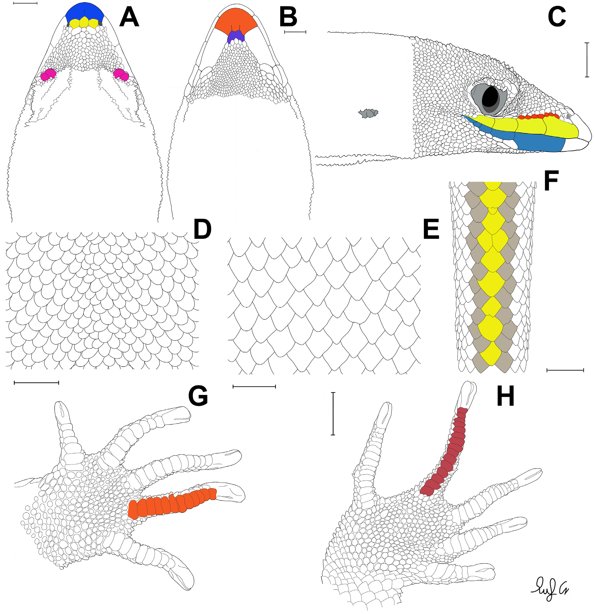
Extended definition. Pseudogonatodes furvus can be distinguished from all of its congeners by the following combination of characters ( Figure 1 View FIGURE 1 ): (1) maximum SVL of 45 mm for males and 43.8 mm for females; (2) dorsal scales homogenous, granular, subimbricate, and larger than occipital scales; (3) 83–104 scales around midbody; (4) posterior edge of rostral indented by three postrostrals, with a median cleft; (5) 4–5 supralabials; (6) 4–5 infralabials; (7) 7–10 loreals; (8) posterior edge of mental scale with two median clefts (W-shaped), indented by (9) 2–4 postmentals, which are larger than subsequent scales; (10) 44–67 ventrals in straight line between anterior level of forelimbs and border of cloaca, (11) 34–39 ventrals between anterior levels of forelimbs and hindlimbs; (12) long digits lacking expanded subdigital third lamella, (13) 11–13 subdigital lamellae under finger IV and (14) 11–15 subdigital lamellae under toe IV; (15) subcaudal pattern with sequence of moderately to heavily enlarged medial scales in contact laterally with one to two scales.
Description of holotype. Meristic and lepidosis data are included on Table 1. Head cone-shaped, long and pointed ( Figure 2 View FIGURE 2 ). Rostral wide, distinctly visible from above, with a posterior medial cleft. Three postrostrals, subequal in size, distinctly larger than adjacent scales on the snout. Nostril bordered by rostral, lateral post-rostral (= supranasal or lateral internasal), two postnasals, and first supralabial. Lower postnasal is about twice as large as the upper one. Scales on snout and anterior loreal scales flat, smooth, subimbricate; decreasing in size and changing into juxtaposed granules on top of head and on posterior part of loreal region. Ten loreal scales. Scales on supraorbital region granular, similar to adjacent scales on parietal region. Most of the anterodorsal ocular scales form a supraciliary flap, with four enlarged scales, of which the second is largest. Five supralabials, suture between fourth and fifth below center of eye. Scales on the temporal region similar to those on top of head (small, granular, juxtaposed). Tympanic membrane small, round to roughly triangular.
Mental large, posteriorly indented by two postmentals and with two short clefts (W-shaped). Postmentals several times larger than adjacent scales on the chin. Scales on chin are mostly granular, juxtaposed; anterior scales polygonal, smooth, some of those close to infralabials elongate. A larger scale borders the first (posteriorly) and second infralabials. Five infralabials, first reaching anterior level of orbit and twice as wide as second infralabial; suture between third and fourth infralabial below center of eye. Scales on upper part and sides of neck granular, similar to dorsals. Anterior scales of the throat granular, posterior flat, smooth, and imbricate; with an abrupt transition between the two areas.
Dorsal scales granular, granules larger than those on parietal region, slightly inclined posteriorly. Ventral scales distinctly larger than dorsals, flat, smooth, imbricate, approximately rhomboid (especially on posterior part of belly); 39 scales in a midventral line between anterior levels of fore- and hindlimbs, 46 scales between throat to vent, and 27 scales in a transverse line at midbody. Abrupt transition between dorsals and ventral scales. Scales around midbody 106. Scales on preanal plate wider than ventrals. Escutcheon scales absent.
Scales on tail flat, smooth, imbricate, small and elongate dorsally and on the sides, larger and wider ventrally. Midventral scale row moderately enlarged, with a sequence 1’2’1*’, where * is a small scale. Scales on anterior and part of upper aspects of forelimbs, and on anterior and ventral aspects of hind limbs flat, smooth, imbricate. Elsewhere on limbs scales granular. Sole of foot with homogeneous squamation. Lamellae under fourth fingers 13, under fourth toes 15. Lamellae under digits subequal in size, with none much larger than the others. Claws enclosed by an ungual sheath composed of five scales, as characteristic for the genus.
Variation. Although the general appearance of the examined specimens is similar to the holotype, several characters of lepidosis exhibit some variation. All the specimens referred have three postnasals on both sides of the head. One specimen ( ICN-R 3502 ) has the same arrangement of postnasals as the holotype (lower twice as large as upper). For two specimens, the lower postnasal is the smallest, about as large as adjacent loreal scales ( MCZ:Herp: R-29700 and CBUMAG:REP: 00295). On the other hand, three specimens show a lower postnasal subequal to the medial one, and larger than adjacent loreals ( CBUMAG:REP:00283, 293, and ICN-R 3501 ). Three to seven scales on the supraciliary flap, that may or may not be symmetrical on both sides of the head. Scales between sutures of third and fourth supralabials approximately below the center of the eye. The supraciliary scales may be enlarged from first to fourth. All examined specimens show four supralabials symmetrical on both sides .
The number of infralabials is variable. One specimen ( CBUMAG:REP:00283) shows five on the left side and four on the right side. The remaining specimens have four symmetric infralabials, with the sutures of second and third ( CBUMAG:REP:00293) or third and fourth ( MCZ:Herp:R-29700, CBUMAG:REP: 00295, and ICN-R 5101 ) below the center of the eye. Mental scale exhibits variation, one specimen ( CBUMAG:REP:0029) has a concave posterior border, without medial clefts, partially indented by three postmentals. The number of postmentals varies between two ( holotype and MCZ:Herp:R-29700), three ( CBUMAG:REP: 00293, 295, and ICN-R 3501–02 ), or four ( CBUMAG:REP: 00283) scales .
Counts of scales around midbody are less in examined specimens compared to the holotype. Ventrals between anterior levels of fore- and hindlimbs are less than in the holotype in MCZ:Herp:R-29700, equal in ICN-R 3502 , and higher in CBUMAG:REP:00283, 293, 00295, and ICN-R 3502 . Number of ventrals between anterior level of forelimbs to cloaca is less than in the holotype in MCZ:Herp:R-29700, and ICN-R 3501–02 , equal in CBUMAG: REP:00293, and higher in CBUMAG:REP:00283 and 00295. Ventrals in the transverse line at midbody of almost all examined specimens are lower than the holotype , except in CBUMAG:REP:00283. The midventral scales of tail are hardly distinct in specimen MCZ:Herp:R-29700, in a sequence of 1’1’, while in CBUMAG:REP:00293 in a sequence of 1*’’1’2’’1’1’’ proximally. Examined specimens exhibit the same number of subdigital lamellae under the fourth fingers as the holotype (except CBUMAG:REP: 00295, which has ten lamellae on the left side and eleven on the right side) . Counts of subdigital lamellae under the fourth toe are asymmetric in most of the specimens, in all cases less than in the holotype.
Morphometry. All measurements and proportions are given in Table 2. The holotype is the largest specimen known for the species, SVL of 45 mm. For the rest of the specimens the SVL ranges from 23.2 to 43.8 mm. In two specimens with original tail, the tail is slightly longer than SVL (TL/SVL = 1.02–1.07). Axilla-groin length about one-third to one-half of SVL (AXG/SVL = 0.35–0.45). Head one-half to two-thirds longer than wide (HW/HL = 0.55–0.71), but short in comparison with body (HL/SVL = 0.20–0.27). Eye-nostril distance about one-fifth to onefourth of HL (EYN/HL = 0.21–0.24), and snout about one third of HL (RL/HL = 0.31–0.41).
Color in ethanol. Dorsal surface of head, back, flanks and limbs uniformly brown in holotype, sides of head, flanks and limbs with dark brown spots in MCZ 29700. In the latter specimen, a pair of dorsolateral, light stripes, bordered by dark brown, at the level of hind limbs. Ventral region is slightly lighter than the dorsum, with some darker spots, especially on the throat and chest. Tail of similar color as body, with some longitudinal rows of dark brown spots. Dorsolaterally, on the proximal part of the tail, the spots may be connected into continuous stripes. A proximal, longitudinal stripe is also present mid-dorsally, in MCZ 29700. For color in life, see available photographs in Rueda-Solano et al. (2014) and Uetz et al. (2019).
Comparisons. A summary of morphological characters of the genus Pseudogonatodes is given in Table 3. Pseudogonatodes furvus is quickly distinguished from all members of the Group I ( P. guianensis and P. lunulatus ) sensu Huey & Dixon (1970) by the following characters: absence of enlarged third lamellae under fourth toe (present in Group I); posterior edge of mental with two median clefts (W-shaped) and postmentals larger than following scales (posterior border of mental slightly concave to heavily concave [U-shaped], postmentals equal in size to following scales); and sole of foot with homogeneous squamation (heterogeneous squamation in P. lunulatus ). Regarding the only member of the Group II ( P. barbouri ), P. furvus is distinguishable by the absence of enlarged third subdigital lamellae under the fourth toe (present in P. barbouri ); dorsals small, granular, conical, and sub-imbricated (large, flat, and imbricate). From Group III, P. furvus differs by having posterior edge of mental with two median clefts (Ushaped without clefts in P. peruvianus Huey & Dixon 1970 , V-shaped with a median cleft in P. gasconi Avila-Pires & Hoogmoed 2000 ); two to four postmentals larger than following scales (six equal in size to following scales for P. gasconi , six to eight slightly larger than following scales for P. manessi Avila-Pires & Hoogmoed 2000 ); subcaudal pattern with sequence of moderately to heavily enlarged midventral scales in contact laterally with one to two scales (no enlarged midventral subcaudal scales in P. gasconi ); and seven to ten loreals (six in P. peruvianus ).
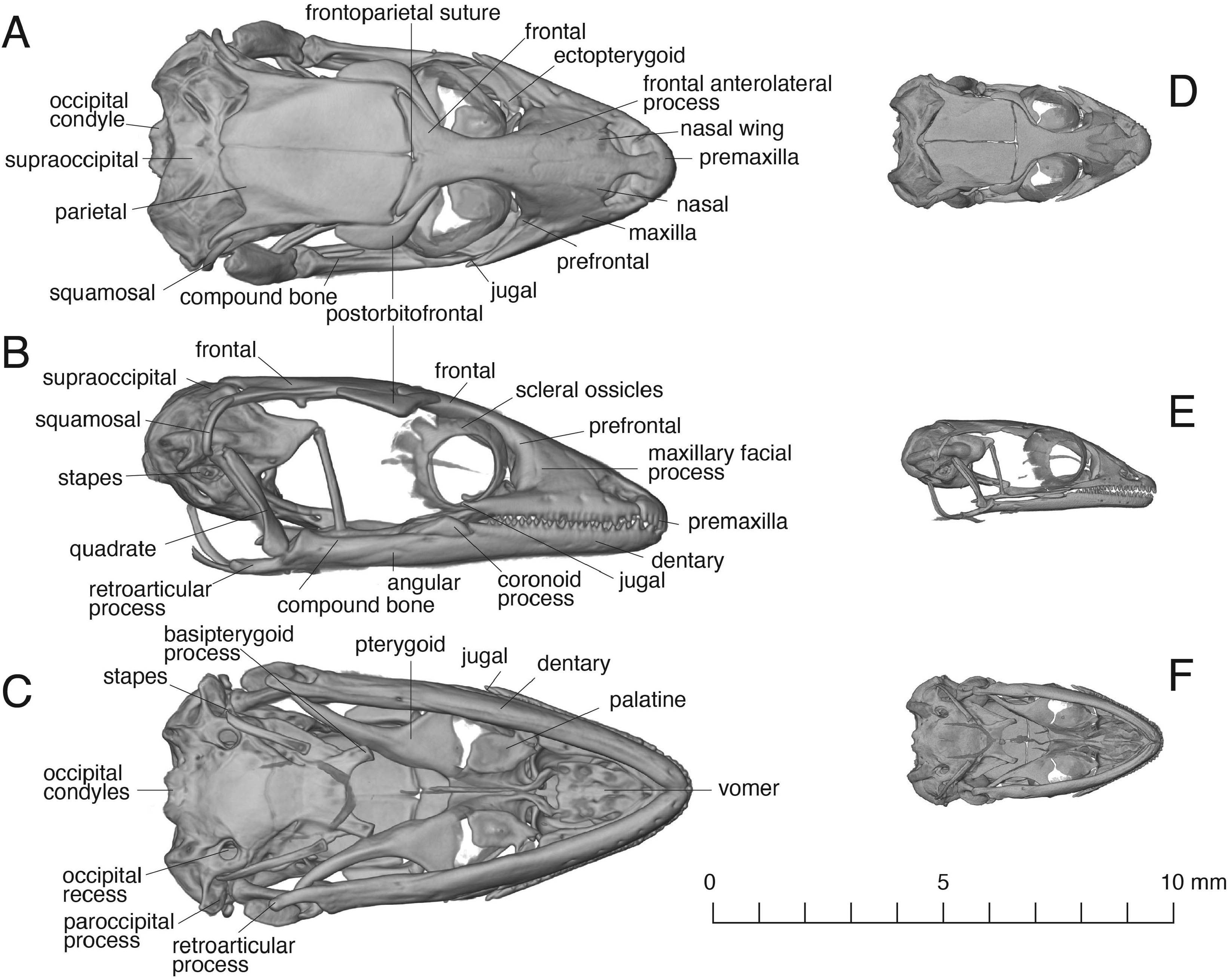
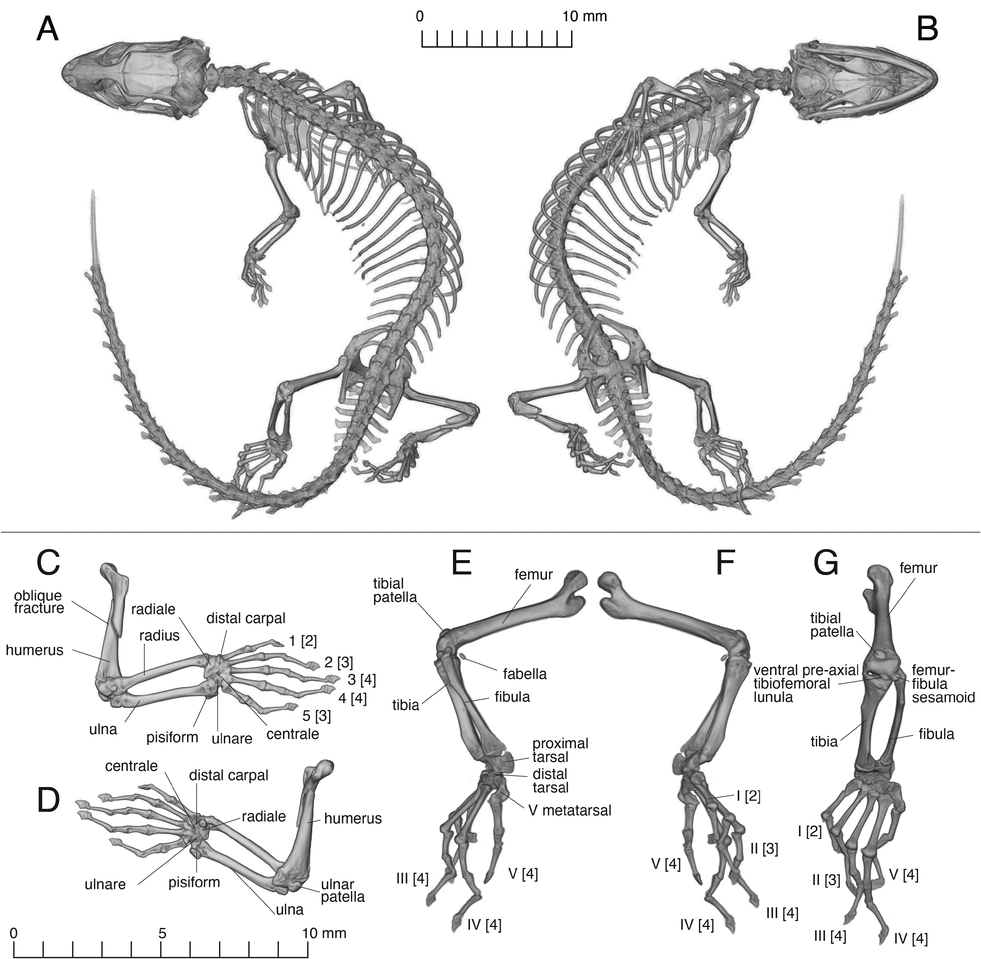
Osteology. The detailed description of P. barbouri ( Bauer et al. 2018) allows us to establish morphological comparisons with P. furvus ( Figures 3 View FIGURE 3 A–C, 4). These preliminary observations indicate that there is a good amount of morphological variation in the skeleton ( Figure 4 View FIGURE 4 ). However, the main differences could be attributed to size differences. Although these two species are miniaturized, the specimen of P. barbouri (MCZ:Herp:R-14385) is nearly half the size of P. furvus (MCZ:Herp:R-29700, Figure 3 View FIGURE 3 D–F).
Osteological differences. The skulls of these species ( Figures 3 View FIGURE 3 , 4 View FIGURE 4 ) differ in degree of overlap of the bones of the snout. The nasal bones cover the anterodorsal surface of the frontal in P. furvus , while in P. barbouri , the anterolateral process of the frontal remains exposed and extends almost to the border of the osseous naris, the nasals are more rectangular in P. furvus and more triangular in P. barbouri . The anterolateral process of the frontal contacts the facial process of the maxilla extensively in P. barbouri , while the anterolateral process of P. furvus barely contacts the maxilla. The frontoparietal suture is nearly transverse in P. barbouri , and this suture is more curved in P. furvus . The posterior margins of the parietals are straight and converge gradually towards the midline in P. barbouri , while in P. furvus the posterior margins of the parietals are more sinuous. The quadrate bone (and the paroccipital process) is attached to the braincase in a mid-position, while the paroccipital process is shifted more anteriorly in P. barbouri (also in Chatogekko , see Gamble et al. 2011), the posterior edge of the osseous nares is mainly bounded by the facial process of the maxilla in P. barbouri , while the same edge is bounded by the nasals and maxilla in P. furvus . The palatines approach each other in the midline in P. furvus reducing extensively the pyriform recess, while in P. barbouri they are more separated. The anterior edge of the pterygoid is more concave in P. barbouri than in P. furvus ; the coronoid eminence is lower in P. barbouri than in P. furvus . The retroarticular process is expanded distally, “spoon-like” in P. barbouri while it maintains a constant width in P. furvus . The semicircular canal ducts and the ampullae bulge more in P. barbouri than in P. furvus . The occipital condyle is less bulging in P. barbouri than in P. furvus . The interclavicle in P. furvus has some short but discrete lateral processes ( Figure 4 View FIGURE 4 ), being cruciform, while the one illustrated for P. guinanensis (MZUSP 94826) is nearly blade-like, with no lateral process ( Daza & Bauer 2012). The intercentrum also has the well-developed medial projections of New World sphaerodactylids ( Bauer et al. 2018), but the lateral projections were not as developed as in P. barbouri .
Osteological similarities. As in many miniaturized taxa, the frontoparietal suture is located in the middle of the skull ( i.e. muzzle unit/ parietal unit length ~ 1:1, Daza et al. 2008). The nasals are separated anteriorly for most of their length and become in contact posterior to the premaxillary nasal process; the postorbitofrontal has a rounded lateral margin and the posterior process is nearly three times the width of the anterior process; the basipterygoid process is not expanded distally, occipital recess visible in ventral view, causing the aperture of the recessus scalae tympani to open ventrally and not laterally ( Daza et al. 2008).
......continued on the next page
Osteological synapomorphies of the genus. In X-rays of several species within the genus Pseudogonatodes ( P. guianensis , P. lunulatus , Pseudogonatodes sp.) it was consistent that toe IV has four phalanges ( Figure 4 View FIGURE 4 ), contrary to all other New World sphaerodactylids, where there are five; this character was originally recognized by Kluge (1995). Characters proposed by Bauer et al. (2018) are also present in P. furvus include the ectopterygoid clasping the anterolateral process of the pterygoid, and the broad lateral wing of the nasals, but these characters can also be found in Coleodactylus (former) and Chatogekko (latter).
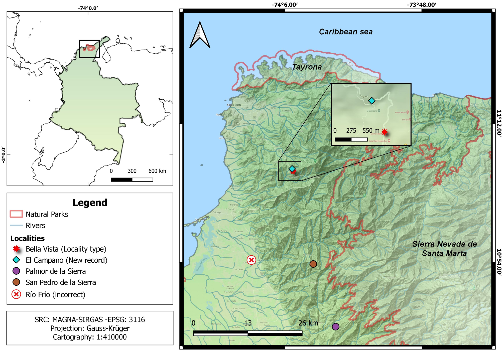
Distribution and conservation status. Pseudogonatodes furvus was previously recorded from three localities on the northwestern and western slopes of the SNSM, from 1400–1600 m asl. Herein, we report a fourth locality with confirmed presence of P. furvus , at Vereda El Campano, Santa Marta, Magdalena ( 11°6’46.07” N, 74°5’30.32” W, 1420 m asl.). Adding this record, the geographic distribution of P. furvus reaches an extent of occurrence (EOO) of 24.05 km 2 and an area of occupation (AOO) of 12 km 2 ( Figure 5 View FIGURE 5 , Appendix 2).
In the last assessment of the conservation status ( Caicedo et al. 2015) of P. furvus , it was categorized as Data Deficient (DD) due to (1) no records since 1980, and (2) threats from illegal land clearance. As this assessment proposed, the EOO and AOO of the species are small, and all records are near the Sierra Nevada de Santa Marta National Park, but the closest point is approximately 3.4 km from the “ San Pedro de La Sierra” sector. Therefore, it is not certain whether some populations can be found within this protected area.
With the preliminary review of vouchers from herpetological collections, we obtained a first approximation of the areas of occurrence and extension of P. furvus . Given these results, we suggest that this species could be categorized within the IUCN criteria as Endangered: EN B2ab (iii), due to the following criteria: (1) AOO < 500 km 2, (2) less than five confirmed localities, and (3) a decrease in habitat availability. We have no direct data for the latter point, but an exponential loss of natural covers in the area in the last 10 years, mainly those outside the SNSM National Park, has been observed ( Granda-Rodríguez et al. 2020). Such loss in natural covers can be a threat to the habitat quality and availability of the species.
No known copyright restrictions apply. See Agosti, D., Egloff, W., 2009. Taxonomic information exchange and copyright: the Plazi approach. BMC Research Notes 2009, 2:53 for further explanation.
|
Kingdom |
|
|
Phylum |
|
|
Class |
|
|
Order |
|
|
SuperFamily |
Gekkota |
|
Family |
|
|
Genus |
Pseudogonatodes furvus Ruthven 1915
| Montes-Correa, Andrés Camilo, Saboyá-Acosta, Liliana P., Jiménez-Bolaño, Juan David, Angarita-Sierra, Teddy, Briceño-Pérez, Vladimir, Núñez, Samuel, Renjifo, Juan Manuel, Schargel, Walter E., Daza, Juan D. & Hoogmoed, Marinus S. 2021 |
Lepidoblepharis intermedius
| Boulenger-Ruthven 1922: 58 |
Pseudogonatodes furvus
| Ruthven 1915: 2 |
Pseudogonatodes furvus
| Ruthven 1915 |