
Microtendipes famiefeus Sasa, 1996
|
publication ID |
https://doi.org/10.11646/zootaxa.4320.3.8 |
|
publication LSID |
lsid:zoobank.org:pub:5C046843-3E95-4D75-A891-50559A12C05E |
|
DOI |
https://doi.org/10.5281/zenodo.3510582 |
|
persistent identifier |
https://treatment.plazi.org/id/6107879D-C448-FD33-E6F7-FB317074916D |
|
treatment provided by |
Plazi |
|
scientific name |
Microtendipes famiefeus Sasa |
| status |
|
Microtendipes famiefeus Sasa View in CoL
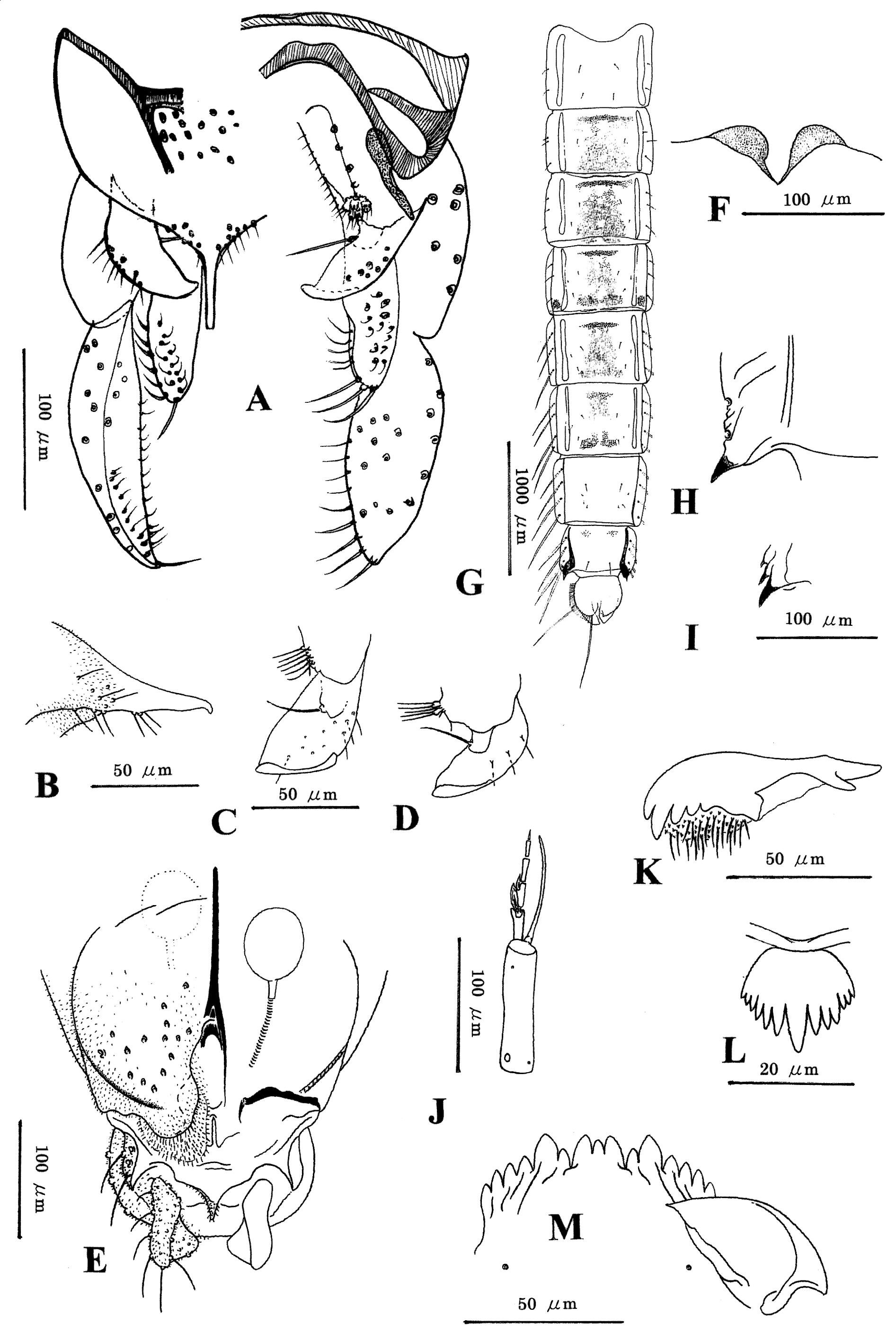
( Figure 3 View FIGURE 3 )
Microtendipes truncatus Kawai & Sasa, 1985: 18 View in CoL [preoccu. Kieffer 1922: 13]; Qi & Wang 2006: 43 View Cited Treatment . Microtendipes famiefeus Sasa, 1996: 53 View in CoL .
Microtendipes tusimadeeus Sasa & Suzuki, 1999: 5 View in CoL . Syn. nov.
? Microtendipes rydalensis View in CoL [ nec Edwards, 1929: 404]: Tanaka, Sasa & Hashizume 2003: 122.
Material examined. Holotype of Microtendipes famiefeus , M (NSMT-I-Dip 4940), labelled, “ No. 255: 11”, JAPAN : Toyama, Lake in the Toyama City Family Park , 21.ix.1993. Holotype of Microtendipes tusimadeeus , M (NSMT-I-Dip 5245), labelled, “ No. 373: 5”, JAPAN : Nagasaki, Tsushima Island, Izuhara, Azugawa River , 23.iii.1998. Non-types. M, Pe /F, L ( SUM), JAPAN : Miyagi, Shiroishi, Kamasaki Hot Spring, Yukawa River , 1.i.1997 (emerged 11 and 19.i.1997); Pe /M, L ( SUM) , Fukushima, Iwaki, Yaguki, Matuyamazawa , 10.viii.1997 (emerged 30.viii.1997); 3 Pe /M ( SUM), as previous except 2.i.1998 (emerged 19–29.i.1998); 3 M, Pe/F ( SUM), as previous except 15.vii.2012 (emerged 3–7.viii.2012); Le/Pe/F ( SUM), as previous except 5.i.2013 (emerged 13.i.2013); M ( SUM), as previous except 27.iii.2013 (emerged 10.iv.2013); 2 Pe/M ( SUM), Iwaki, Obisa River , 5.i.1990 (emerged 15 and 20.ii.1990); Pe /M ( SUM) , Fukushima, Naraha, Kido River , 24.xii.1991; Pe /M ( SUM) , Fukushima, Hirono, Asami River , 25.iii.2006 (emerged 1.iv.2006); 2 M ( SUM) , Tochigi, Nakagawa, Naka River , 4.v.1996; M ( SUM) , Tochigi, Nasukarasuyama, Naka River , 4.v.1999; M ( SUM) , Shizuoka, Shimizu, Yanbara River , 28.iv.2000; 5 M ( EJNU), CHINA : Anhui, Mt. Huang, Fuxi stream, 26.v.2012; M ( EJNU) , Liaoning, Benxi, Xiaodonggou village , 6.vii.2015; Pe ( EJNU) , Guangdong, Shantou, Jinxi stream, 14.x.2016; 2 Pe ( EJNU) , Yunnan, Anning, Qinglongxia , 23.x.2016.
Description. Male (n = 16). Total length 3.0–4.3, 3.6 mm.
Coloration. Thorax yellowish green with scutal vittae indistinct. Abdomen green, occasionally with dark segments VII–IX. Wing without any marking. Legs entirely pale yellow.
Head. Temporals 10–17, 12. AR 1.3–1.7, 1.4 (15). Clypeus with 12–17, 14 setae. Lengths of palpomeres 1–5 (µm): 40–55, 46 (15); 40–60, 50 (15); 190–255, 226 (15); 145–215, 183 (15); 230–380, 303 (15), respectively. Pm4/Pm3 0.75–0.86, 0.81 (15); Pm5/Pm4 1.5–1.8, 1.7 (15). Pm3 apically with 4–5, 4 sensilla clavata, longest 18–23, 19 µm long.
Thorax. Lateral antepronotals 0–2, 1 (15); acrostichals 0–2, 2; dorsocentrals 5–11, 7; prealars 3–4, 3; scutellars 7–12, 9 (15).
Wing. Length 1.9–3.0, 2.5 mm. VR 1.1–1.2, 1.2. Veins R, R1 and R4+5 with 14–29, 21; 11–19, 16; and 18–50, 31 setae, respectively. Squama with 5–10, 7 setae.
Legs. Mid ta1 with 3–5, 4 (15) sensilla chaetica, distalmost located 0.35–0.62, 0.46 (15) from base. Lengths and proportions of leg segments as in Table 3.
Hypopygium ( Figure 3A View FIGURE 3 ). Anal tergite with 1–8, 4 median setae on each end of tergal bands; anal point ( Figure 3B View FIGURE 3 ) parallel-sided, apically truncated and curved ventrad. Superior volsella ( Figures 3C, D View FIGURE 3 ) relatively broad, curved ventrally, with one basal seta arising from large tubercle and 3–10, 5 dorsolateral setae. Median volsella well developed, composed of tubercles bearing 5–12, 8 (14) apical setae. Gonostylus 130–165, 150 (12) µm long, 3.6–4.2, 3.9 (12) times as long as broad at middle.
Female (n = 3). Total length 2.5–3.6 (2) mm.
Coloration. Similar to male.
Head. Temporals 9–10, 9. Antenna with terminal flagellomere 140–155, 150 µm long, shorter than preceding 2 flagellomeres together; AR 0.36–0.42, 0.40. Clypeus with 16–18, 17 setae. Lengths of palpomeres 1–5 (µm): 50– 60, 55; 50–65, 58; 205–265, 237; 185–235, 210; 320–390, 350, respectively. Pm4/Pm3 0.88–0.90, 0.89; Pm5/Pm4 1.6–1.7, 1.7. Pm3 with 4 sensilla clavata, longest 18–20, 19 µm long.
Thorax. Lateral antepronotals 1 (2); acrostichals 0–2, 1; dorsocentrals 10–13, 11; prealars 3–4, 3; scutellars 7– 10, 8.
Wing. Length 2.0–3.2 (2) mm. VR 1.2 (2). Veins R, R1 and R4+5 with 18–34 (2), 16–24 (2) and 29–51 (2) setae, respectively. Squama with 8–12, 9 setae.
Legs. Forefemur externally with 2 rows of proximally directed setae. Mid ta1 with 6–7 (2) sensilla chaetica, distalmost located 0.47–0.50 (2) from base. Lengths and proportions of leg segments as in Table 3.
fe ti ta1 ta2 ta3 ta4 ta5 LR BR Male P1 836–1167 761–1091 1066–1523 482–736 406–619 330–508 152–228 1.3–1.6 2.1–2.9
1040 923 1346 638 537 453 200 1.5 2.4 P2 914–1294 812–1142 533–787 279–381 203–305 152–206 76–102 0.66–0.73 3.2–5.0
1121 988 681 332 257 175 92 0.69 3.8 P3 964–1345 838–1218 660–990 381–533 305–431 178–254 102–127 0.73–0.82 3.7–5.0
1158 1060 829 471 381 221 113 0.78 4.2
Female P1 990–1421 761–1091 1345–1802 609–812 533–711 457–609 178–228 1.7–1.8
P2 990–1447 863–1244 609–863 279–406 203–305 127–178 76–102 0.69–0.71
P3 1015 –1497 888–1345 711–1041 381–558 330–457 178–228 102–129 0.77–0.80 Genitalia ( Figure 3E View FIGURE 3 ). Sternite VIII with 6–16, 11 setae on each side. Gonocoxite IX with 1 seta. Segment X with 3–6, 4 setae on each side. Notum 115–150, 130 µm long, 1.5–2.1, 1.7 times as long as ramus. Labium without microtrichia. Seminal capsule 55–58 (2) µm long, 1.1–1.2 (2) times as long as broad, and 0.37–0.46 (2) times as long as notum.
Pupa (n = 11). Total length 3.8–4.9, 4.3 mm.
Coloration. Exuviae pale brown with infuscated thorax.
Cephalothorax. Cephalic tubercles ( Figure 3F View FIGURE 3 ) dome-shaped, 25–50, 39 (8) µm long, 0.25–0.56, 0.47 (8) times as long as basal width in mounted exuviae. Thorax weakly pebbled on dorsum.
Abdomen ( Figure 3G View FIGURE 3 ). Tergites I, VII and IX without spinules; II–V each with more or less extensive spinulation; VI with anterior and posterior spinule patches; VIII with anterolateral spinules. Tergites II–V each with anterior transverse band of pale spines. Tergite II with row of 47–81, 67 (10) caudal hooklets; its row 0.46– 0.64, 0.54 (5) times as long as tergal width. Segment V with 3 Lt-setae on each side, VI–VIII each with 4 Lt-setae. Anal comb ( Figures 3H, I View FIGURE 3 ) with one strong tooth and 0–5, 1 weak tooth. Anal lobe 215–260, 237 (8) µm long, 1.7– 1.9, 1.8 (8) times as long as broad, with fringe of 29–42, 36 lateral taeniae; with dorsal seta located 0.18–0.27, 0.21 (8) from apex. Male genital sac 1.1–1.3, 1.1 (5) times as long as anal lobe.
Fourth instar larva (n = 3). Body length 5.3 (1) mm.
Coloration. Generally white except dark brown postoccipital margin in alcoholic specimen.
Head. Length 345–400 (2) µm long; cephalic index 0.78 (1). Antenna ( Figure 3J View FIGURE 3 ) 0.42–0.44 (2) times as long as head capsule; lengths of first to sixth segments (µm): 88–95, 91; 23–28 (2); 20–23 (2); 15 (1); 11 (1); 6 (1). AR 1.1 (1). First segment with ring organ located 0.08–0.11, 0.09 from base; blade 78 (1) µm long, barely reaching apex of terminal segment; and accessary blade 15 (1) µm long. Second and third segments each with Lauterborn organ 20 (2) µm long. Third segment with style 8–10 (2) µm long. Premandible ( Figure 3K View FIGURE 3 ) 75–85, 82 µm long, with 5 teeth. Labral lamella with 13–15 (2) teeth. Pecten epipharyngis with one large middle tooth and 6 pairs of lateral teeth becoming smaller laterally ( Figure 3L View FIGURE 3 ). Mandible 125–135, 128 µm long; seta subdentalis 28–30 (2) µm long. Mentum ( Figure 3M View FIGURE 3 ) 113–125, 119 µm wide; median tooth trifid, pale, 33–40, 35 µm wide. Ventromental plate 53–60, 58 µm long, 93–95, 94 µm wide, with 16–19 (2) striae; distance between both plates 0.51–0.56 (2) times as broad as width of mentum. Postmentum 138–170, 157 µm long.
Body. With 8 anal setae.
Remarks. In the original description of M. famiefeus Sasa , the author ( Sasa 1996: 54) wrote that the species is separable from M. truncatus Kawai & Sasa as the male has no antepronotal seta and the hypopygium has dorsal appendages with a conspicuous ridge along the outer margin and stout ventral appendages. After re-examination of the holotype male, it had become clear that the male possesses two distinct setal pits on the antepronotum. Generally, the ridge of the superior volsella is not stable in the appearance, which is variable depending on the mounting orientation. Indeed, Kawai & Sasa (1985: 18, fig. 3) drew slightly the apical ridge of the volsella in the original description of M. truncatus . Not only superior volsellae but also inferior volsellae may be deformed when the specimen is compressed by the cover glass. Therefore, Microtendipes famiefeus is considered to be conspecific with M. truncatus Kawai & Sasa , which is a junior primary homonym of M. truncatus Kieffer, 1922 described from Cameroon in central Africa.
Microtendipes tusimadeeus Sasa & Suzuki, 1999 View in CoL was established on the basis of three male specimens collected from Tsushima Island in western Japan. By the comparison between the holotypes of M. tusimadeeus View in CoL and M. famiefeus View in CoL , however, it was proved that there is no major difference between them. Microtendipes tusimadeeus View in CoL is a junior synonym of M. famiefeus View in CoL .
The male of M. famiefeus View in CoL much resembles that of European M. rydalensis ( Edwards, 1929) View in CoL in the yellowish coloration on the body and legs, the hypopygium with broad superior volsellae and well-developed median volsellae, but differs in the hypopygial anal point with a truncate apex. In the latter, the anal point is pointed apically ( Pinder 1976). The closer examination of the specimens deposited in SUM revealed a more distinct difference between the anal points of both the males. In the lateral view, the anal point is narrow and suddenly bent ventrad at the apex in M. famiefeus View in CoL , whereas broad and gently curved ventrad along its entire length in M. rydalensis View in CoL ( Pinder 1976: 179, fig. 1). The immature forms, pupa and larva, are also very similar to those of M. rydalensis View in CoL , but barely separable by the cephalic tubercles of the pupa and the pecten epipharyngis of the larva. The cephalic tubercles are relatively small, dome-shaped in M. famiefeus View in CoL , whereas broadly rounded in M. rydalensis View in CoL ( Langton & Visser 2003, fig. 123g). The pecten epipharyngis is armed with a large median tooth and 6 pairs of small lateral teeth, becoming smaller laterally in M. famiefeus View in CoL , whereas in M. rydalensis View in CoL , it has three large media teeth and 2 or 3 pairs of small lateral teeth ( Pinder 1976: 179, fig. 5d; Epler et al. 2013: 506, fig. 10.41, G).
Microtendipes famiefeus View in CoL is placed in the M. rydalensis View in CoL group, because the pupa has anterior transverse bands consisting of pale spines on the abdominal tergites II–V, 4 Lt-setae on the abdominal segment VIII, and an anal tergite without spinules ( Pinder & Reiss 1986), and the larva possesses a mentum with a trifid median tooth, a pecten epipharyngis with one large middle tooth and 12 small lateral teeth, and premandibles with 5 teeth ( Pinder & Reiss 1983).
Tanaka et al. (2003) recorded a chironomid midge under the name M. rydalensis View in CoL from a rice paddy area in Gunma, central Japan. Actually, the species may be M. famiefeus View in CoL , although it is a record without morphological accounts. Recently M. famiefeus View in CoL was recorded, under the name of M. truncatus Kawai & Sasa View in CoL , from Fujian, Guizhou, Yunnan and Shaanxi Provinces in China ( Qi & Wang 2006). The first author also collected the species from Anhui, Guangdong and Yunnan Provinces. Microtendipes famiefeus View in CoL may be widely distributed in China.
| SUM |
Stellenbosch University |
No known copyright restrictions apply. See Agosti, D., Egloff, W., 2009. Taxonomic information exchange and copyright: the Plazi approach. BMC Research Notes 2009, 2:53 for further explanation.
|
Kingdom |
|
|
Phylum |
|
|
Class |
|
|
Order |
|
|
Family |
|
|
SubFamily |
Chironominae |
|
Genus |
Microtendipes famiefeus Sasa
| Niitsuma, Hiromi 2017 |
Microtendipes tusimadeeus
| Sasa 1999: 5 |
Microtendipes rydalensis
| Tanaka 2003: 122 |
| Edwards 1929: 404 |