
Tripartiella orthodens Basson & Van As , 1987
|
publication ID |
https://doi.org/10.11646/zootaxa.3681.2.6 |
|
publication LSID |
lsid:zoobank.org:pub:C6752FC1-4A02-4DA6-8798-3B864561C21D |
|
DOI |
https://doi.org/10.5281/zenodo.6158937 |
|
persistent identifier |
https://treatment.plazi.org/id/653B5414-FF8E-FF9B-75F2-B3BDFDFEF91B |
|
treatment provided by |
Plazi |
|
scientific name |
Tripartiella orthodens Basson & Van As , 1987 |
| status |
|
Tripartiella orthodens Basson & Van As, 1987
Host: Pelteobagrus nitidus
Locality: Shapingba, Chongqing
Site: Gills
Date of sampling: February, 2009
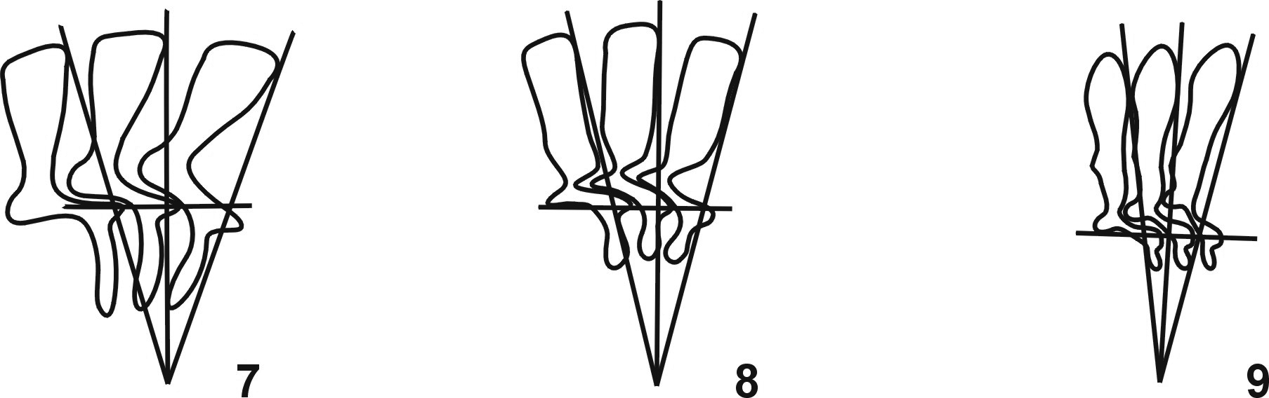
Description ( Figs. 3, 4 View FIGURES 1 – 6 , 8 View FIGURES 7 – 9 ). The following morphological description were based on 18 specimens measured (n = 18). Small to medium freshwater Tripartiella with a disc-shaped body, diameter 20.0–30.0 (25 ± 2.5); adhesive disc 16.0–26.0 (21 ± 2.3); width of border membrane 1.5–2.0 (1.8 ± 0.2); diameter of denticle ring 9.0–16.0 (11 ± 1.7); number of denticles 24 - 27; number of radial pins per denticle usually 4–5; span of denticle 5.0–7.0 (6.0 ± 0.7); length of denticle 2.0–3.0 (2.3 ± 0.4); blade length 3.0–4.0 (3.4 ± 0.4), generally narrow with anterior and posterior margins parallel. Distal margin rounded, nearly parallel with border membrane, and higher than tangent point; anterior and posterior surfaces straight and smooth and almost parallel with each other with the anterior surface just touching the Y +1 axis; blade apophysis and posterior projection present but not very distinct. Central part barely developed with a round point fitting tightly into preceding denticle and far away from the Y-1 axis. Shapes of the central part above and below the X-axis almost similar and 1.0–3.0 (1.5 ± 0.4) in width. Ray connection hardly conspicuous and hardly distinguishable from the ray. Ray short, straight, erect and nearly attached to the Y axis with a blunt point of ray; ray apophysis absent and length of ray was 1.0–1.5 (1.1 ± 0.2); ratio between denticle above and below X axis more than two. Macronucleus C-shaped, external diameter 15.0–22.5 (17.8 ± 1.1) and internal diameter 12.5–17.5 (15.6 ± 1.3). Micronucleus elliptical, 1.0–2.5 (1.7 ± 0.2) in length and 0.5–1.5 (1.2 ± 0.3) in width, situated in + Y position. Adoral ciliary spiral turns about 230° - 270° around peristomial disc.
Remarks. Tripartiella orthodens was first found and described from Tilapia rendalli swierstrai by Basson and Van As (1987) in the Sabie River of the Komati River system, South Africa. Tripartiella orthodens is characterized by the erect ray and the morphology and morphometric data of the present population from gills of Pelteobagrus nitidus fall well within the range of the original description with the only exception the smaller body size (20.0– 30.0µm in present population vs. 27.6–36.5µm in original population). This species has not been reported since its first description from Tilapia rendalli swierstrai by Basson and Van As in 1987, and thus this report is the first record of Tripartiella orthodens in Asia.

No known copyright restrictions apply. See Agosti, D., Egloff, W., 2009. Taxonomic information exchange and copyright: the Plazi approach. BMC Research Notes 2009, 2:53 for further explanation.
|
Kingdom |
|
|
Phylum |
|
|
Class |
|
|
Order |
|
|
Family |
|
|
Genus |