
Choniomyzon libiniae
|
publication ID |
https://doi.org/ 10.5281/zenodo.158250 |
|
DOI |
https://doi.org/10.5281/zenodo.6270730 |
|
persistent identifier |
https://treatment.plazi.org/id/68349C5A-CB75-C509-0750-D8ADE434710F |
|
treatment provided by |
Plazi |
|
scientific name |
Choniomyzon libiniae |
| status |
|
Choniomyzon libiniae , sp nov.
Material examined
Holotype: adult female ( MZUSP 15713), collected on Libinia spinosa H. Milne Edwards (Crustacea, Decapoda , Majidae ) from Anchovas Beach, São Sebastião Island, SP, Brazil, 15 December 2001.
Paratypes: eggs, nauplii, copepodid I, and copepodid II ( MZUSP 15714, 15715, 15716, 15717), collected on Libinia spinosa (Crustacea, Decapoda , Majidae ) from Anchovas Beach, São Sebastião Island, SP, Brazil, 15 December 2001; one male and one female (collection of Dr. Carlos F. Rocha), collected on Libinia spinosa (CCrustacea, Decapoda , Majidae ).
Description
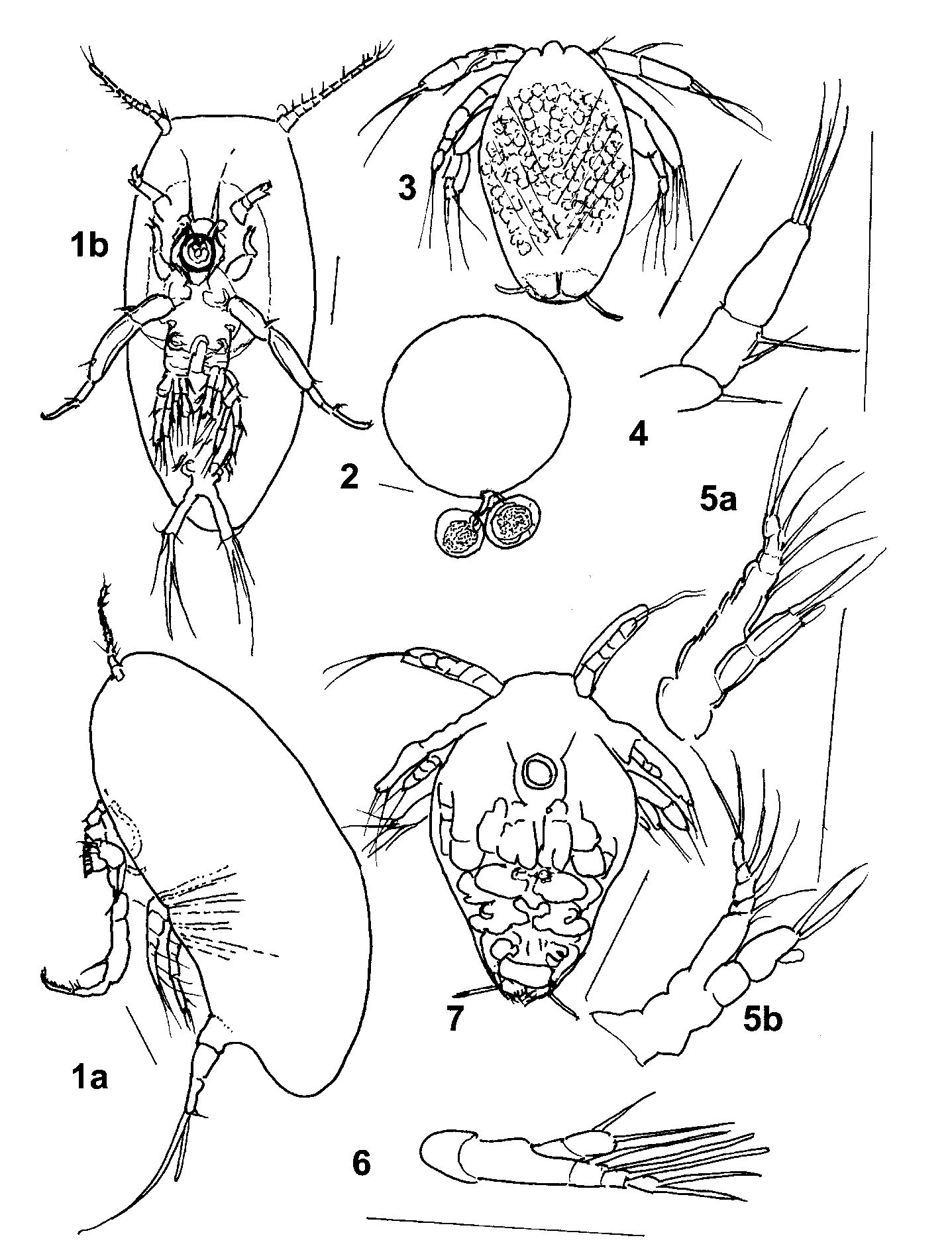
Eggs: The female nicothoid ( Figs. 1 View FIGURE 1 a, 1b) attaches her eggs (0.12 mm in diameter) to the eggs (0.59 mm in diameter) of the host. Two eggs (pink in color) were attached by a short cord to each host´s egg (Fig. 2). From the copepod eggs, nauplii emerge.
Nauplii (Figs. 3–7): Cyclopoidlike. Length 0.12–0.24 mm, ovoid in shape. Muscles parallel when seen laterally; Vshaped (Fig. 3) when observed dorsally, as in all cyclopoid nauplii. Antennule trimerous (Fig. 4) with s. f.: 1: 2: 3. Antenna (Figs. 5a, 5b) with coxa and basis unarmed, not articulated with 5–jointed exopod (s. f.: 1: 1: 1: 1: 2); endopod 2– jointed with last joint bearing 2 terminal setae and a small, lateral ovoid aesthetasc (Figs. 5a, 5b) which is very transparent, and hardly visible. Mandible (Fig. 6) with separate coxa and basis; exopod 3jointed and not articulated to basis; exopod with thick aesthetasclike setae (s. f. 1: 1: 1: 2); endopod articulated with basis, 2jointed (s. f.: 0: 2). Caudal armature: 2 short thick setae. Inside mature nauplius, next stage (the copepodid) is visible (Fig. 7).
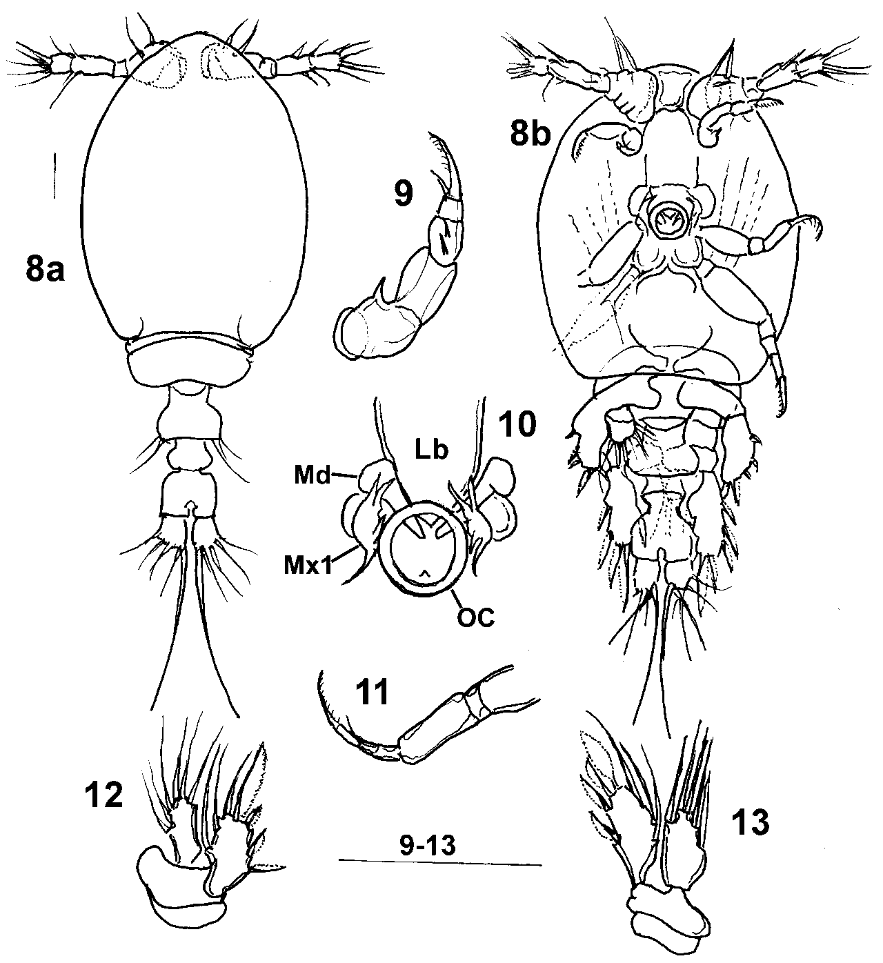
Copepodid I ( Figs. 8 View FIGURE 8 – a –13): Cyclopiform, length 0.265–0.280 mm, well chitinized. Prosome oval: comprising cephalon fused with first thoracic segment, and one free segment. Urosome threesegmented. Caudal rami about as long as wide and bearing 5 setae each; 2 dorsal, 2 ventral plus very long internal seta ( Figs. 8a, 8 View FIGURE 8 – a b). Antennule ( Fig. 8 View FIGURE 8 – a b) 4– jointed: first joint indistinctly and incompletely subdivided; s. f. 2 + 1: 1: 4: 3 + 1 or 2 aesthetascs. Antenna (Fig. 9) 5–jointed (s. f. 0: I: 0: 2: 2); terminal pinnate setae, hooklike. Mandible (Fig. 10) bladelike, projecting into mouth, surrounded by sucker or conelike projection with row of setules and transparent rim, the oral disc. Maxillule (Fig. 10 Mx1) very transparent, base with anterior and posterior branches or lobes and 4 thick setae.
Maxilla (Mx2) ( Fig. 8 View FIGURE 8 – a b) trimerous, with hooklike terminal pinnate spine or seta. Maxilliped (Fig. 11) 6jointed bearing hooklike terminal pinnate spine; fifth joint with one seta. First leg (Fig. 12): coxa and basis not ornamented, exopod with one joint (s. f.: III + I + 3); endopod onejointed, with 6 setae. Second leg (Fig. 13): coxa and basis without ornamentation, endopod with 5 setae; exopod onejointed (s. f.: III + I + 3). Posterior corners of first urosome segment each bearing two setae, one shorter than other ( Fig. 8a View FIGURE 8 – a ).
– Antennule of male. – Antennule of copepodid III. 28 – Antenna of male. FIGURE 29 – Oral cone of male. FIGURE 30 – Male urosome. FIGURE 31 – Male: second leg. FIGURE 32 – Male: first leg. FIGURE 33 – Male: deformed first leg. FIGURE 34 – Male maxilliped. FIGURE 35 – Male maxilla. FIGURE 36 – Copepodid III: part of urosome and caudal ramus. FIGURE 37 – Copepodid III maxilliped. FIGURE 38 – Copepodid III first leg. FIGURE 39 – Copepodid III second leg. All scale bars equal to 0.10 mm.
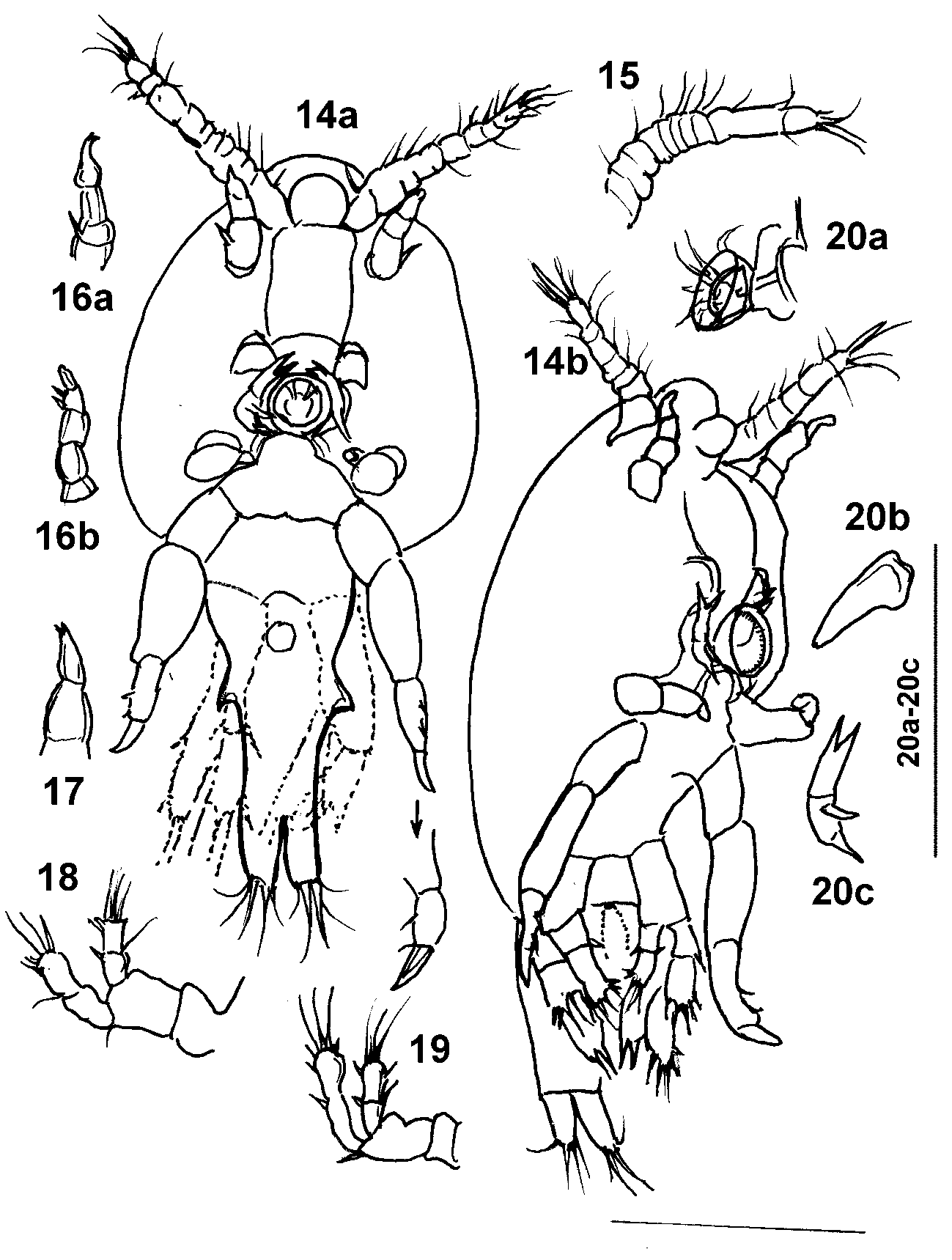
Copepodid II ( Figs. 14 View FIGURE 14 a, 14b): Body globular, reduced towards urosome, less sclerotized than first copepodid; length 0.273–0.298 mm. Articulations not or hardly visible. Widest part of urosome, provided with 2 thornlike lateral protuberances. Antennule with ten joints (s. f.: 1: 0: 1: 1: 1: 1: 1: 1: 2 + 1: 4); boundaries between joints indistinct (Fig. 15). Antenna (Figs. 16a, 16b) 4 or 3–jointed; second joint with spinelike protuberance composed of two or three setae: last joint with cylindrical terminal protuberance and 2 lateral flanged spines. Mandibles ( Fig. 14 View FIGURE 14 ) bladelike, well sclerotized, protruding into sucker or oral disc, lateral to labrum. Oral disc, circular, cuplike, with thin transparent rim. Maxillule (Fig. 20): very thin, transparent structure, similar to maxillule of adult, with coxobasis and 2 lobes, one bearing thick pointed seta, protuberance with thin seta and anterior lobe with 2 setae visible over border of sucker or oral disc. Maxilla (Fig. 17) 2 or 3–jointed, ending in 2 minute spines. Maxilliped ( Fig. 14 View FIGURE 14 a) indistinctly 4–jointed, with 2 lateral spines on third joint and last joint in form of thick spine. First leg (Fig. 18): coxobasis with one seta; endopod 2–jointed (s. f.: 1 + I: III + 3); exopod 2–jointed (s. f.: 1: II + 3). Second leg (Fig. 19): coxobasis with one outer seta; endopod indistinctly 2–jointed (s. f.: 1 + I: 1 + 3 + I); exopod 2–jointed (s. f.: I: II + 3 + 1).
Female copepodid ( Figs. 21a, 21 View FIGURE 21 b): Size 0.335 mm. General form of copepodid II with very conspicuous lateral transparent margins (wings), visible dorsally and ventrally. Segmentation of appendages distinct. Antennule (Fig. 27): 10 or 11 segments. Antenna, oral cone, mandible, maxillule and maxilla, maxillipeds (Fig. 37) similar to those of copepodid II. First and second legs with 2jointed endopods and exopods (Figs. 38–39). First leg with spine on coxobasis; endopod with s. f.: 1: 6; exopod, s. f.: I: II + 5. Second leg (Fig. 39) with bare coxobasis and endopod with s. f.: 1: 5; exopod with spine on first segment and spinelike distal and external protuberance, 2 spinelike external distal protuberances on second segment, plus 5 setae. Urosome (Fig. 36) showing two lateral round protuberances, characteristic of female urosome.
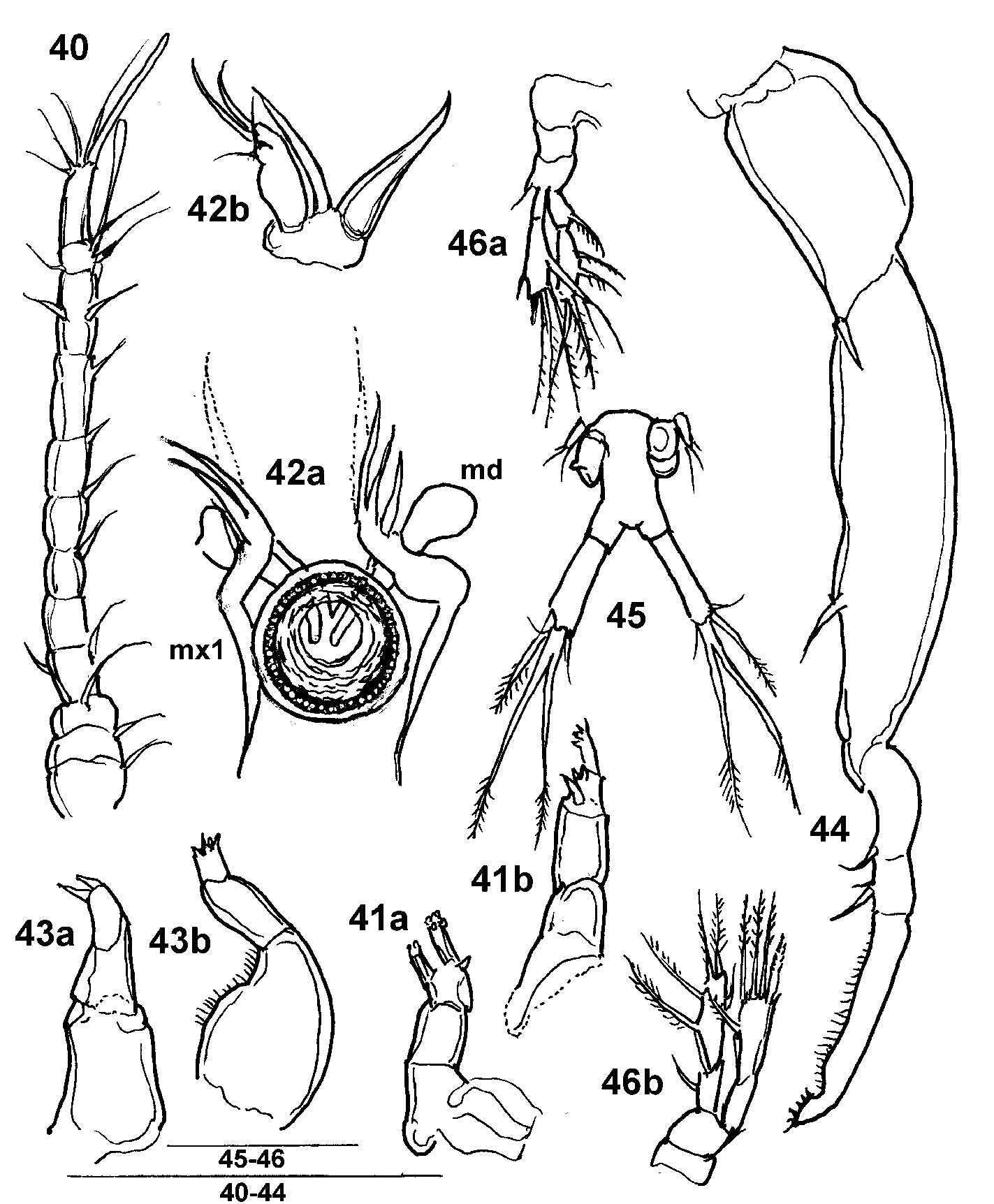
Adult female ( Figs. 1 View FIGURE 1 a, 1b): Length 0.62–0.83 mm (5 specimens); body ovalshaped, without divisions in prosome, rounded and wider in front. Lateral "winglike" fold protuberant on both sides of head. Margin of fold ornamented by transparent rim conspicuous in young females (semi lateral view), in gravid females hardly or not visible. Urosome narrow, short and overlapped by posterior region of prosome; indistinctly divided into 2 segments. Antennule ( Fig. 40 View FIGURE 40 ) 12–jointed (s. f.: 1: 1: 1: 2: 0: 1: 1: 1: 1: 3 + ae: 2: 4 + ae). Antenna (A2) (Figs. 41a, 41b) 3–jointed; first and second joints unarmed, third bearing 2 thick, fingerlike structures one of which with 2 small pointed spines distally and a terminal denticulate or comblike protuberance. Mandible: bladelike, tapering distally. Maxillule (Mx1) (Fig. 42b) situated lateral to oral cone with long pointed posterior lobe or seta, middle protuberance ending in pointed structure, and frontal lobe or joint indistinctly divided into two which projects over border of cone and is armed with 2 thick pointed setae and two thinner, smaller setae. In frontal view (Fig. 42a), it is possible to see mandibles (Md) protruding into the mouth of the specimen in form of two blades. Maxilla (Mx2) (Figs. 43a, 43b) 3–jointed: last joint with four short spiny protuberances – two of which longer. Maxilliped (Mxp) (Fig. 44) 5–jointed with first, second and third joints with seta or spine each, fourth joint with small lateral seta and spine, last joint with margin provided with row of thin setules and terminal comblike structure ornamented with minute spines. First leg (P1) (Fig. 45): coxa unarmed, basis with external seta; exopod bimerous with s. f. = I + 0: I + I + 4; endopod bimerous, with s. f. = 0 + 1: 1 + 2 + 3. Second leg (Fig. 46): coxa unarmed; basis I + 0; exopod bimerous, with s. f. = I + 0: I + I + 3 + 1; endopod I + 1: 1 + 2 + 2. Third leg singlejointed with two longer and one very short small seta. Urosome marked off from body by two cuplike protuberances (genital openings). Urosome cylindrical, not terminal, placed posterior to genital cups, with 2 or 3 indistinct segments, last of which divided into two branches which end in the elongate caudal rami, about three times longer than wide. Caudal rami ornamented with two lateral fine setae, one on each side; a long bladelike bipinnate seta and two double as long setae coalesced at base and bipinnate.
Male (Fig. 22): Size 0.320–0.350 mm. Characterized by transparency and by presence of winglike protuberances on each side of body. Antennule (Fig. 26) has 10 segments. Antenna (Fig. 28) has long terminal fingerlike protuberance, 4 times as long as wide. Maxilla of male (Fig. 35) with terminal segment 5 times as long as wide. Oral cone, maxillule, maxilliped (Fig. 34) similar to those of CII. First legs of male (Fig. 32) with spine or seta on the proximal segment of the endopod and exopod. Last exopod segment bearing 2 spines and 3 setae, and last endopod segment with 4 setae. Second legs (Fig. 31) with seta on first endopod segment and 4 setae on last. First segment of exopod with 2 spines; second segment with 4 setae and spinelike protuberance.
Legs of male (Fig. 33) frequently with deformed rami. Spine and protuberance with 2 minute setae representing third pair of legs on urosome (Fig. 30). Urosome unsegmented, containing 2 pearshaped spermatophores. Caudal rami bearing 2 very fine lateral setae and 3 longer setae of which the 2 longest are coalesced at base.
In addition to the above mentioned forms of Choniomyzon , the following stages were also present in the samples and will be treated in more detail in a later paper: young female, ovigerous female and spent female (a female which has laid her eggs) (Figs. 23 to 25).
Diagnosis
The new species is characterized by the ovoid form and the indistinctly 3segmented urosome, which is not terminal. The first and second legs do not show a buttonlike ornament as in C. panuliri Pillai. The antenna of the adult female bears fingerlike projections, one or two of which have denticulate margins. The maxilla has four terminal spines and the maxillipeds have a terminal denticulate margin.
Etymology
The name of the species is a reference to its host, Libinia spinosa .

| MZUSP |
Museu de Zoologia da Universidade de Sao Paulo |
No known copyright restrictions apply. See Agosti, D., Egloff, W., 2009. Taxonomic information exchange and copyright: the Plazi approach. BMC Research Notes 2009, 2:53 for further explanation.
|
Kingdom |
|
|
Phylum |
|
|
Class |
|
|
Order |
|
|
Family |
|
|
Genus |