
Tribolonotus parkeri, Rittmeyer, Eric N. & Austin, Christopher C., 2017
|
publication ID |
https://doi.org/ 10.11646/zootaxa.4268.1.4 |
|
publication LSID |
lsid:zoobank.org:pub:BFA3EAB8-7D6C-4497-8486-A1CBB09CCC26 |
|
DOI |
https://doi.org/10.5281/zenodo.6032291 |
|
persistent identifier |
https://treatment.plazi.org/id/6A0187ED-FFF5-1F7A-F0AF-2CE26E34FF42 |
|
treatment provided by |
Plazi |
|
scientific name |
Tribolonotus parkeri |
| status |
sp. nov. |
Tribolonotus parkeri sp. nov.
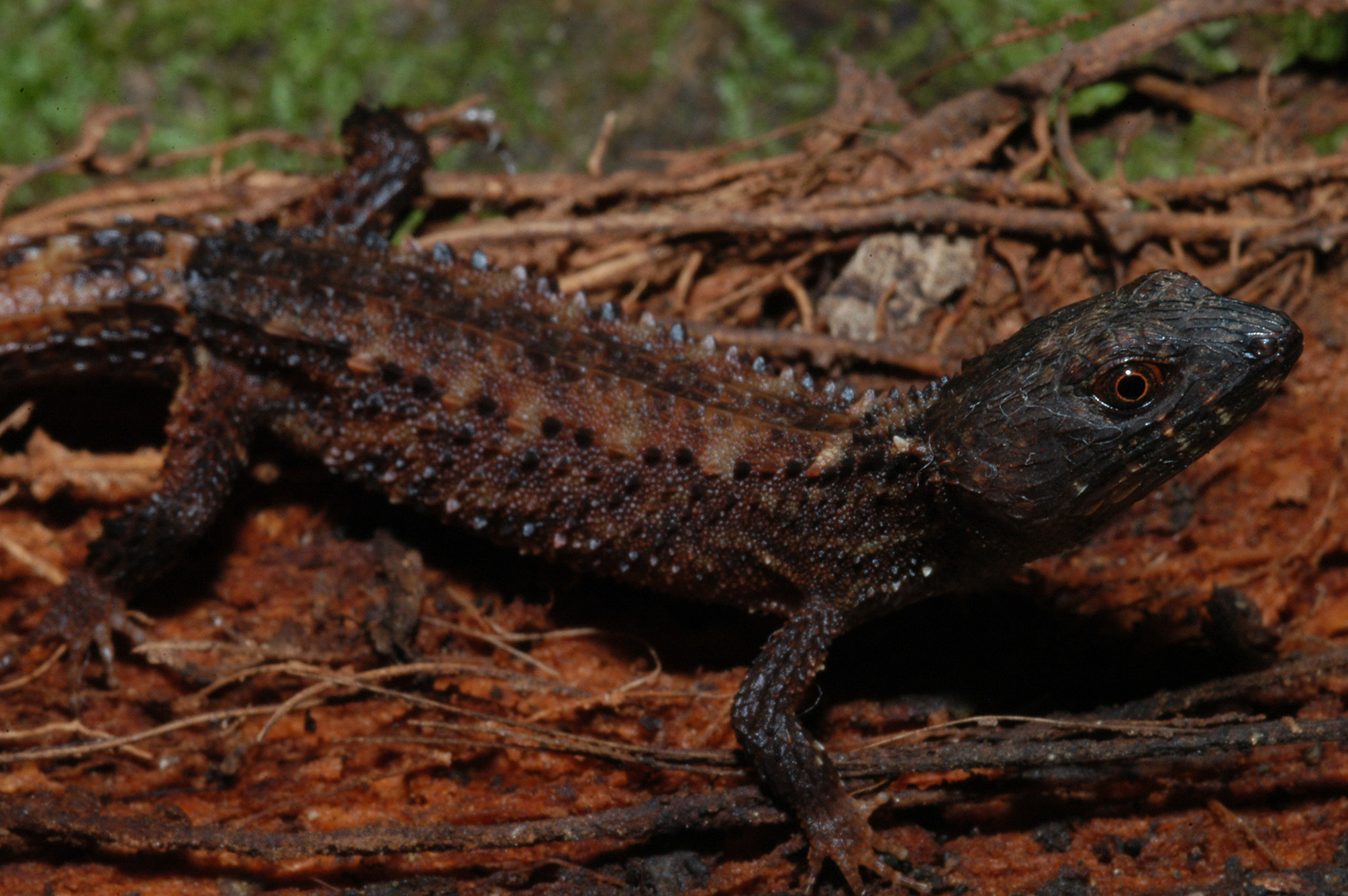
( Figs. 5 View FIGURE 5 , 6 View FIGURE 6 A,C)
Holotype. LSUMZ 93510 View Materials (field number CCA 2734 View Materials ), collected by Christopher C. Austin, Nova Area , Southeast of bridge at Ramunfun River, Buka Island, North Solomons Province , Papua New Guinea, 5.3893°S, 154.6518°E, WGS84, 7 m elevation, 30 November 2005. GoogleMaps
Paratypes. AMS R18811, collected by Fred Parker , Buka Island, North Solomons Province, Papua New Guinea, 5.4110°S, 154.6794°E, WGS84 GoogleMaps ; LSUMZ 93500–3 View Materials , collected by Christopher C. Austin, Nova Area , near Chi Chi Hav Village, Buka Island, North Solomons Province , Papua New Guinea, 5.3908°S, 154.6409°E, WGS 84, 149 m elevation, 29 November 2005; LSUMZ 93504–9, 93511–3 , same collection details as holotype GoogleMaps ; MCZ 67706–67710 collected by Fred Parker , Buka Island, North Solomons Province, Papua New Guinea, 5.4110°S, 154.6794°E, WGS84, 28 January 1962 GoogleMaps ; MCZ 67711–6 About MCZ , same data except collected 31 January 1962 GoogleMaps ; MCZ 73850– 4 About MCZ , same data except collected 8 March 1963 GoogleMaps ; MCZ 73855–61 About MCZ , same data except collected 9 March 1962 GoogleMaps ; MCZ 92491 collected by Fred Parker , Kubu, Buka Island, North Solomons Province, Papua New Guinea, 5.4110°S, 154.6794°E, WGS84, 25 May 1966 GoogleMaps .
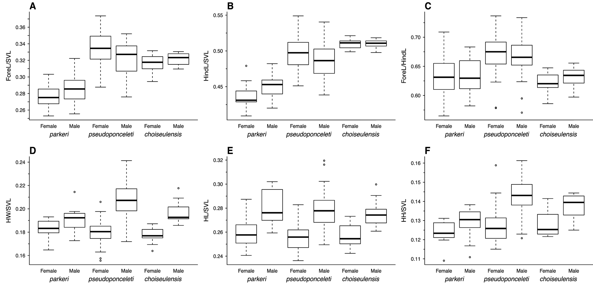
Diagnosis. A small (maximum SVL 48.5 mm) scincid lizard of the genus Tribolonotus diagnosable from congeneric species by the following combination of characters: 1) two longitudinal rows of 20–23 enlarged vertebral scales, separated from parietal plates by granular scales and commencing at the posterior of the nape; 2) 42–47 ventral scale rows from the mental to the vent; 3) a longitudinally elongate first supralabial scale, separating the nasal and second supralabial scales; 4) two primary temporal scales; 5) 10–13 finger III lamellae and 20–23 toe IV lamellae; and 6) moderately short limbs (ForeL/SVL = 0.253–0.322, mean = 0.281; HindL/SVL = 0.409–0.482, mean = 0.445). Dorsolateral and lateral scales mostly granular, but with several longitudinal rows of spinose scales, approximately one for every enlarged vertebral scale. Tail relatively long, up to approximately 190% of SVL when complete. Head rugose, with up to five strong keels per scale; triangular from dorsal aspect and distinct from neck, slightly larger in males than in females (male HW/SVL = 0.173–0.214, female HW/SVL = 0.165–0.193; male HL/ SVL = 0.259–0.302, female HL/SVL = 0.241–0.287; Table 2, Fig. 2 View FIGURE 2 ). Two primary temporal scales; five supralabial scales, the first narrow and elongate, with no contact between the nasal and second supralabial scale, and five or six infralabial scales. Plantar pores, up to slightly less than the diameter of the toes, variably present in males (absent in approximately half of males examined), absent in females. When present, arranged in parallel rows of up to nine pores at the base of toe IV, frequently with a small gap after the distal two or three pores, and up to three at the base of toe III. Palmar pores rarely present in males, absent in females. When present, only one to two pores present at the base of finger IV. A pair of abdominal glands present just anterior to insertion of the hind limbs in both sexes. Dorsal coloration brown with extensive pale, yellowish-tan markings dorsally and laterally, typically triangular in shape and connecting to form vague paravertebral lines. Ventral coloration pale tan.
Comparisons. Tribolonotus parkeri sp. nov. is differentiated from T. blanchardi , T. gracilis and T. novaeguineae by the presence of two longitudinal rows of enlarged vertebral scales (versus one in T. blanchardi , and four in T. gracilis and T. novaeguineae ). Tribolonotus gracilis and T. novaeguineae also exhibit a much larger maximum SVL than T. parkeri sp. nov., while T. blanchardi exhibits a somewhat smaller maximum SVL (maximum SVL = 48.5 mm in T. parkeri sp. nov., versus 103 mm in T. gracilis , 103 mm in T. novaeguineae , and 40 mm in T. blanchardi ).
T. choiseulensis sp. nov. T. parkeri sp. nov. T. pseudoponceleti
Female Male Female Male Female Male
N 1 1 1 0 1 6 1 6 5 7 4 4
SVL 40.16±3.45 44.86±2.63 44.07±2.05 43.46±2.88 53.3±4.61 60.32±5.56 (31.1–44.4) (40.7–47.8) (40.7–48) (39.4–48.5) (43.5–64.7) (47.7–73.4)
ForeL/SVL 0.317±0.011 0.322±0.008 0.276±0.015 0.286±0.017 0.335±0.018 0.324±0.018 (0.294–0.332) (0.31–0.331) (0.253–0.303) (0.255–0.322) (0.288–0.374) (0.276–0.352)
F3/SVL 0.065±0.003 0.065±0.002 0.062±0.006 0.066±0.005 0.065±0.004 0.063±0.005 (0.061–0.068) (0.061–0.069) (0.054–0.072) (0.056–0.076) (0.056–0.076) (0.054–0.083)
HindL/SVL 0.51±0.008 0.51±0.006 0.437±0.016 0.452±0.015 0.498±0.025 0.487±0.025 (0.499–0.521) (0.498–0.518) (0.409–0.479) (0.42–0.482) (0.451–0.549) (0.438–0.54)
T4/SVL 0.133±0.006 0.13±0.009 0.122±0.008 0.131±0.008 0.128±0.008 0.122±0.008 (0.124–0.143) (0.121–0.147) (0.111–0.145) (0.123–0.153) (0.112–0.149) (0.108–0.14)
ForeL/HindL 0.621±0.02 0.631±0.019 0.632±0.04 0.633±0.03 0.672±0.031 0.665±0.031 (0.586–0.647) (0.597–0.656) (0.565–0.709) (0.582–0.683) (0.578–0.737) (0.57–0.734)
F3/T4 0.486±0.016 0.503±0.027 0.511±0.034 0.5±0.037 0.509±0.03 0.518±0.039 (0.456–0.512) (0.45–0.533) (0.462–0.566) (0.435–0.558) (0.449–0.585) (0.427–0.667)
HW/SVL 0.178±0.007 0.197±0.01 0.183±0.007 0.191±0.01 0.18±0.01 0.208±0.015 (0.164–0.187) (0.186–0.218) (0.165–0.193) (0.173–0.214) (0.156–0.206) (0.172–0.241)
HL/SVL 0.257±0.01 0.276±0.012 0.26±0.013 0.281±0.014 0.256±0.011 0.279±0.016 (0.242–0.273) (0.261–0.3) (0.241–0.287) (0.259–0.302) (0.236–0.283) (0.249–0.32)
HH/SVL 0.128±0.007 0.137±0.007 0.124±0.005 0.129±0.007 0.127±0.008 0.143±0.01 (0.122–0.141) (0.125–0.144) (0.109–0.131) (0.111–0.138) (0.115–0.159) (0.121–0.161)
HW/HL 0.692±0.03 0.716±0.041 0.705±0.043 0.682±0.058 0.706±0.031 0.745±0.04 (0.661–0.743) (0.639–0.777) (0.582–0.761) (0.588–0.78) (0.636–0.761) (0.678–0.834)
HH/HL 0.498±0.024 0.499±0.026 0.478±0.034 0.461±0.036 0.496±0.026 0.513±0.031 (0.459–0.536) (0.451–0.536) (0.385–0.528) (0.391–0.508) (0.439–0.596) (0.444–0.579) Tribolonotus parkeri sp. nov. is further distinguished from T. annectens in having two primary temporal scales (versus three in T. annectens ), and in having more spinose scales in longitudinal rows paralleling the vertebral scales (approximately one scale for every enlarged mid-dorsal scale in T. parkeri sp. nov. versus approximately one scale for every two vertebral scales in T. annectens ). In T. parkeri sp. nov., the enlarged vertebral scale rows extend anteriorly to the posterior of the nape (granular scales separate the enlarged vertebral scales from the parietal plates) versus extending to the parietal plates in T. brongersmai Cogger 1972 and T. schmidtii Burt 1930 , and are fewer in number (20–23 scales per longitudinal row in T. parkeri sp. nov. versus 29–32 in T. brongersmai and 29– 35 in T. schmidtii ). Tribolonotus parkeri sp. nov. also typically has more subdigital lamellae (20–23 toe IV lamellae in T. parkeri sp. nov. versus 16–20 in T. schmidtii ). Tribolonotus ponceleti Kinghorn 1937 is much larger than T. parkeri sp. nov. (maximum SVL= 144 mm in T. ponceleti versus 48.5 mm in T. parkeri sp. nov.); the two are further distinguished by the separation of the nasal and second supralabial scales by the longitudinally elongate first supralabial scale in T. parkeri sp. nov. versus contact between the nasal scale and second supralabial scale in T. ponceleti .
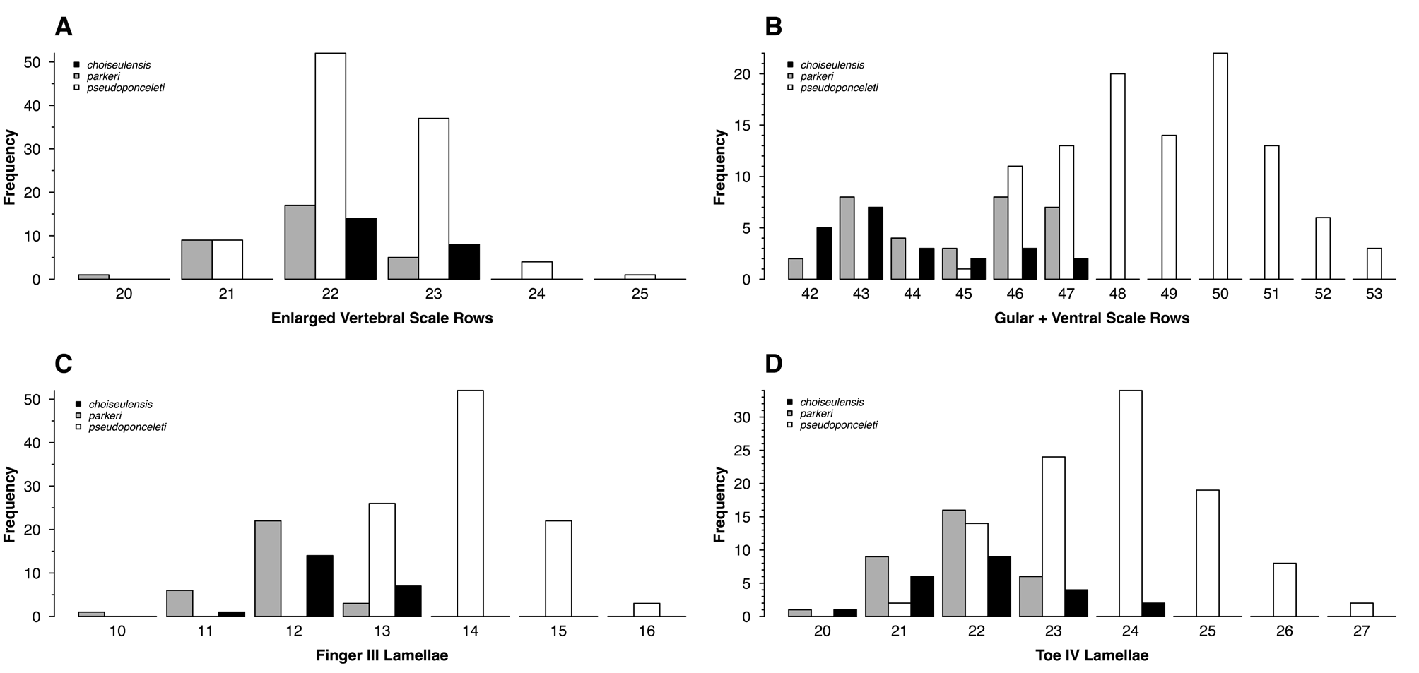
Tribolonotus parkeri sp. nov. is most similar to T. choiseulensis sp. nov. and T. pseudoponceleti Greer & Parker 1968 . However, it can be diagnosed from T. pseudoponceleti based on its smaller number of ventral scale rows (42–47, mean = 44.875, mode = 45 in T. parkeri sp. nov. versus 45–53, mean = 49.008, mode = 50 in T. pseudoponceleti ; Table 3, Fig. 7 View FIGURE 7 ), smaller number of subdigital lamellae (F3L = 10–13, mean = 11.844, mode = 12 in T. parkeri sp. nov. versus 13–16, mean = 14.027, mode = 14 in T. pseudoponceleti ; T4L = 20–23, mean = 21.844, mode = 22 in T. parkeri sp. nov. versus 21–27, mean = 23.885, mode = 24 in T. pseudoponceleti ; Table 3, Fig. 7 View FIGURE 7 ), and shorter legs (ForeL/SVL = 0.253–0.322, mean = 0.281 in T. parkeri sp. nov. versus 0.276–0.374, mean = 0.330 in T. pseudoponceleti ; HindL/SVL = 0.409–0.482, mean = 0.445 in T. parkeri sp. nov. versus 0.438–0.578, mean = 0.495 in T. pseudoponceleti ; Table 2, Fig. 2 View FIGURE 2 ). Tribolonotus parkeri sp. nov. is also much smaller than T. pseudoponceleti (maximum SVL = 48.5 mm in T. parkeri sp. nov., 76 mm in T. pseudoponceleti ), and has more extensive light dorsal markings, which frequently connect to form paravertebral lines in T. parkeri sp. nov., but are typically restricted to non-connected, paravertebral spots in T. pseudoponceleti . Tribolonotus parkeri sp. nov. is distinguished from T. choiseulensis sp. nov. by its shorter limbs (ForeL/SVL = 0.253–0.322, mean = 0.281 in T. parkeri sp. nov. versus 0.294–0.332, mean= 0.319 in T. choiseulensis sp. nov.; HindL/SVL=0.409–0.482, mean = 0.445 in T. parkeri sp. nov. versus 0.498–0.521, mean = 0.510 in T. choiseulensis sp. nov.; Table 2, Fig. 2 View FIGURE 2 ), and by color pattern: T. parkeri sp. nov. has more extensive light dorsal markings of pale triangles, often merging to form paravertebral lines lateral to the enlarged vertebral scales whereas T. choiseulensis sp. nov. has a dorsal pattern of faint, indistinct yellowish tan markings in the form of paired, separated paravertebral spots.
T. choiseulensis sp. nov. T. parkeri sp. nov. T. pseudoponceleti
N 2 1 3 2 1 0 1
F3L 12 12 14
(11–13) (10–13) (13–16) T4L 22 22 24
(20–24) (20–23) (21–27) DSR 22 22 22
(22–23) (21–23) (21–25) VSR 43 43 50
(42–47) (42–47) (45–53)
Description of holotype. An adult male, SVL 41.1 mm, total length 110.0 mm, with two longitudinal rows of 21 strongly keeled, enlarged vertebral scales, beginning at the posterior of the nape ( Figs. 6 View FIGURE 6 A). Lateral and dorsolateral scales granular, but with several rows of larger, spinose scales laterally, approximately one per every enlarged mid-dorsal scale, but continuing to the parietal scales. Ventral scales weakly keeled, 46 scale rows from mental to vent. Head relatively large and broad (HW = 7.7 mm; HL = 11.5 mm; HH = 4.8 mm; HW/HL = 0.670; HH/HL = 0.417), distinct from neck, and rugose, with up to five keels per head scale. Two primary temporal scales, five infralabial scales, and five supralabial scales, the first supralabial narrow and elongate, with no contact between the nasal and second supralabial scales. Limbs moderately short (ForeL = 12.3 mm; ForeL/SVL = 0.299; HindL = 18.0 mm; HindL/SVL = 0.438). Digits moderately long (Fing3 = 2.7 mm; Toe4 = 5.5 mm), with 11 rounded subdigital lamellae under finger III and 21 rounded subdigital lamellae under toe IV. Ten plantar pores present, arranged in two parallel rows: one row of eight at the base of toe IV, extending linearly towards toe V with the proximal three pores shifted slightly towards the median of the foot and a small gap between the two distal pores and the remaining pores, and a second row of two at the base of finger III. One very small palmar pore present at base of finger IV. One pair of abdominal glands visible externally just anterior to the insertion of the hind limbs.
Dorsal coloration brown, with paired light, yellowish tan markings, triangular in shape, with vague borders but most distinct anteriorly and connecting to form a more distinct chevron at the base of the tail ( Fig. 6 View FIGURE 6 A). Ventral coloration pale, yellowish in color, with darker brown markings along the edges of some scales ( Fig. 6 View FIGURE 6 B). Underside of limbs and tail mottled with pale yellow and dark brown. Mental, post-mental, and infralabial scales colored dark brown with some lighter streaking.
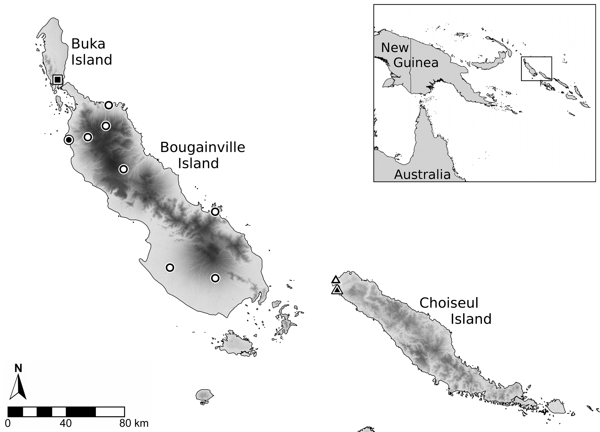
Distribution. Tribolonotus parkeri sp. nov. is only known from Buka Island, North Solomons Province, Papua New Guinea (geologically and biogeographically part of the Solomon Archipelago; Fig. 1 View FIGURE 1 ).
Etymology. The specific epithet was chosen to honor Fred Parker in recognition of his substantial contributions to herpetology in Papua New Guinea, and his collections of much of the type series of the species.

No known copyright restrictions apply. See Agosti, D., Egloff, W., 2009. Taxonomic information exchange and copyright: the Plazi approach. BMC Research Notes 2009, 2:53 for further explanation.