
Campylothorax mitrai Bellini & Meneses, 2012
|
publication ID |
https://doi.org/10.11646/zootaxa.4300.2.1 |
|
publication LSID |
lsid:zoobank.org:pub:92ECE1FD-6C96-492E-AB44-62AB9D02FEA6 |
|
DOI |
https://doi.org/10.5281/zenodo.6017078 |
|
persistent identifier |
https://treatment.plazi.org/id/6F205733-3420-3A10-CC8A-FF5B43A0FF25 |
|
treatment provided by |
Plazi |
|
scientific name |
Campylothorax mitrai Bellini & Meneses, 2012 |
| status |
|
Campylothorax mitrai Bellini & Meneses, 2012
Figs 30–61 View FIGURES 30 – 34 View FIGURES 35 – 39 View FIGURES 40 – 44 View FIGURES 45 – 46 View FIGURES 47 – 55 View FIGURES 56 – 61
Campylothorax mitrai Bellini & Meneses, 2012: 451 , figs 1–13, Alagoas state, Brazil (orig. descr.). Cipola & Oliveira, 2016: 493 (key). Santos et al. 2016: 1, 12, 22 (cit.). Soto-Adames, 2016: 12, 26 (compared).
Type material. Holotype male plus six female and two males paratypes were studied, Brazil, Alagoas State, Rio Largo municipality, Universidade Federal de Alagoas, Centro de Ciências Agrárias, ( 09°27'50"S; 35°50'02"W), Atlantic Forest Biome , 19.xi.2010, pitfall-traps, I.P.S. Santos coll. Material deposited at CM/ MNRJ. GoogleMaps
Other material studied. Two males and three females on slides and 14 specimens in alcohol (INPA), same data as type series. Four males and eight females on slides ( DBEZ / UFRN and INPA) , same data as type series, excepty Parque Municipal de Maceio , 09°36'44"S; 35°45'40"W. One female and one juvenile on slides and three specimens in alcohol ( INPA) GoogleMaps , same data as type series, excepty Bairro Serraria , 09°35'18"S; 35°44'02"W, 11- 13.i.2014 GoogleMaps .
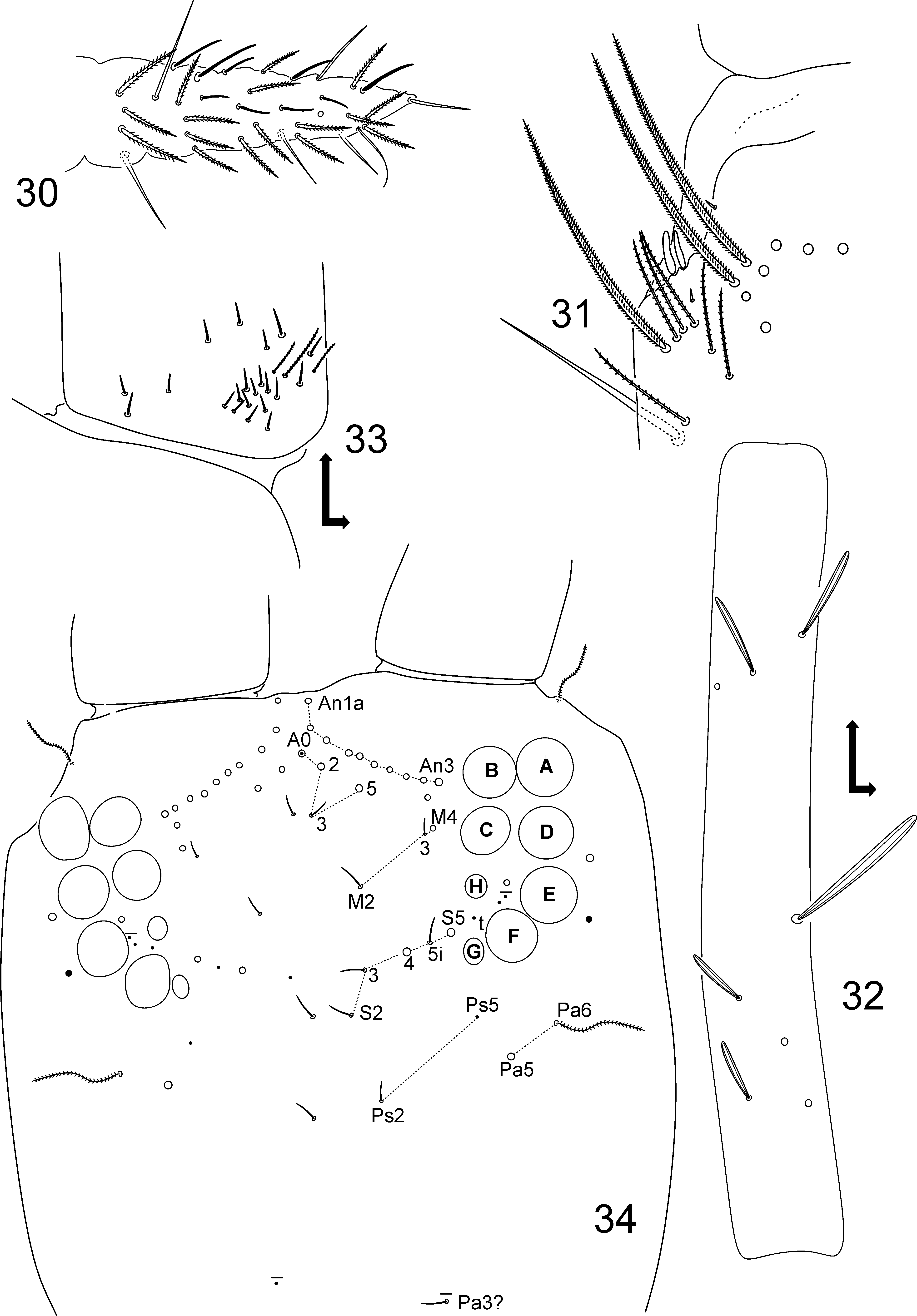
Redescription. Habitus typical of Campylothorax , body bent between Th. III Abd. I. Measurements and color pattern as in the original description. Brownish heavily ciliate apically truncate or rounded scales covering Ant. I and II, basal halves of Ant. III and IV, head, thorax, abdomen, legs and furcula. Collophore with few scales on posterior face. Head and trunk with reduced number of mac.
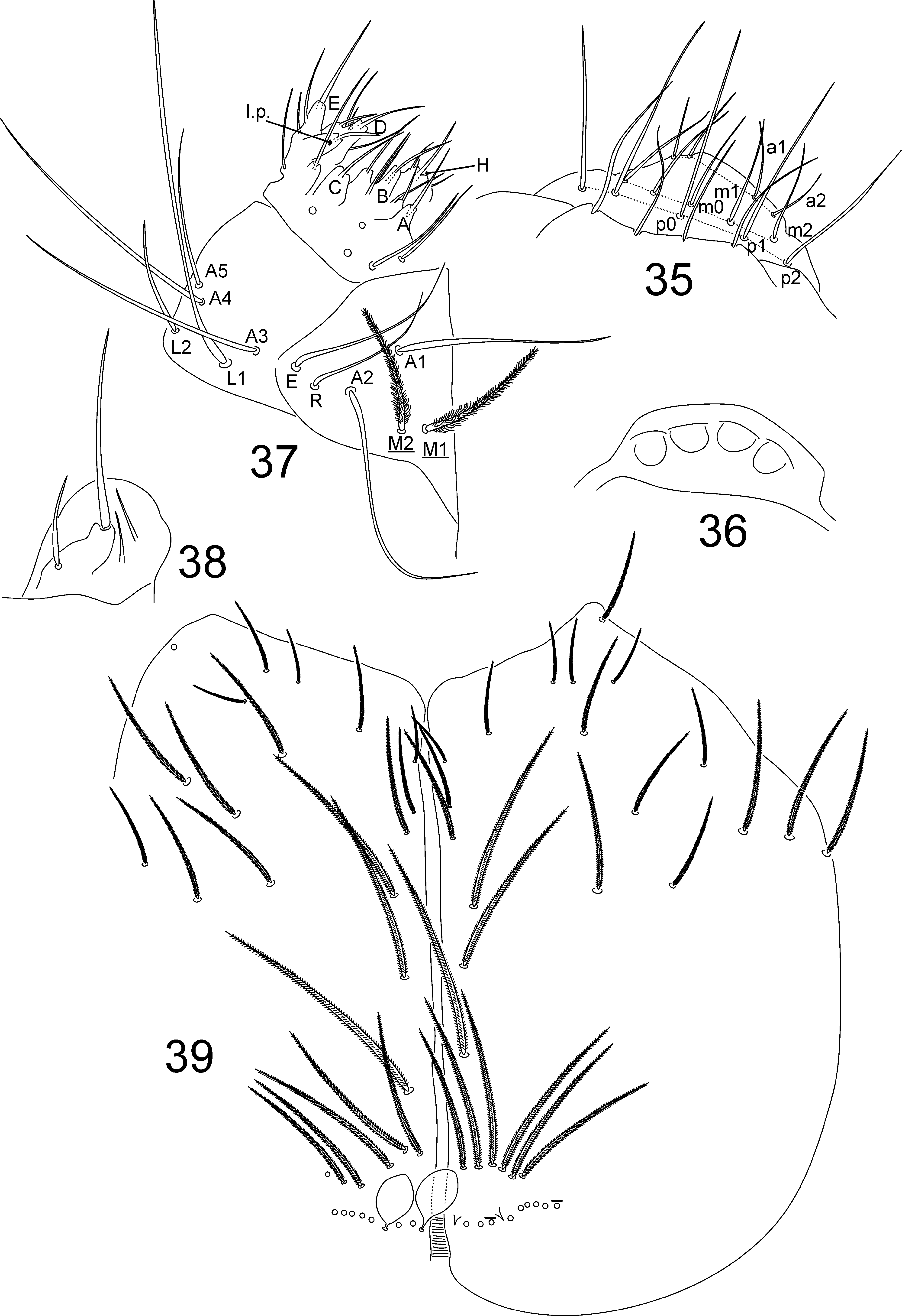
Head ( Figs 30–39 View FIGURES 30 – 34 View FIGURES 35 – 39 ). Antennae longer than body, antennal ratio as I: II: III: IV = 1: 1: 1.23: 1.21 in holotype. Ant. IV annulated, without apical bulb and with at least three types of chaetae: blunt sensilla, smooth acuminate chaetae and ciliate chaetae ( Fig. 30 View FIGURES 30 – 34 ). Ant. III sense organ with two rod-like sensilla, two surrouding guard sensilla (third one not seen but possibly present), plus some ciliate and smooth chaetae ( Fig. 31 View FIGURES 30 – 34 ). Ant. I with 7 dorsal mac of different sizes ( Fig. 32 View FIGURES 30 – 34 ); dorso-proximal lateral region with about 21 smooth mic ( Fig. 33 View FIGURES 30 – 34 ). Eyes 8+8, G and H vestigial, A-F subequal, with 3–4 interocular chaetae ( Fig. 34 View FIGURES 30 – 34 ). Dorsal chaetotaxy with 10 antennal mac (An), 4 anterior (A), 3 medio-ocellar (M), 5 sutural (S), 2 post-sutural (Ps) and 3–2 postoccipital anterior (Pa) chaetae; anterior and post-ocular ( Pa6) bothriotricha present ( Fig. 34 View FIGURES 30 – 34 ). Four prelabral smooth chaetae ( Fig. 35 View FIGURES 35 – 39 ). Labral formula 5 ( p0–2), 5 ( m0–2), 4 ( a1–2), all smooth chaetae, chaetae on posterior row larger than others ( Fig. 35 View FIGURES 35 – 39 ). Labral papillae without spine like projections ( Fig. 36 View FIGURES 35 – 39 ). Labial basolateral and basomedian fields with M1–2 ciliate, R, E, L1–2, A1–5 smooth; A2 larger than A1, L2 smaller than L1 but not minute; R normal in size ( Fig. 37 View FIGURES 35 – 39 ). Labium with five smooth proximal chaetae; labial palp with five main papillae (A–E), and with 0, 5, 0, 4, 4 guard chaetae, respectively; papilla E with lateral process (l.p.) finger-shaped; H (main hypostomal chaeta) with two accessorial hypostomal chaetae ( Fig. 37 View FIGURES 35 – 39 ). Maxillary outer lobe with one apical appendage and one subapical chaeta, both smooth, apical appendage longer; sublobal plate with two smooth appendages ( Fig 38 View FIGURES 35 – 39 ). Ventral chaetotaxy as in Fig. 39 View FIGURES 35 – 39 , all chaetae ciliate, scales present; cephalic groove with 7+7 marginal chaetae, three anterior smaller; two posterior chaetae present or absent ( Fig. 39 View FIGURES 35 – 39 ).
Thorax chaetotaxy ( Figs 40–41 View FIGURES 40 – 44 ). Th. II with 1 S-microchaeta ( ms), 1 anterolateral sens ( al), and 7 posterior mac ( p1–4 plus 3 secondary chaetae) ( Fig. 40 View FIGURES 40 – 44 ). Th. III with 1 anterolateral sens ( al) and 4 central mac ( a4–5, p2–3) ( Fig. 41 View FIGURES 40 – 44 ).
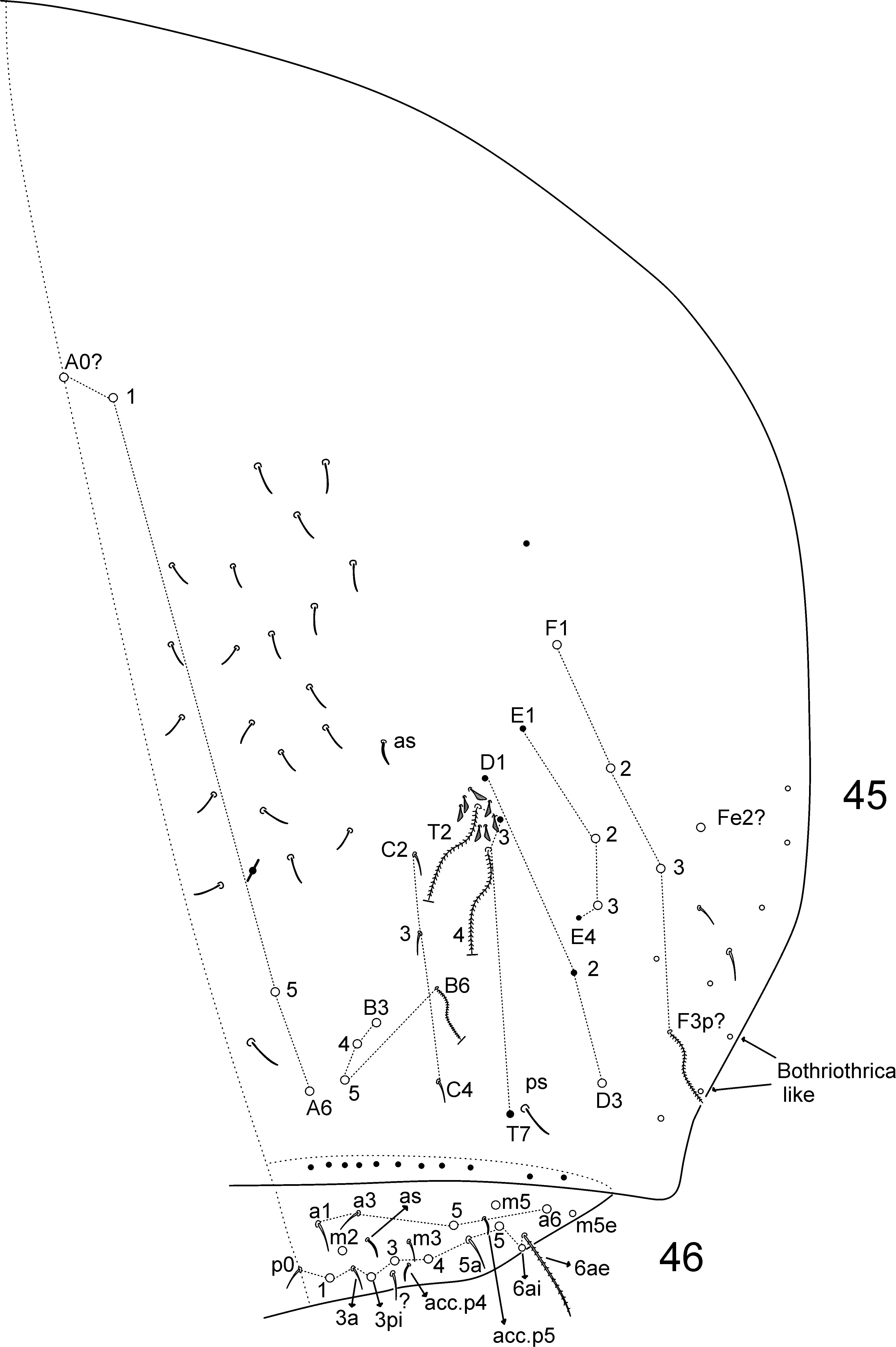
Abdomen chaetotaxy ( Figs 42–46 View FIGURES 40 – 44 View FIGURES 45 – 46 ). Abd. I with 1 S-microchaeta ( ms); 1 anterior ( a6), 4 medial ( m2–4, 6), and 1 posterior ( p6) mic ( Fig. 42 View FIGURES 40 – 44 ). Abd. II with 1 anterosubmedial sens ( as), bothriotricha m2 and a5 present surrounded by 4 fan-shaped chaetae each; mac m3 and m5 present plus 5 lateral mic ( a6–7, m6, p6–7) ( Fig. 43 View FIGURES 40 – 44 ). Abd. III with 1 S-microchaeta ( ms), 1 anterosubmedial sens ( as), bothriotricha m2, m5 and a5 present, surrounded by 2, 4 and 6 fan-shaped chaetae respectively; mac m3, am6, pm6, p6 and a7 (which can be mes) present ( Fig. 44 View FIGURES 40 – 44 ). Abd. IV with 1 anterosubmedial sens ( as) and 1 posterior sens ( ps) plus about 21 other central sens; primary bothriotricha T2 and T4 plus secondary B6 and F3p? and 2 bothriotricha-like lateral chaetae present; T2 and T4 surrounded by 4 and 3 fan-shaped chaetae respectively; row ‘A’ with 4 ( A0?–1, A5–6), ‘B’ with 3 ( B3–5), ‘D’ with 1 ( D3), ‘E’ with 2 ( E2–3), ‘F’ with 3 ( F1–3), and ‘Fe’ with 1 ( Fe2?) mac; 11+11 posterior chaetae ( Fig. 45 View FIGURES 45 – 46 ). Abd. V with 1 anterosubmedial sens ( as) and 2 accessory sens ( acc.p4–p5), 2 anterior ( a5–6), 2 medial ( m2, m5), and 5 posterior ( p1, p 3i –5) mac ( Fig. 46 View FIGURES 45 – 46 ).
Legs ( Figs 47–55 View FIGURES 47 – 55 ). Subcoxa I external face with one posterior chaeta and two pseudopores ( Fig. 47 View FIGURES 47 – 55 ), internal face with about 17 ciliate chaetae ( Fig. 48 View FIGURES 47 – 55 ); subcoxa II external face with one anterior row of 11 plus two proximal chaetae, posterior row with five chaetae plus two pseudopores ( Fig. 49 View FIGURES 47 – 55 ); subcoxa III external face with three anterior and 11 posterior chaetae plus two pseudopores ( Fig. 50 View FIGURES 47 – 55 ). Trochanter I external face with 18–19 proximal chaetae ( Fig. 51 View FIGURES 47 – 55 ). Trochanteral organ with approximately 23 spine-like smooth chaetae ( Fig. 52 View FIGURES 47 – 55 ). Tibiotarsi I–III with about 13 erect spine-like weakly ciliate inner mac ( Fig. 53 View FIGURES 47 – 55 ). Ungues with four inner teeth, one pair basal, one unpaired median, and one minute unpaired distal plus a pair of outer lateral teeth and one smaller dorsal tooth. Unguiculi weakly truncate, all lamellae smooth, smaller in the first and second pairs of legs ( Figs 54–55 View FIGURES 47 – 55 ). Tenenthairs smooth, subequal or slightly longer in length than the ungues ( Figs 54–55 View FIGURES 47 – 55 ). Pretarsal chaetae absent ( Figs 54– 55 View FIGURES 47 – 55 ). Tibiotarsus III lacking smooth inner distal chaeta ( Fig. 55 View FIGURES 47 – 55 ).
Collophore ( Fig. 56 View FIGURES 56 – 61 ). Anterior side with three large ciliate chaetae plus six surrounding ciliate smaller chaetae ( Fig. 56 View FIGURES 56 – 61 ); lateral flap with about 23–24 smooth chaetae ( Fig. 56 View FIGURES 56 – 61 ); posterior side densely covered by ciliate chaetae and some scales, modified distal chaetae not seen (unclear) ( Fig. 56 View FIGURES 56 – 61 ).
Retinaculum, genital plate and furcula ( Figs 57–61 View FIGURES 56 – 61 ). Retinaculum rami with four teeth; corpus with one weakly ciliate chaeta ( Fig. 57 View FIGURES 56 – 61 ). Male genital plate as in Fig. 58 View FIGURES 56 – 61 , circinate and with 19 surrounding plus two guard smooth chaetae. Manubrium without spines, manubrial plate with four ciliate chaeta plus three pseudopores ( Fig. 59 View FIGURES 56 – 61 ). Dens smooth and cylindrical with about 46 internal and 16 ciliate spiniform mac of different sizes ( Fig. 60 View FIGURES 56 – 61 ). Mucro long and rectangular with five teeth, four apical and one proximal ( Fig. 61 View FIGURES 56 – 61 ).
Remarks. The redescription of C. mitrai corrects mistakes and lack of information of the original description. The most important corrections are related to the absence of pretarsal chaetae, presence of ciliate spiniform mac of different sizes on dens and presence of a fifth tooth on proximal mucro. Concerning the dental spiniform mac, most of the central ones are long and can easily be confused with other chaetae; they are more easily distinguished from other chaetae at proximal dens. The proximal mucronal tooth can be reduced or well-marked in different specimens, which led to the original mistake.
After redescription the most diagnostic features of C. mitrai among other species of the genus are the combination of: body darkly pigmented, S2–3 chaetae on dorsal head as mic, Th. II with 7 posterior mac, Th. III with 4 central mac, and 3+3 large chaetae on anterior side of collophore. The species resembles especially C. sabanus in color pattern, but they differ in: pre-labral chaetae (smooth in C. mitrai vs. ciliate in C. sabanus ), chaeta L 2 in labial region extremely reduced in C. sabanus (normal in C. mitrai ), dorsal head chaetae S2–3 (mic in C. mitrai vs. mac C. sabanus ), Abd. IV posterior central mac ( 5 in C. mitrai vs. 6 in C. sabanus ) and unguiculus with smooth edges in C. mitrai vs. with one finely serrated edge in C. sabanus ( Mari Mutt 1987a, Soto-Adames 2016).


No known copyright restrictions apply. See Agosti, D., Egloff, W., 2009. Taxonomic information exchange and copyright: the Plazi approach. BMC Research Notes 2009, 2:53 for further explanation.
|
Kingdom |
|
|
Phylum |
|
|
Class |
|
|
Order |
|
|
SuperFamily |
Entomobryoidea |
|
Family |
|
|
Genus |
Campylothorax mitrai Bellini & Meneses, 2012
| Bellini, Bruno Cavalcante & Cipola, Nikolas Gioia 2017 |
Campylothorax mitrai
| Santos 2016: 1 |
| Soto-Adames 2016: 12 |
| Bellini 2012: 451 |