
Saropathes margaritae, Molodtsova, 2005
|
publication ID |
https://doi.org/10.5281/zenodo.5402885 |
|
persistent identifier |
https://treatment.plazi.org/id/713C8792-FFE8-6229-5069-FDAAFC6E73F6 |
|
treatment provided by |
Marcus |
|
scientific name |
Saropathes margaritae |
| status |
sp. nov. |
Saropathes margaritae View in CoL n. sp.
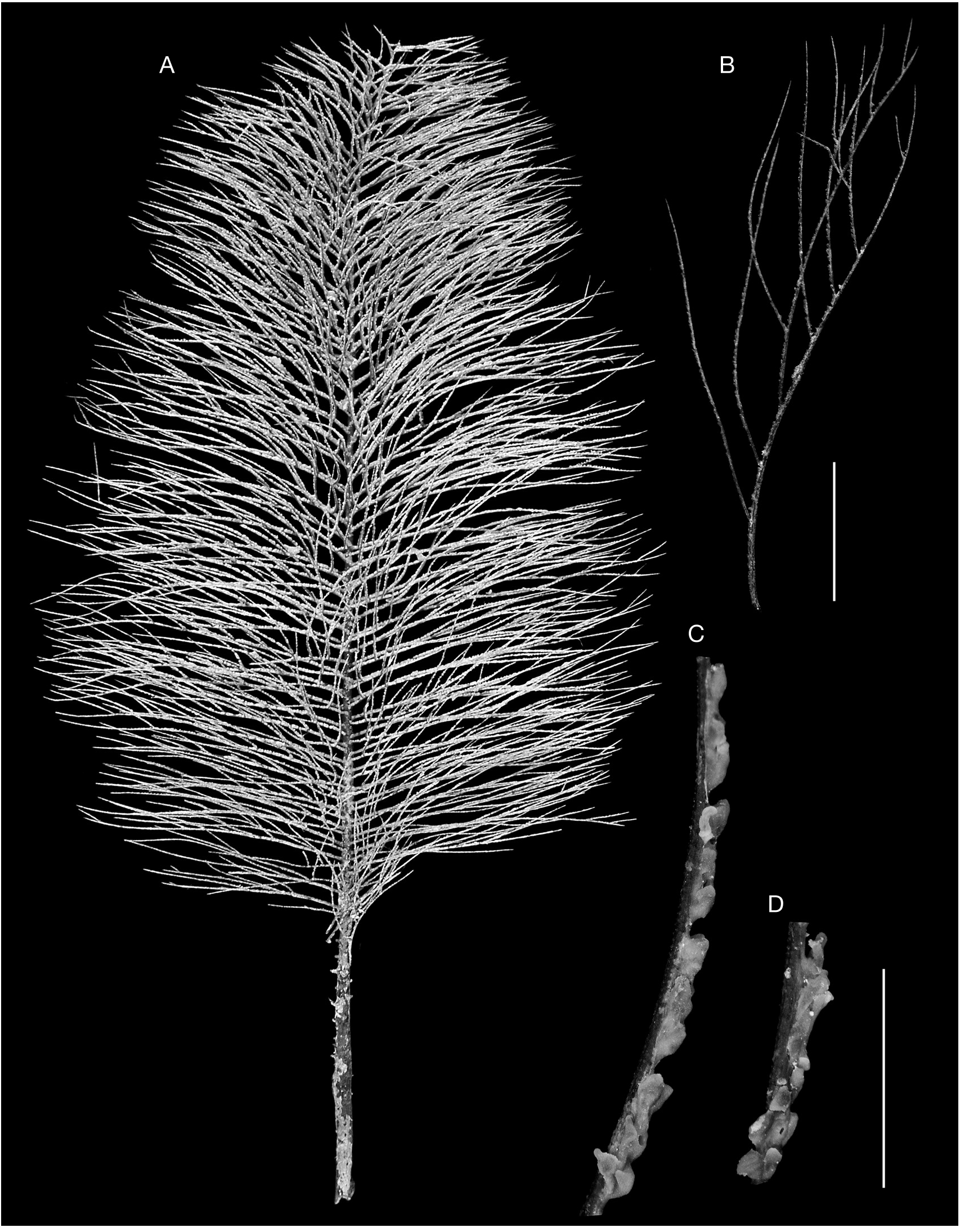
( Figs 1-3 View FIG View FIG View FIG )
HOLOTYPE. — NORFOLK 1, stn CP 1721, 26. VI. 2001 ( MNHN).
TYPE LOCALITY. — South of New Caledonia, Norfolk Ridge, RV Alis, NORFOLK 1, stn CP 1721, 23°19’S, 168°01’E, 416- 443 m.
ETYMOLOGY. — The species is named after Margarita (diminutive Gretchen), a character of Goethe’s “Faust”.
DISTRIBUTION. — The species is known only from the type locality, the north part of the Norfolk Ridge.
DIAGNOSIS. — Corallum monopodial and pinnulate to the third order. Primary pinnules arranged in four bilateral rows and also in alternating groups of two each. Pinnules are up to 9.5-10 cm in length with base diameter 0.9-1.4 mm, spaced 2.8-6.0 mm apart in each row. Subpinnules (up to eight on each pinnule) are up to 7.5 cm, arranged uniserially on anterior (polyp) side of lower order pinnules and spaced 0.7- 2.0 cm apart. Spines triangular compressed, smooth, acute, up to 0.12 mm high on polypar side of axis; abpolypar spines usually shorter, 0.03 to 0.08 mm, less regular in form, often bifurcated. Polyps 1.5-3.0 mm in transverse diameter, arranged in one series with 3 to 3.5 polyps per cm.
DESCRIPTION OF THE HOLOTYPE
The holotype is a colony 40 cm tall with a maximum width of about 20 cm ( Fig. 1A View FIG ). The corallum is broken somewhere above the basal plate and the lower part is missing. In the lower part of the colony, the stem is compressed and elliptical in cross section, with a maximum diameter of 7 mm. The stem is almost upright, slightly curved backwards in the upper part of the corallum. The lowermost 4.5 cm of the stem lacks pinnules and its surface is covered by a series of feebly marked grooves. For the next 4 cm, the bases of broken primary pinnules, arranged in four rows are visible.
The primary pinnules ( Fig. 1B View FIG ) are arranged in four rows with two lateral rows set in planes at an angle of 135-140°, and two anterolateral rows at an angle of 80°. The outer lateral primary pinnules are spaced 2.8-6.0 mm (mainly 3.0- 4.5 mm) apart in each row; the distance between pinnules in the row increases toward the apex of the corallum. In the middle of the colony there are, in average, 15 to 16 primary pinnules per 10 cm in each row. Each anterolateral primary pinnule is positioned 1.0- 1.5 mm below the adjacent lateral primary pinnule on the same side of the axis, generating bilateral groups consisting of one lateral and one anterolateral pinnules that are arranged alternately along the stem. Lateral primary pinnules are 5.0- 5.5 cm long on the lower part of the corallum; in the middle of the colony their length increases to 9.5-10.0 cm; and in the apical part of the colony it does not exceed 3.5 to 4.0 cm. Both lateral and anterolateral pinnules curve backward and slightly incline distally, with the distal angle formed by the lateral pinnules varying from about 65-80° in the lower part of corallum to 50-55° toward the apex of the colony. The diameter of the base of the primary pinnules ranges from 0.9 to 1.4 mm.
Lateral primary pinnules bear up to eight secondary pinnules. The distance between secondaries ranges from 0.7 to 2.0 cm (occasionally up to 2.7 cm). The length of secondary pinnules does not exceed the distance between the attachment of the secondary and the apex of the corresponding primary pinnule; therefore, the length of the secondary pinnules diminishes distally. The longest secondary pinnules do not exceed 7.5 cm. Secondary pinnules form a rather narrow distal angle (15-18°) with the primary on which they arise, and are always curved distally toward the apex of the primary pinnule. The length of the most distal secondaries usually does not exceed 1.0-2.0 cm. Most of secondary pinnules are simple but some can bear one to three tertiary pinnules that are also curved distally. No quaternary pinnules were observed. Anterolateral primary pinnules usually bear two to four uniserially arranged simple secondaries of the same structure as those on the lateral primaries.
Spines are arranged in longitudinal rows. The number of rows visible in lateral view increases from three to four in the distal parts of tertiary pinnules to seven to nine at the basal parts of the primary and secondary pinnules. Along the distal parts of the pinnules the spines are mostly triangular, laterally compressed, inclined distally, acute and close to equal around the axis ( Fig. 2A View FIG ). On pinnules 0.11-0.12 mm in diameter ( Fig. 2B View FIG ) they are 0.03-0.05 mm high, slightly larger on the polypar side of the axis. More proximally ( Fig. 2C, D View FIG ) the difference between polypar and apolypar spines is obvious and on a pinnule 0.2- 0.36 mm in diameter the abpolypar spines are 0.03-0.08 mm high and polypar spines are 0.06- 0.11 mm high. Polypar spines ( Fig. 3A View FIG ) are triangular to conical, laterally compressed and generally inclined distally; however, some can be inclined in other directions ( Fig. 2D View FIG ). Abpolypar spines ( Fig. 3 View FIG B-E) are of irregular form, from conical, inclined distally, with blunt apex and hooklike to forked, with two to three secondary spines. They often form dense clusters at the base of primary spines so the longitudinal rows are hardly distinguishable ( Fig. 3D, E View FIG ). Distances between polypar spines (measured between bases of adjacent spines of the same row) range from 0.18 to 0.4 mm; those for abpolypar ones are 0.12 to 0.24 mm. There are, on average, three polypar and four to five abpolypar spines per mm in each row.
The polyps ( Fig. 1C, D View FIG ) are arranged in a single row on the anterior, convex side of the pinnules and anterior side of the distal part of the stem. They are 2 to 3 in transverse diameter with interpolypar distances ranging from 0.3 to 0.75 mm. There are 3 to 3.5 polyps per cm. Some developing polyps about 1.4 mm long can be found between full-grown polyps. The tentacles in preserved specimens are short, up to 0.8 mm long.
REMARKS
The new species differs from the closely related Saropathes scoparia ( Totton, 1923) in having shorter rigid primary pinnules that are curved backward (in contrast with forward curving long primary and secondary pinnules of S. scoparia ); in a lower order of subpinnulation (three vs four in S. scoparia ) and in a larger number of shorter secondary pinnules on each of primary (up to eight vs six in S. scoparia ). These two species also differ in the form and size of the spines. The polypar and abpolypar spines in S. margaritae n. sp. are of obviously different size and structure, whereas in S. scoparia the spines are subequal around the axis. The maximum height of the polypar spines in the new species is 0.12 mm, whereas in the type species of the genus, the maximum height of the spines is almost one half, being no more than 0.075 mm. There is no great difference in size and arrangement of polyps between the two species.


| VI |
Mykotektet, National Veterinary Institute |
| MNHN |
Museum National d'Histoire Naturelle |
| RV |
Collection of Leptospira Strains |
No known copyright restrictions apply. See Agosti, D., Egloff, W., 2009. Taxonomic information exchange and copyright: the Plazi approach. BMC Research Notes 2009, 2:53 for further explanation.