
Ishimosorex, Zazhigin & Voyta, 2022
|
publication ID |
https://doi.org/ 10.26879/1209 |
|
publication LSID |
lsid:zoobank.org:pub:1726FDAE-2EE5-4145-A124-6D24287C0514 |
|
DOI |
https://doi.org/10.5281/zenodo.11105188 |
|
persistent identifier |
https://treatment.plazi.org/id/67243F45-1EBF-46CC-A2C7-E6B73680B820 |
|
taxon LSID |
lsid:zoobank.org:act:67243F45-1EBF-46CC-A2C7-E6B73680B820 |
|
treatment provided by |
Felipe |
|
scientific name |
Ishimosorex |
| status |
gen. nov. |
Genus ISHIMOSOREX gen. nov.
zoobank.org/ 67243F45-1EBF-46CC-A2C7-E6B73680B820
Type species. Ishimosorex ishimiensis sp. nov., by monotypy, see below.
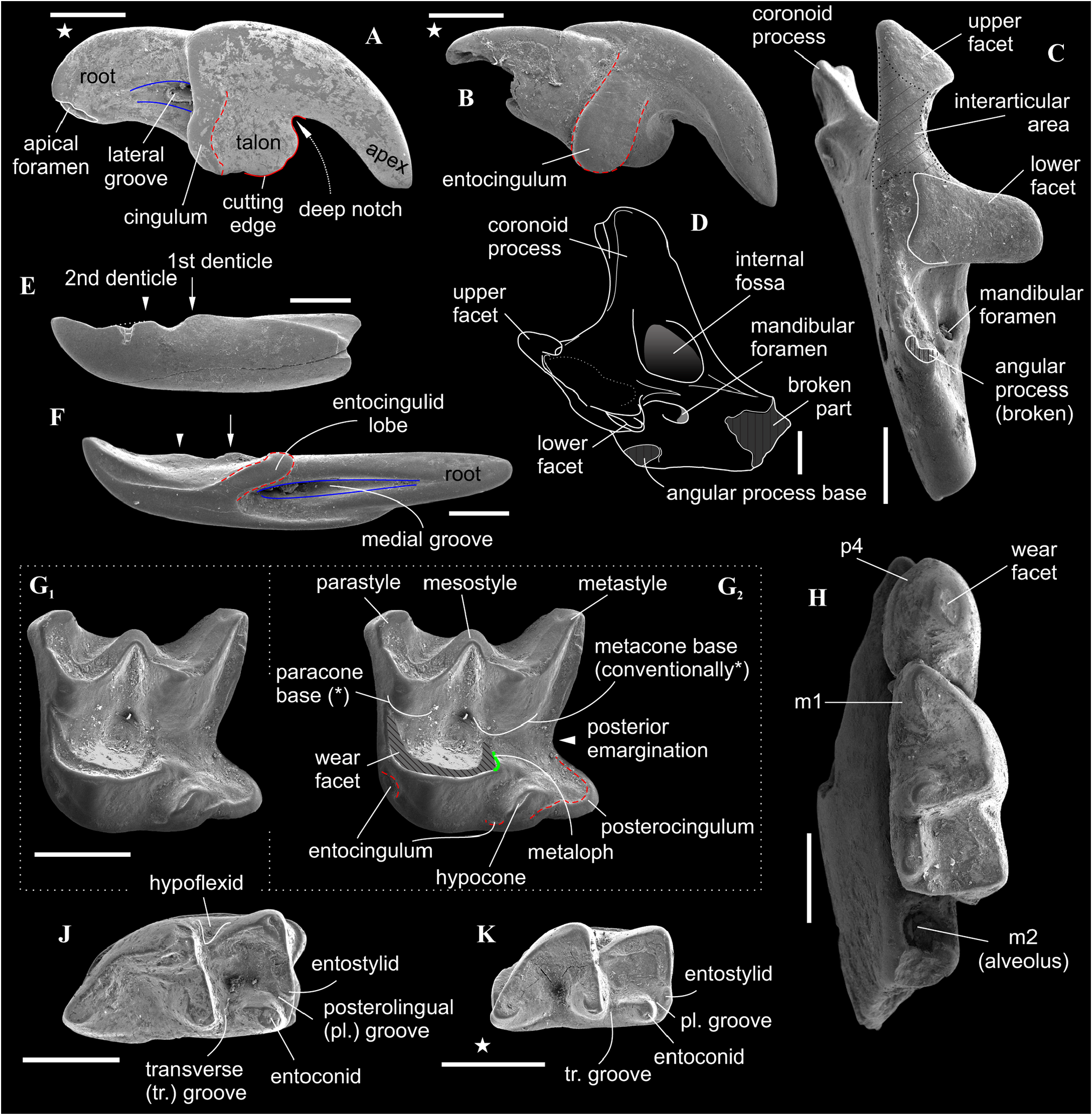
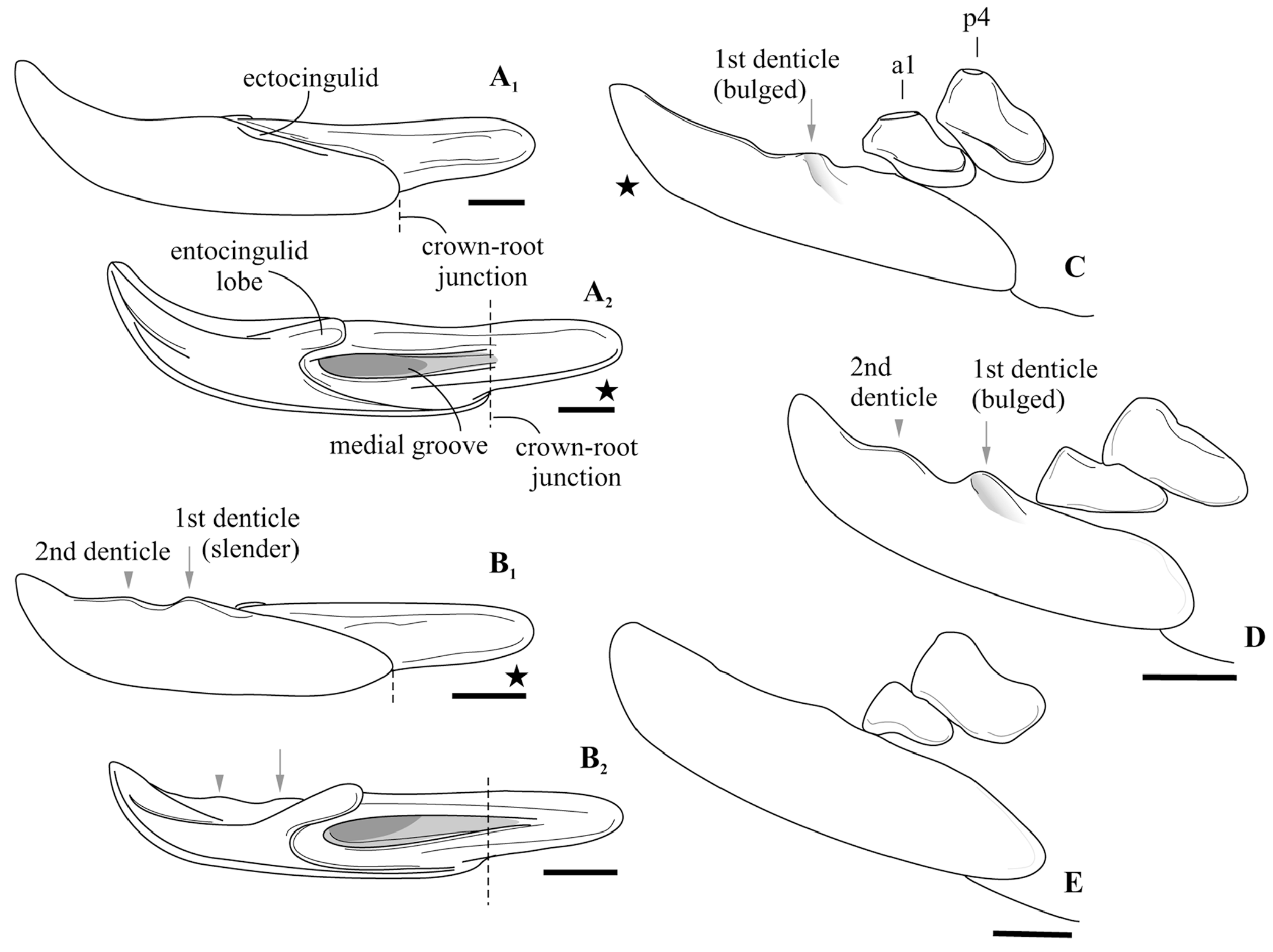
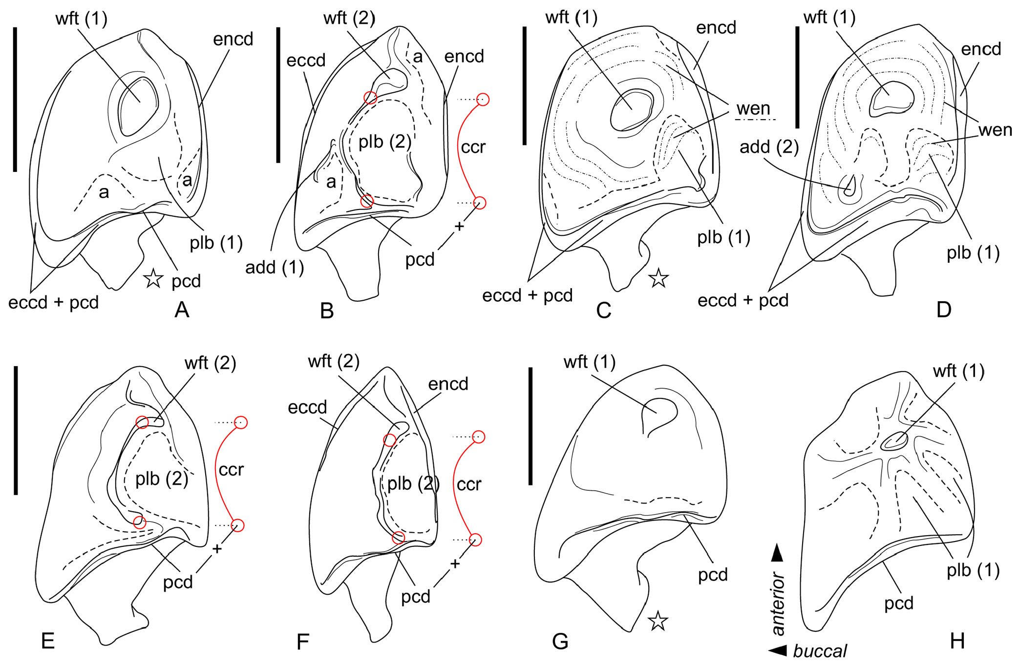
Diagnosis. Small-sized anourosoricin shrew. I1 with an elongated and relatively low crown, and short slightly curved root; the hatchet-like (bladelike) talon with expressed narrow, deep notch (like a carnassial notch of lower molars). M1 has a subquadrate occlusal shape with a long and narrow hypoconal flange; the mesostyle is well-developed and protruded buccally; the metaloph is short and separated from the metacone base. i1 with the moderately upturned tip and two slender denticles (bicuspulate) on the cutting edge. p4 has a bulbous-like shape with the round (spot-shaped) wear facet and the very weak expressed central crest; the ecto- and entocingulids are wide and well developed. The hypoconid of m1 and m2 is buccally protruded and overhangs the base of the crown; the entostylid is presented as a small bulge and does not reach the entoconid level by height; entocristid is short. The interarticular area of the condylar process is moderately wide.
Differential diagnosis. Ishimosorex gen. nov. differs from Paranourosorex in presence of the deep notch and the long crown of I1 and its relatively short root ( Figure 2A View FIGURE 2 ); in the equal lengths of the lingual and buccal sides of M1; in the deeper posterior emargination of M1 and more posterior protrusion of the hypoconal flange relative the hypocone position ( Figure 2G View FIGURE 2 ); in presence of two denticles on the cutting edge of i1 and its long medial groove ( Figure 3 View FIGURE 3 ); in the wider interarticular area of the condylar process and the relatively large fossa of the temporal muscle of the mandibular ramus.
Ishimosorex gen. nov. differs from Crusafontina in the general proportion of M1 (subsquare vs. trapeziform outline shape), more expressed Wshaped line of the M1 buccal crests, more backwardly protruded the hypoconal flange of M1 ( Figure 2G View FIGURE 2 cf. M1 of C. endemica from Puente Minero 2, Teruel Basin by van Dam, 2004: figure 2-3); in the slender basal (first) denticle of i1 ( Figure 3 View FIGURE 3 ); in the bulbous-like crown of p4 with the extremely weak expression of the central crest ( Figure 4 View FIGURE 4 ); in a prominent buccal shifting of the protoconid of m1– m2 from the longitudinal axis of the crown and more central position of the entoconid of m1–m2 ( Crusafontina has a more central position of the protoconid of m1 and a lingually shifted the entoconids of m1–m2 relatively to a basal crown outline; see van Dam, 2004: figure 2).
Ishimosorex gen. nov. differs from Amblycoptus and Kordosia in the general proportion of I1 with the thin talon ( Amblycoptus and Kordosia both show lateromedial inflation of the talon and expressed bulge of the buccal cingulum); in the proportion and outline shape of M1 (similar to Crusafontina buccal crest condition, see above); in the presence of a denticulation of the cutting edge of i1 ( Figure 3 View FIGURE 3 ); in more inflated the anterior part of p4 crown (‘subrectangle’ shape vs ‘subtriangle’ shape in Amblycoptus and Kordosia ; Figure 4 View FIGURE 4 ); in lesser exaenodonty of p4 crown; in a more buccal shifting of the protoconid and hypoconid of m1 from the longitudinal axis of the crown ( Figure 2 View FIGURE 2 ; also see van Dam, 2004: figure 5). The extreme dimily of Ishimosorex gen. nov. is doubtful but cannot be test due to lack of data.
Ishimosorex gen. nov. differs from Darocasorex in the general proportion of M1 outline shape ( Darocasorex shows an anteroposterior compression of M1); in absence of the ectocingulid of i1; in absence of expressed crests of p4 ( Darocasorex has the developed buccal and short lingual crests); in a more longitudinal direction of the oblique crestid of m1 and m2 (see van Dam, 2010).
Ishimosorex gen. nov. differs from Anourosorex in the general proportion of I1 and the buccal cingulum condition ( Anourosorex has a short inflated ectocingulum); in more expressed Wshaped buccal crests and significantly lesser developed of the parastyle of M1 ( Anourosorex has an extremely developed inflated and buccally protruded parastyle; see Storch and Qiu, 1991); in the bicuspulate i1; in the absence of the expressed and sharp central crest of p4 ( Anourosorex has very developed, sharp and high central crest); in presence of the ecto- and entocingulids of p4; in the crown proportion of m1–m2 (similar to Amblycoptus and Kordosia see above).
Etymology. After the Ishim river, located in North Kazakhstan, the provenance of the fossil remains, and from Latin sorex, shrew.
Stratigraphic and geographic range. As for the only included species.
No known copyright restrictions apply. See Agosti, D., Egloff, W., 2009. Taxonomic information exchange and copyright: the Plazi approach. BMC Research Notes 2009, 2:53 for further explanation.