
Apistophonte wasiniensis Gheerardyn and Fiers
|
publication ID |
https://doi.org/10.5281/zenodo.174137 |
|
DOI |
https://doi.org/10.5281/zenodo.5668681 |
|
persistent identifier |
https://treatment.plazi.org/id/72289B3F-FFE6-FFFA-FECE-FAC6FD97F9E0 |
|
treatment provided by |
Plazi |
|
scientific name |
Apistophonte wasiniensis Gheerardyn and Fiers |
| status |
gen. nov. |
Apistophonte wasiniensis Gheerardyn and Fiers gen. n., sp. n.
Type locality.—Western Indian Ocean, Kenyan coast, Wasini Island ( 4°40’S, 39°23’E), red (terrigenous?) sediment, water depth 3– 4 m.
Material.—(a) From type locality: holotype Ψ dissected on 4 slides ( COP 4727a–d); allotype ♂ dissected on 3 slides ( COP 4728a–c); paratypes are 2 ΨΨ and 1 ♂ dissected on slides ( COP 4729– COP 4731) and 6 ΨΨ and 4 ♂♂ preserved in 75% alcohol ( COP 4732); collected 28 February 2002 by M. Raes.
(b) Western Indian Ocean, Kenyan coast, Kisite Island ( 4°43’S, 39°22’E), coral sand, water depth 3– 6 m.: paratypes, 3 ΨΨ preserved in 75% alcohol ( COP 4733); collected 28 February 2002 by M. Raes.
Etymology.—The specific name wasiniensis refers to the type locality of this species.
Description of female
Total body length 299–406 µm (n=9; average=361 µm; measured from anterior margin of rostrum to posterior margin of caudal rami). Largest width measured at posterior margin of cephalothorax: 88 µm.
Rostrum ( Fig. 2 View FIGURE 2 A) large with straight lateral margins; broad triangular; fused to cephalothorax; with a pair of sensilla anteriorly; dorsal surface pitted.
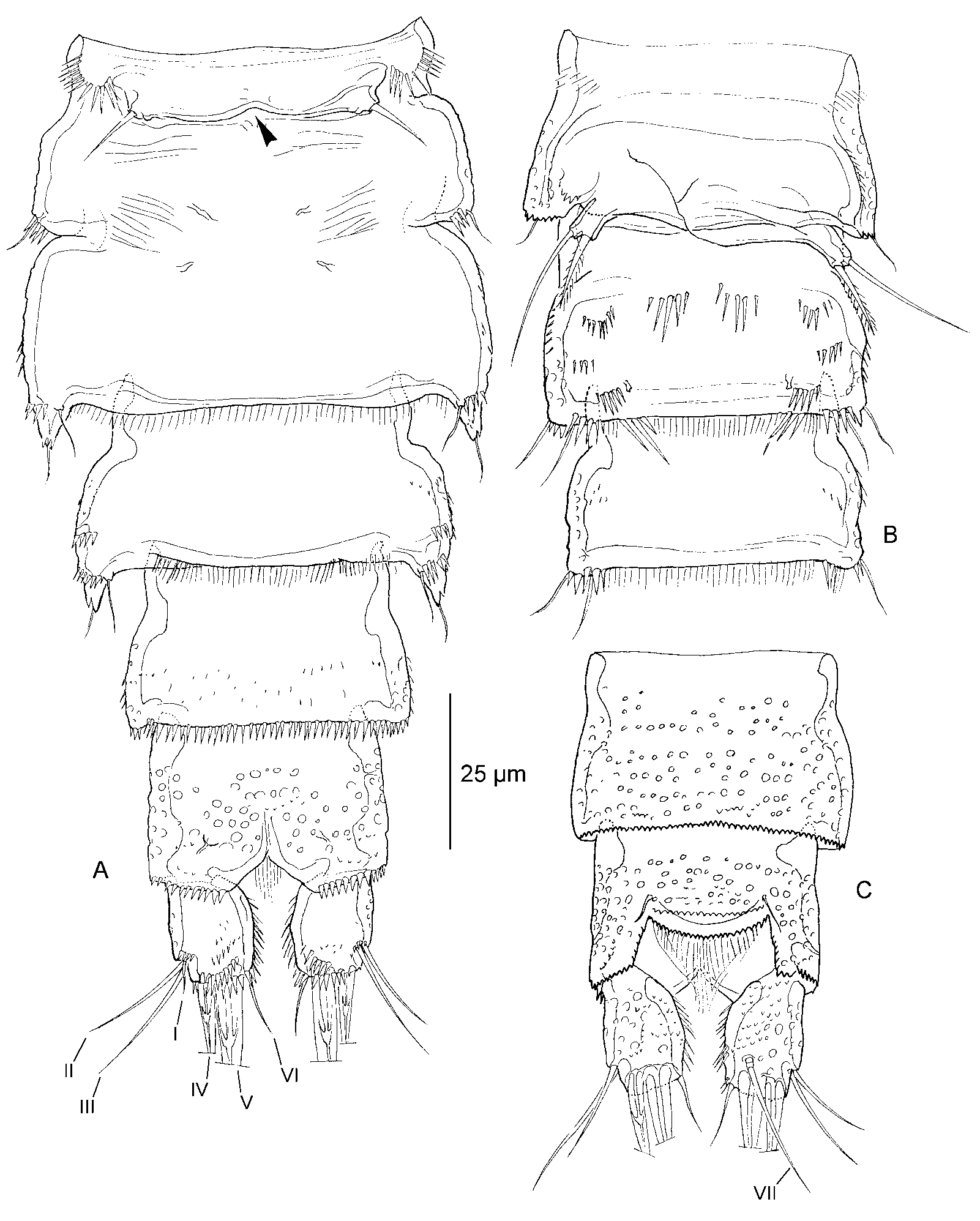
Habitus ( Fig. 1 View FIGURE 1 A–B). Body fusiform prehensile. Cephalothorax with parallel margins. Free prosomites slightly less wide as cephalothorax. Genital doublesomite and following urosomite ventrolaterally expanded. Urosome gently tapering towards the anal somite. Second and third urosomite fused to form genital doublesomite. Original division between first and second somite of genital doublesomite is marked serrate dorsally.
Integument of the cephalothorax pitted; with symmetrical pattern of smooth areas; regularly ornamented with small sensilla. Surface of pleurotergites and dorsal surface of anal somite pitted entirely. Posterodorsal margin of cephalothorax smooth. Posterodorsal margin of the free somites serrate. Posterolateral angles of cephalothorax slightly extended. Posterodorsal margins of cephalothorax and free somites (except penultimate urosomite) bearing a number of small sensilla. Free prosomites and first urosomite additionally bearing 1 pair of sensilla dorsally. Anal operculum not protruding backwardly; flanked by 2 sensilla; with serrate margin.
Ventral surface ( Fig. 4 View FIGURE 4 A) of genital doublesomite smooth, except for some striae in anterior part; bearing spinular row laterally from P6 vestiges. Genital doublesomite and following 2 somites bearing few spinules laterally. Ventral surface of fourth urosomite smooth; of fifth urosomite with some small spinules in posterior part; of anal somite pitted. Posteroventral margins of genital doublesomite and following urosomites bearing row of slender to strong spinules.
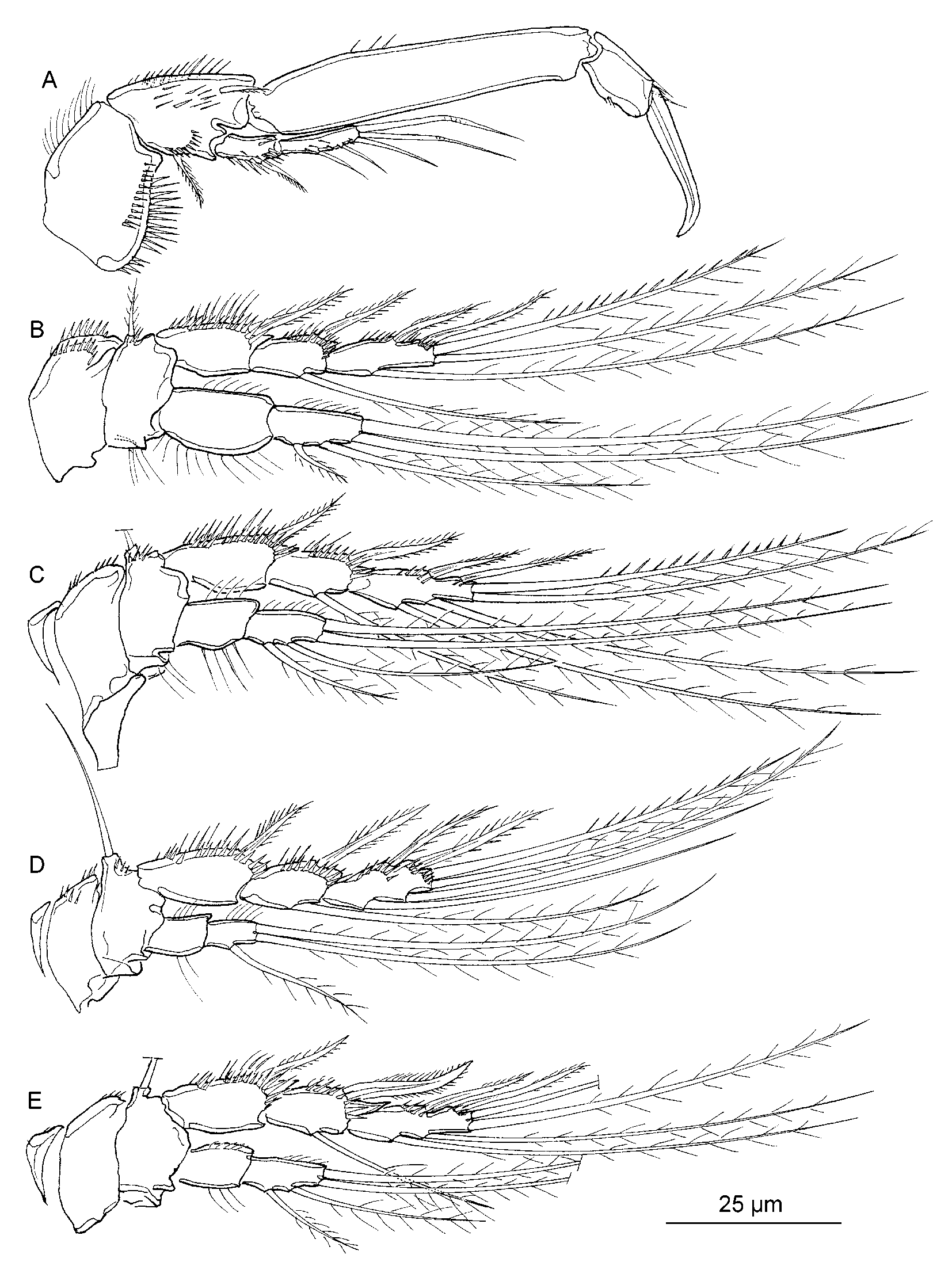
Caudal rami ( Fig. 4 View FIGURE 4 A, 4C) almost 1.5 times as long as wide; cylindrical with slightly convex inner margin; bearing spinules along the inner margin and several spinular rows on the ventral surface; with some small denticles and pits dorsally. Seta I, II and III inserted in distal fourth of outer margin. Seta I rudimentary. Seta IV and V not fused; seta IV pinnate, seta V naked. Seta VII inserted in the distal fourth. Antennule ( Fig. 2 View FIGURE 2 A) 6segmented; majority of setae long and slender. Segment 1 and 2 bearing few pits dorsally, ventral surface smooth; segment 3–6 smooth. Segment 1 short, slightly longer than wide; bearing small, blunt process along outer margin; with spinular row along inner margin. Segment 2 with distinct, posteriorly directed hook along outer margin. Armature formula: 1[1], 2[7 + 1 pinnate], 3[7], 4[1 + (1 + ae)], 5[1], 6[9 + acrothek]. Apical acrothek consisting of a small aesthetasc fused basally to 2 setae.
Antenna ( Fig. 2 View FIGURE 2 F). Allobasis bearing 2 spinular rows; with 1 short, unipinnate abexopodal seta, inserted in distal third. Exopod 1segmented and small, well developed; bearing 4 subequal bipinnate setae, the dorsal one being more slender and less dense pinnate. Endopod with 2 rows of spinules and 1 subapical frill; with following armature: 2 spines (1 unipinnate) and slender seta subapically, 2 clawlike spines, 3 geniculate setae and small, slender seta apically.
Mandible ( Fig. 2 View FIGURE 2 B). Biting edge formed by several blunt teeth and seta. Palp uniramous; endopod and exopod represented by 3 and 1 smooth seta(e), respectively. Medial seta plumose.
Maxillule ( Fig. 2 View FIGURE 2 G). Praecoxal arthrite bearing spinular row on posterior surface; with 5 setae/spines apically; with 1 small, obliquely positioned seta along the outer margin and 2 small setae along the inner margin. Coxal endite with 1 seta and 1 curved spine. Basal endite with 2 naked setae and 1 curved spine. Endopod obsolete, represented by 3 setae. Exopod 1segmented with 2 apical setae.
Maxilla ( Fig. 2 View FIGURE 2 H). Syncoxa with 3 endites; with 1 row of spinules along outer margin and 2 along inner margin. Praecoxal endite small, with 1 seta. Proximal coxal endite with 1 strong, pinnate spine and 2 slender, naked setae. Distal coxal endite with 1 strong, pinnate spine and 2 slender, naked setae. Allobasis drawn out into strong, slightly curved claw; bearing 2 setae. Endopod obsolete, represented by 2 naked setae.
Maxilliped ( Fig. 2 View FIGURE 2 E). Syncoxa with 2 spinular rows; apically bearing pinnate seta and rudimentary seta next to it. Basis with some spinules along the slightly convex outer margin. Endopod clawshaped, unarmed, with short, naked seta at base.
P1 ( Fig. 3 View FIGURE 3 A). Coxa cylindrical with 1 inner and 2 outer spinular rows. Basis with 1 pinnate seta along outer margin; medial, unipinnate seta arising on anterior surface; spinules on anterior surface, along inner and outer margin. Exp1 bearing 1 unipinnate outer seta, spinular row along the outer margin and a few spinules on the anterior surface; exp2 bearing 3 naked outer setae and 2 geniculate apical setae, with a few spinules on the anterior surface. Enp1 2.5 times as long as exp, with few spinules along the inner margin; enp2 with 1 strong, smooth claw and 1 minute, naked accessory seta.
P2–P4 ( Fig. 3 View FIGURE 3 B–D). Setal formulae in table 1. Exopods 3segmented and endopods 2 segmented. Praecoxae small and triangular. Coxae and bases with spinules along the outer margin. Inner margin of basis in P2 and P3 with some slender long hairs. Outer margin of basis with short, pinnate (P2) or long, naked (P3–P4) seta. P2 endopod reaching to the proximal third of exp3. P3 endopod reaching just beyond the middle of exp2. P4 endopod slightly longer than exp1. Segments of endopods and exopods with pattern of spinules as figured.
P5 ( Fig. 2 View FIGURE 2 I) with separate exopod and baseoendopod; both covered anteriorly with few small spinules; the margins bearing strong and long spinules. Basal seta arising from a cylindrical setophore. Proximal setae of endopodal lobe bipinnate; subapical and apical seta naked. Baseoendopod reaching to middle of exopod. Exopod with ovate shape; about 2 times as long as wide; bearing 5 plumose setae.
P6 vestiges ( Fig. 4 View FIGURE 4 A) bearing 1 seta. Copulatory pore minute, situated in middle of anterior somite.
Description of male
Total body length 280–387 µm (n=6; average=326 µm; measured from anterior margin of rostrum to posterior margin of caudal rami). Largest width measured at posterior margin of cephalothorax: 78 µm.
Habitus ( Fig. 1 View FIGURE 1 C) as in female; except for the fully separated second and third urosomite, and the lack of ventrolateral extensions in the second to fourth urosomites ( Fig. 4 View FIGURE 4 B). Ventral surface of third urosomite bearing several short rows of long spinules. Posteroventral margin of third urosomite with slender hairs and some long spinules near the lateral sides.
Antennule ( Fig. 2 View FIGURE 2 C–D) 8segmented; subchirocer. Segment 1 and 2 as in female. Armature formula: 1[1], 2[8 + 1 pinnate], 3[5 (?)], 4[2], 5[10 (?) + 1 pinnate + (1 + ae)], 6[0], 7[1], 8[7 + acrothek]. Apical acrothek consisting of a small aesthetasc fused basally to 2 setae.
Antenna, mouthparts and P1 as in female.
Endopods of P2–P4 as in female. Exopods of P2 and P4 as in female; except that the inner seta on exp2 is shorter than the corresponding seta in the female (reaching not far beyond the distal margin of exp3). P3 exopod ( Fig. 3 View FIGURE 3 E) as in female; except for a curved, stronger outer spine on exp2, the distal outer corner of exp2 being more strongly developed and the inner seta on exp2 being shorter than the corresponding seta in the female (reaching not far beyond the distal margin of exp3).
P5 ( Fig. 2 View FIGURE 2 J). Endopodal lobe of P5 obsolete; without a seta. Exopod small; slightly longer than wide; bearing 3 plumose setae.
P6 vestiges ( Fig. 4 View FIGURE 4 B) asymmetrical. One vestige functional; one vestige fused to somite. Both produced into a cylindrical process bearing 1 pinnate inner and 1 naked outer seta.
Variability.—Among the 12 females and 6 males studied, no variability in setal formulae was observed.
Known range.—To date, A. wasiniensis is only known from Wasini and Kisite Islands along the Kenyan coast.


| COP |
Coleccion Ornitologica Phelps |
No known copyright restrictions apply. See Agosti, D., Egloff, W., 2009. Taxonomic information exchange and copyright: the Plazi approach. BMC Research Notes 2009, 2:53 for further explanation.
|
Kingdom |
|
|
Phylum |
|
|
Class |
|
|
Order |
|
|
Family |
|
|
Genus |