
Propephonte duangitensis Gheerardyn and Fiers
|
publication ID |
https://doi.org/10.5281/zenodo.174137 |
|
DOI |
https://doi.org/10.5281/zenodo.5668686 |
|
persistent identifier |
https://treatment.plazi.org/id/72289B3F-FFEE-FFE2-FECE-FC46FB7DF8D8 |
|
treatment provided by |
Plazi |
|
scientific name |
Propephonte duangitensis Gheerardyn and Fiers |
| status |
gen. nov. |
Propephonte duangitensis Gheerardyn and Fiers gen. n., sp. n.
Type locality.—Western Pacific Ocean, Papua New Guinea, Madang Province, Hansa Bay (Duangit Reef) ( 4°10’S, 144°53’E), coral sand and coral rubble from the east side, water depth 40– 46 m.
Material.— Holotype Ψ dissected on 1 slide ( COP 1940); allotype ♂ dissected on 1 slide ( COP 1941); paratypes are 1 Ψ dissected on 3 slides ( COP 4726a–c) and 1 ♂ preserved in 75% alcohol ( COP 1942); all collected 28 May 1979 by J. Pierret.
Etymology.—The specific name duangitensis refers to the type locality of this species.
Description of female
Total body length 326–350 µm (measured from anterior margin of rostrum to posterior margin of caudal rami). Largest width measured at posterior margin of cephalothorax: 88 µm.
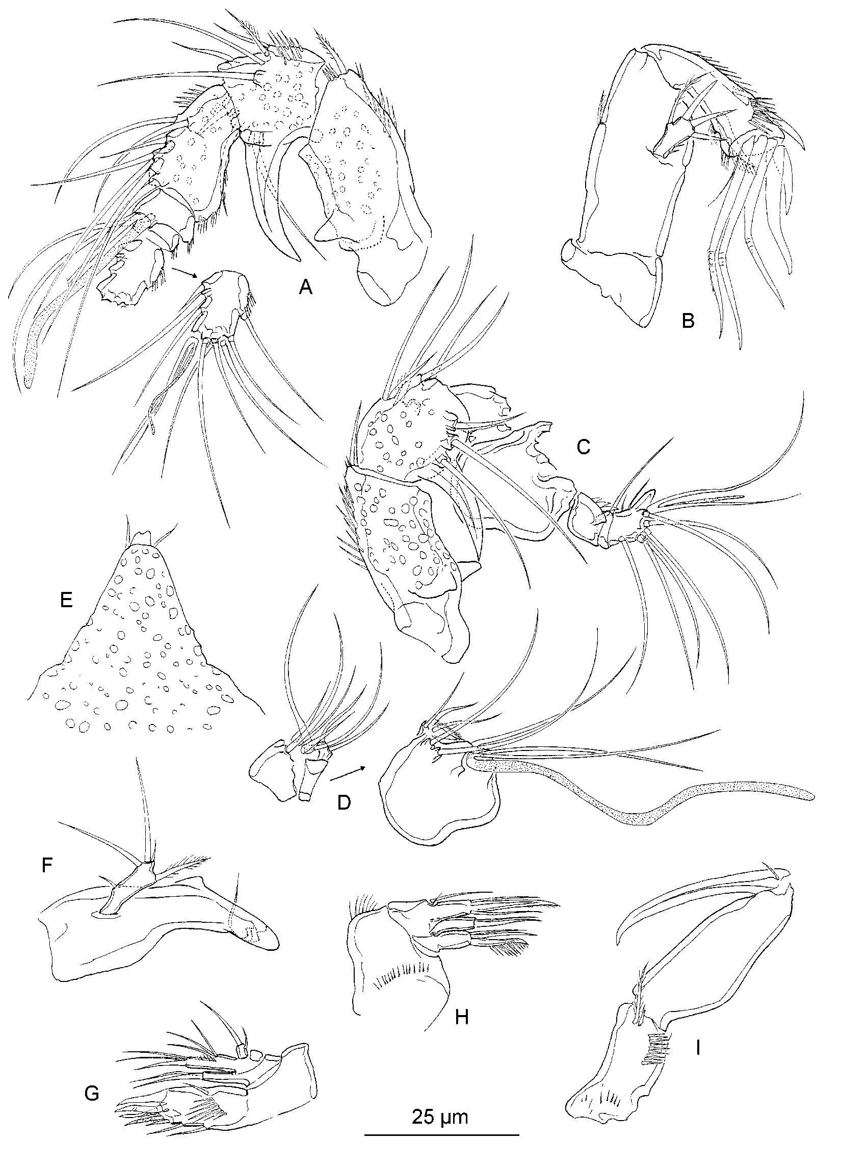
Rostrum ( Fig. 6 View FIGURE 6 E) strongly prominent and triangular; fused to cephalothorax; rather narrow, with slightly concave margins; tip small, slightly bifid; with pair of sensilla anteriorly; dorsal surface pitted.
Habitus ( Fig. 5 View FIGURE 5 A–B). Body fusiform prehensile, slightly depressed. Cephalothorax with parallel margins, only tapering in anterior quarter. Free prosomites and first urosomite as wide as cephalothorax; second to fourth urosomites expanded ventrolaterally. Urosome gently tapering towards the anal somite. Posterolateral angles of cephalothorax lobate. Pleural areas of free prosomites well developed and rounded, bearing spinules along margin. Second and third urosomite fused to form genital doublesomite. Genital doublesomite with transverse serrate surface ridge dorsally and laterally, indicating original segmentation; fused ventrally.
Integument of cephalothorax pitted; regularly ornamented with small sensilla. Pleurotergites of prosomites and urosomites, and dorsal surface of anal somite and caudal rami entirely pitted. Rows of closely arranged pits transforming into rows of small denticles. Posterodorsal margin of cephalothorax smooth; of the free somites serrate. Posterodorsal margins of cephalothorax and free somites (except penultimate urosomite) bearing a number of small sensilla. Anal operculum well developed and slightly protruding backwardly; flanked by 2 sensilla; with serrate margin.
Ventral surface (Fig. 9A) of the genital doublesomite striated anteriorly, smooth posteriorly. Ventral surface of following 2 urosomites smooth; of anal somite pitted. Posteroventral margins of genital doublesomite and following urosomites bearing a row of spinules.
Caudal rami (9A–B) almost twice as long as wide; cylindrical; surface of the caudal rami without processes. Ventral surface and outer margin of the caudal rami spinulose. Inner margin slightly tapering towards the distal margin and bearing strong spinules. Seta I, II and III inserted in distal third of outer margin. Seta IV and V not fused. Seta VII inserted in the distal third.
Antennule ( Fig. 6 View FIGURE 6 A) 6segmented; majority of setae long and slender. Segment 1–3 pitted dorsally, smooth ventrally. Segment 4–6 smooth. Segment 1 elongate, almost 2.5 times as long as wide; dorsally with blunt process on the proximal half; outer margin bears blunt thorn proximally. Segment 2 with large, posteriorly directed hook along outer margin. Inner margin of first to third segment and outer margin of third to sixth segment with spinules. Armature formula: 1[1 pinnate], 2[7 + 1 pinnate], 3[7], 4[1 + (1 + ae)], 5[1], 6[9 + acrothek]. Apical acrothek consisting of a small aesthetasc fused basally to 2 setae.
Antenna ( Fig. 6 View FIGURE 6 B). Allobasis with 1 short, unipinnate abexopodal seta, inserted in distal half. Exopod 1segmented and small, but well developed; bearing 3 subequal setae apically, and 1 bipinnate, slender and slightly longer seta subapically. Endopod with 2 rows of spinules and 2 subapical frills; with following armature: subapically 2 spines (one is unipinnate) and a small, slender seta, apically 2 clawlike spines, 3 geniculate setae (the outermost pinnate) and 1 slender seta.
Mandible ( Fig. 6 View FIGURE 6 F). Biting edge formed by several blunt teeth and a seta. Palp uniramous; endopod and exopod represented by 3 and 1 smooth seta(e), respectively. Medial seta plumose.
Maxillule ( Fig. 6 View FIGURE 6 G). Praecoxal arthrite bearing a spinular row on the posterior surface; apically with 6 setae/spines; with 1 small, obliquely positioned seta along the outer and 2 slender setae along the inner margin. Coxal endite with 1 seta and 1 curved spine. Basal endite with 2 setae and 1 curved spine. Endopod obsolete, represented by 3 setae. Exopod 1segmented with 2 apical setae.
Maxilla ( Fig. 6 View FIGURE 6 H). Syncoxa with 2 endites; with a spinular row along the inner and along the outer margin. Praecoxal endite absent. Proximal coxal endite with 1 strong, pinnate spine and 2 slender, naked setae. Distal coxal endite with 1 curved spine and 1 slender seta. Allobasis drawn out into strong, slightly curved, distally pinnate claw; bearing 2 setae. Endopod obsolete, represented by 2 setae (one of which is very short).
Maxilliped ( Fig. 6 View FIGURE 6 I). Syncoxa with spinular row along the outer margin and some spinules proximally; apically bearing pinnate seta and small seta next to it. Basis with slightly convex outer margin. Endopod clawshaped, unarmed, with short, naked seta at base.
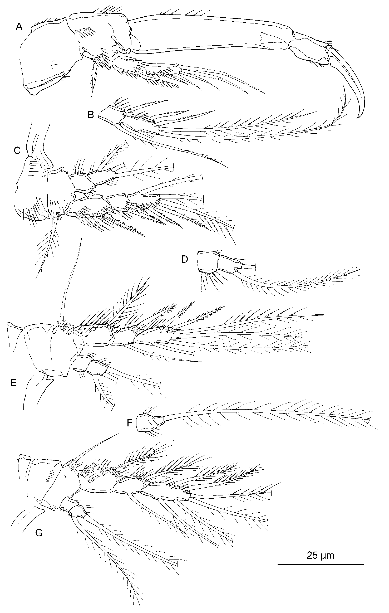
P1 ( Fig. 7 View FIGURE 7 A). Coxa and basis cylindrical, each about as long as broad; with several spinular rows. Basis with slender, plumose outer seta; inner unipinnate seta arising on anterior surface. Exopod 2segmented, outer margins and anterior surfaces with spinules. Exp1 with a strongly armed outer spine; exp2 with 3 naked outer setae and 2 geniculate apical setae. Enp1 about 2.5 times as long as exp; enp2 with a strong, smooth claw and 1 minute, naked accessory seta.
P2–P4 ( Fig. 7 View FIGURE 7 B–7G). Setal formulae in table 1. Exopods 3segmented and endopods 2 segmented. Praecoxae small and triangular; devoid of integumental structures. Coxae and bases with spinules along the outer margin. Outer margin of basis with long, plumose (P2) or long, naked (P3–P4) seta. Proportional lengths of the endopods rather short; reaching to middle of exp 2 in P2, to the distal margin of exp 1 in P3 and to middle of exp 1 in P4. Outer spine of exp1 of P3 and outer exopodal spines of P4 ornamented with slender, long spinules. Segments of endopods and exopods with pattern of spinules as figured.
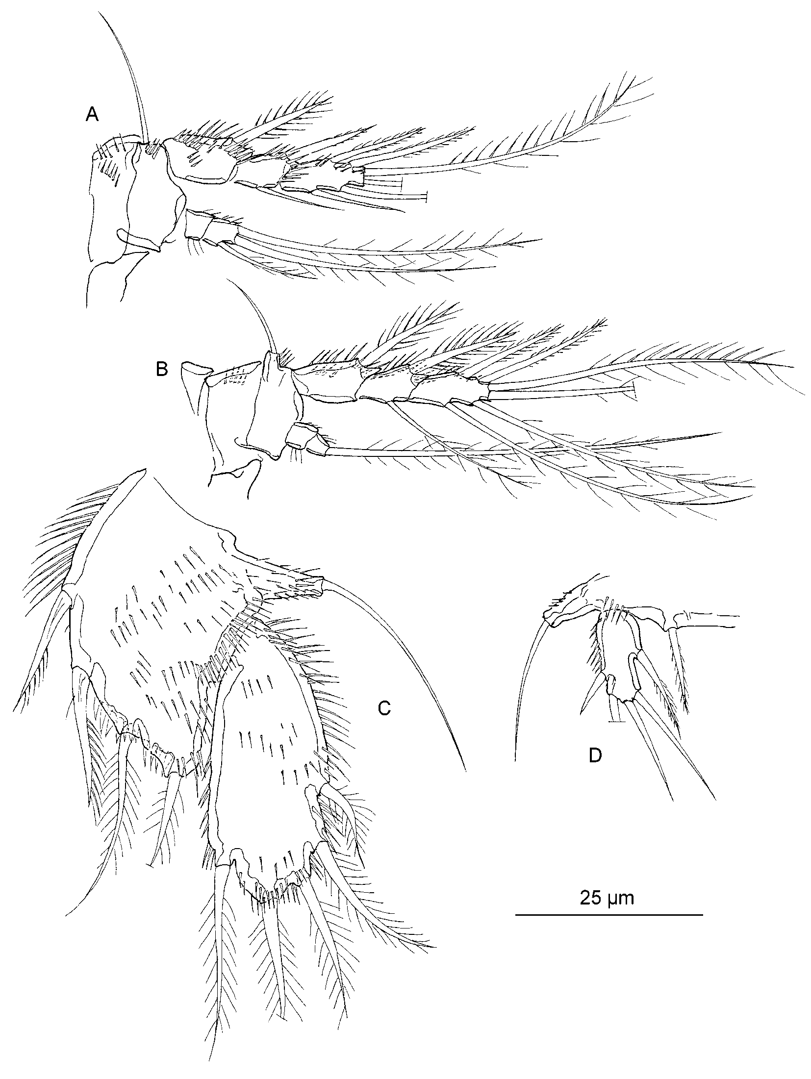
P5 ( Fig. 8 View FIGURE 8 C) with separate exopod and baseoendopod; the margins bearing long, slender spinules or stout, short spinules. Anterior surface furnished with rows of spinules. Proximal setae of endopodal lobe unipinnate; subapical and apical seta plumose. Baseoendopod reaching to middle of exopod. Exopod ovate shape; about 2 times as long as wide; bearing 5 plumose setae distally.
P6 vestiges (Fig. 9A) each bearing 1 small, naked seta. Copulatory pore minute, situated in middle of anterior somite.
Description of male
Total body length 309–350 µm (measured from anterior margin of rostrum to posterior margin of caudal rami). Largest width measured at posterior margin of cephalothorax: 80 µm.
Habitus ( Fig. 5 View FIGURE 5 C). More slender than female; especially with respect to the urosome. Second and third urosomite fully separated. Ventrolateral extensions of second to fourth urosomite are absent. Ventral surface of third urosomite with 2 rows of long spinules; anterior one along the entire surface, posterior one with a large gap in the middle (Fig. 9C).
Antennule ( Fig. 6 View FIGURE 6 C–D) 8segmented; subchirocer. Segment 1 and 2 as in female. Armature formula: 1[1], 2[8 + 1 pinnate], 3[6], 4[2], 5[9 (?) + (1 + ae)], 6[0], 7[1], 8 [8 + acrothek]. Apical acrothek consisting of a small aesthetasc fused basally to 2 setae.
Antenna, mouthparts and P1 as in female.
Swimming legs P2–P4 as in female ( Fig. 8 View FIGURE 8 A–B), except enp2 of P4 has lost the inner seta.
P5 ( Fig. 8 View FIGURE 8 D) pair of legs medially fused. Endopodal lobe of P5 obsolete; bearing 1 pinnate seta. Exopodite oblong; bearing 5 setae and a row of spinules along the outer margin.
P6 vestiges (Fig. 9C) asymmetrical; 1 vestige functional; 1 vestige fused to somite; outer distal corner with 1 pinnate inner and 1 naked outer seta, each on small pedestal.
Variability.—The female holotype has a left P2 enp ( Fig. 7 View FIGURE 7 D) with only one apical seta, a right P2 exp2 ( Fig. 7 View FIGURE 7 C) without an inner seta and a left P3 enp ( Fig. 7 View FIGURE 7 F) with a very small second segment, bearing only one seta. The allotype bears only two setae on the right endopod of P3, which contrasts with the other paratypes and the left endopod of the same specimen.
Known range.—To date, P. duangitensis is known from the type locality only.

| COP |
Coleccion Ornitologica Phelps |
No known copyright restrictions apply. See Agosti, D., Egloff, W., 2009. Taxonomic information exchange and copyright: the Plazi approach. BMC Research Notes 2009, 2:53 for further explanation.
|
Kingdom |
|
|
Phylum |
|
|
Class |
|
|
Order |
|
|
Family |
|
|
Genus |