
Tyrannomyrmex alii, Sadasivan & Kripakaran, 2017
|
publication ID |
https://doi.org/10.11646/zootaxa.4344.2.3 |
|
publication LSID |
lsid:zoobank.org:pub:A128394F-4586-4867-82D7-2DFD2099FEE5 |
|
DOI |
https://doi.org/10.5281/zenodo.6024681 |
|
persistent identifier |
https://treatment.plazi.org/id/7869165B-5966-F82E-E199-FBA1FEE307B4 |
|
treatment provided by |
Plazi |
|
scientific name |
Tyrannomyrmex alii |
| status |
sp. nov. |
Tyrannomyrmex alii sp. nov.
( Fig. 2–11 View FIGURE 2 View FIGURE 3 View FIGURE 4 View FIGURE 5 View FIGURE 6 View FIGURE 7 View FIGURE 8 View FIGURE 9 View FIGURE 10 View FIGURE 11 )
Material. Holotype worker: Vallakadavu , Periyar Tiger Reserve, Idukky District, Kerala State, India, 23 May 2016 at 1100m above M.S.L, collected by Kalesh Sadasivan, tray-sifting loose soil under a decaying log, at base of a tree in forest floor of a primary evergreen forest. Holotype number NCBS-AV849 , deposited in the Research Collections Facility at the National Center for Biological Sciences ( NCBS), Bangalore, India .
Paratypes: all with same data as holotype. 2 workers ( NCBS-AP 036 and NCBS-AV 848) and a single alate gyne ( NCBS-AP 037) ( NCBS); 1 worker (insect collection of Zoological Survey of India, Calicut, Kerala); 1 worker (Gandhi Krishi Vigyan Kendra, Bangalore).
Additional material examined:. Fifteen live workers were observed and studied in the field, but not collected.
Diagnosis. Tyrannomyrmex alii are monomorphic, terrestrial myrmicine ants with character states of Tyrannomyrmex as defined by Alpert (2013) following Bolton (2003). The new species is easily distinguished from T. dux by its ill-defined petiolar peduncle, which occupies less than one third the length of the petiole (vs. peduncle long, occupying at least half of the total length of the petiole in T. dux ). In T.rex the fovea on lateral pronotum are widely separated and in T.legatus the foveae are contiguous, while T.alii represents a state in between ( Fig.12 View FIGURE 12 ). Tyrannomyrmex legatus and T. dux both have erect setae on the entire mesosoma, while on T.alii erect setae are confined to the dorsal pronotum, and on T. rex they are only on the anterior face of the pronotum.
Measurements. Workers are monomorphic with only minor size variation.
Holotype worker: HW 0.55, HL 0.70, EL 0.05, SL 0.55, ML 0.96, PRNW 0.40, PTL 0.35, PTW 0.24, PTH 0.26, PPTL 0.20, PPTW 0.25, PPTH 0.20, GL 0.91, TL 2.90–3.15, CI 78.6, SI 100.
Paratype workers (n=3): HW 0.51–0.55, HL 0.65–0.70, EL 0.05–0.04, SL 0.50–0.55, ML 0.96–0.98, PRNW 0.40–0.42, PTL 0.35, PTW 0.23–0.24, PTH 0.26, PPTL 0.20–0.21, PPTW 0.25, PPTH 0.20–0.21,GL 0.74–0.91, TL 2.90–3.15, CI 76.9–78.5, SI 90–100.
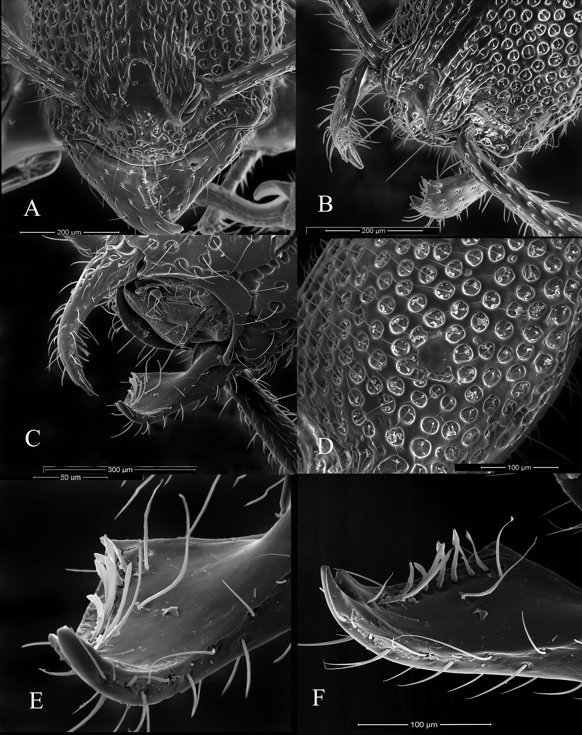
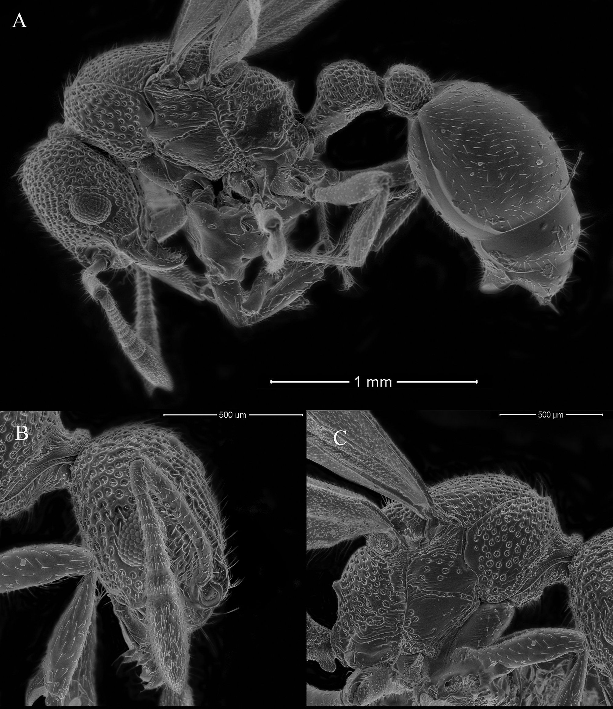
Head: Vertically ovoid, longer than wide (CI 76.9–78.5), wider superiorly and tapering anteriorly, lateral borders are convex, more so in the upper lateral third, vertex convex and medial aspect of the posterior margin slightly concave. Mandibles triangular, basal margin edentate and separated from the masticatory border by a distinct angle, masticatory border edentate except for two close-set teeth–the apical and pre-apical, the apical being longer and sharper than the pre-apical. Palp formula 2,2. Clypeus with anterior surface on lateral view and anterior border convex and protrudes above the mandibles, devoid of any carinae but having irregular small foveae, the posterior border is narrowly inserted between the frontal lobes. Frontal lobes are distinct and the rounded lateral border covers the antennal sockets. Antennae 11 segmented, with an ill-defined 3-segmented club, scape when extended reaches or falls just short of the posterior head margin (SI 90–100). Eyes small and consisting of only three ommatidia; each ommatidium of size of single fovea. Eyes situated just anterior to the mid-length of the lateral head border ( Fig.5 View FIGURE 5 ).
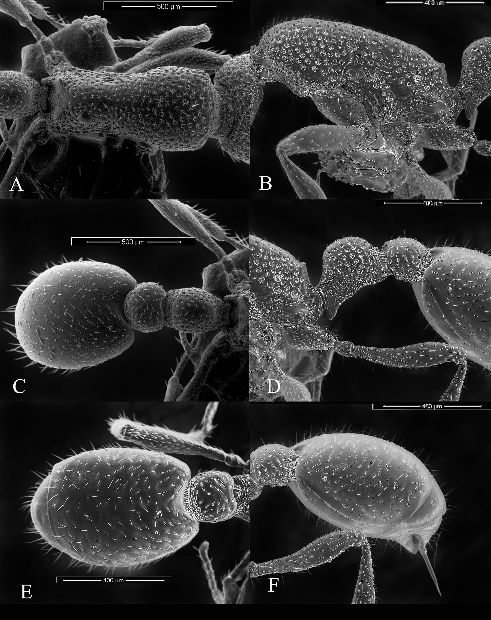
Mesosoma: In lateral view, the dorsum of mesosoma has a continuous, weak convexity with mild depression, almost indiscernible, at the pro-mesonotal suture. It further becomes a short convexity over the metanotal region and reaches the propodeal region where it ends in a small blunt triangular tooth and then it slopes down the propodeal declivity, which is a gentle concavity, to end on the broad and rounded metapleural lobes. In dorsal view the mesosoma is broadest just anterior to the promesonotal suture, where its anterior convex margin meets the lateral margin; this junction on the anterolateral pronotum is marked by a distinct angulation. There is a shallow constriction at the promesonotal suture and after this the mesometanotal region is almost trapezoidal in dorsal view. No sutures are evident on the mesosoma, except the promesonotal suture laterally. Propodeal spiracle is ovoid with its long axis parallel to the propodeal declivity. The metapleural gland orifice is not obvious on lateral view under low magnification. But on careful observation from the dorsal aspect at 200 x or more using SEM it was observed that a crescent shaped dorsally opening cleft was present in the propodeal lobe that probably represents the gland orifice. The end of a single, prominent dorsally directed long filiform seta, arising well below the cleft orifice, marked its anterior end. The region lying immediately anterodorsal to this cleft was devoid of sculpture, and this area extended anteriorly to the vertical level of the propodeal spiracle ( Fig. 7 View FIGURE 7 ).
Petiole: In dorsal view, the petiole is longer than wide and longer than the postpetiole. In lateral view the shape is elliptical. Petiole with ill-defined peduncle and in lateral view the anterodorsal margin curves upwards and abruptly expands into the node. The petiole bears on its ventral surface a quadrangular subpetiolar process whose thin laminar wall forms an acute angle with the peduncle anteriorly ( Fig. 7 View FIGURE 7 ).
Postpetiole: In dorsal view shorter than the petiole, more or less as wide as long and rounded with anterior concave and posterior convex margins. In lateral view, it is shorter than the petiole and almost spherical and is dorsoventrally compressed. The postpetiolar sternite bears a transverse keel forming an acute angle anteriorly in lateral view ( Fig. 7 View FIGURE 7 ).
Gaster: Elliptical in dorsal view with concave anterior margin to accommodate the post-petiole, and flattened dorsoventrally. Tip of the abdomen bears a robust and well-developed sting ( Fig. 7 View FIGURE 7 ).
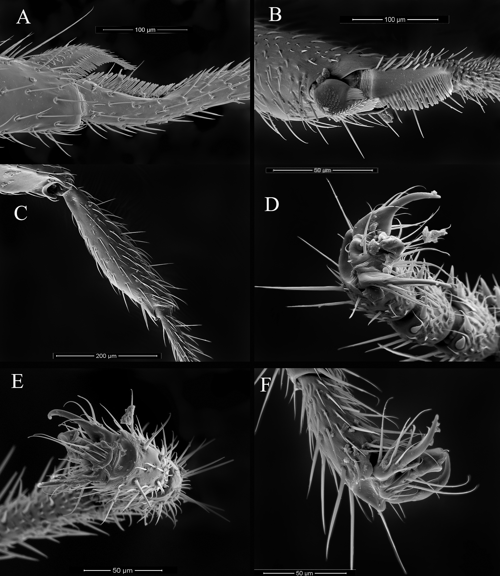
Legs: Forelegs with calcar of strigil (protibial spur) in SEM having squamiform setae on the outer half of its medial (ventral) surface, lanceolate setae on its dorsal edge and digitiform setae on its ventral cleaning edge. Comb of strigil on probasitarsus has digitiform setae on its entire cleaning edge. The middle and hind legs are without tibial spurs. Claws are furnished with arolium on all the legs ( Fig. 8 View FIGURE 8 ).
Sculpture and Pilosity: Head is heavily foveate and bears numerous short erect hairs on its entire surface. Clypeus has irregularly shaped foveae except on its upward extension between the frontal lobes. There is a median band of un-sculpted area on the head extending from the apex of the median portion of clypeus to about the midpart of the head in full-face view.
Mesosoma is also heavily foveate throughout but more so on the lateral pronotum. The foveae are close to each other, especially on the lateral pronotum, but they never touch. On the dorsum the distance between the foveae is about the diameter of the individual foveae or slightly more. But on the lateral pronotum the distance is usually less than the diameter but more than the radius of the individual foveae. The spaces between the foveae are smooth. In the median portion of the dorsum of the mesosoma along the whole length the foveae are more distant from each other than on the lateral pronotum. The inter-foveal distance is more than the diameter of the individual foveae and sometimes twice as much. The petiolar node is foveate and the inferior half and the peduncle is areolate. The postpetiole is predominantly foveate and the ventral part is areolate. Gaster is with fine punctations where the pilosity arises but otherwise un-sculptured and smooth on the first tergite and sternite. The posterior edges of other gastric tergites have punctations from which the erect hairs arise. There is no evidence of any micro-reticulations. Microsculpture is as shown in Figures 5 View FIGURE 5 and 7 View FIGURE 7 .
Hairs on the antennae and mandibles are mostly sub-erect to sub-decumbent. Median clypeus bears a series of 6–8 evenly spaced prominent long erect setae on the inferior margin of which two are at the level of the antennae and the rest along the inferior border. There are otherwise numerous short thin sub-decumbent bristle hairs arising from the irregular foveae on the entire mid-clypeus except its projection superiorly between the frontal lobes. Similar hairs are seen on the outer aspect of mandible. Head is otherwise covered in sub-decumbent hairs. Inner aspect of the mandible on SEM at 1500x revealed modified setae arising from circular pits in the submarginal area of the masticatory border. These are cylindrical at the base and almost fill the pits from which they originate. They become flat and later assume the shape of a scimitar towards their ends ( Fig. 6 View FIGURE 6 ).
These are very different from the normal body hairs and setae seen along the clypeal border. Whole mesosoma in workers covered in short hairs that arise from the foveae, but only those on the pronotum are erect and suberect hairs are seen towards the mesonotal region. Hairs on the mesonotum and metanotum are decumbent and appressed respectively (n=5). Short sub-decumbent to decumbent hairs are seen all over the postpetiole and legs. Gaster and postpetiole have suberect to subdecumbent hairs. Short hairs on the petiolar dorsum are not erect. Generally, the hairs on dorsum of head, mesosoma and petiole are regularly arranged and all other regions have irregularly directed hairs.
Color: Orange red, the head and pronotum is darker; mesonotum and metanotum are brownish. Petiole, postpetiole and gaster are more yellowish. Legs, mandible and antennae are yellowish orange. All hairs are white ( Fig. 2 View FIGURE 2 and 4 View FIGURE 4 ).


| NCBS |
Yale University |
No known copyright restrictions apply. See Agosti, D., Egloff, W., 2009. Taxonomic information exchange and copyright: the Plazi approach. BMC Research Notes 2009, 2:53 for further explanation.
|
Kingdom |
|
|
Phylum |
|
|
Class |
|
|
Order |
|
|
Family |
|
|
SubFamily |
Myrmicinae |
|
Tribe |
Solenopsidini |
|
Genus |