
Parasorex depereti ( CROCHET , 1986)
|
publication ID |
https://doi.org/ 10.2478/if-2019-0027 |
|
persistent identifier |
https://treatment.plazi.org/id/7B4187F3-FFA8-FFF8-FF21-FCC0B9AEFB4B |
|
treatment provided by |
Diego |
|
scientific name |
Parasorex depereti ( CROCHET , 1986) |
| status |
|
Parasorex depereti ( CROCHET, 1986)
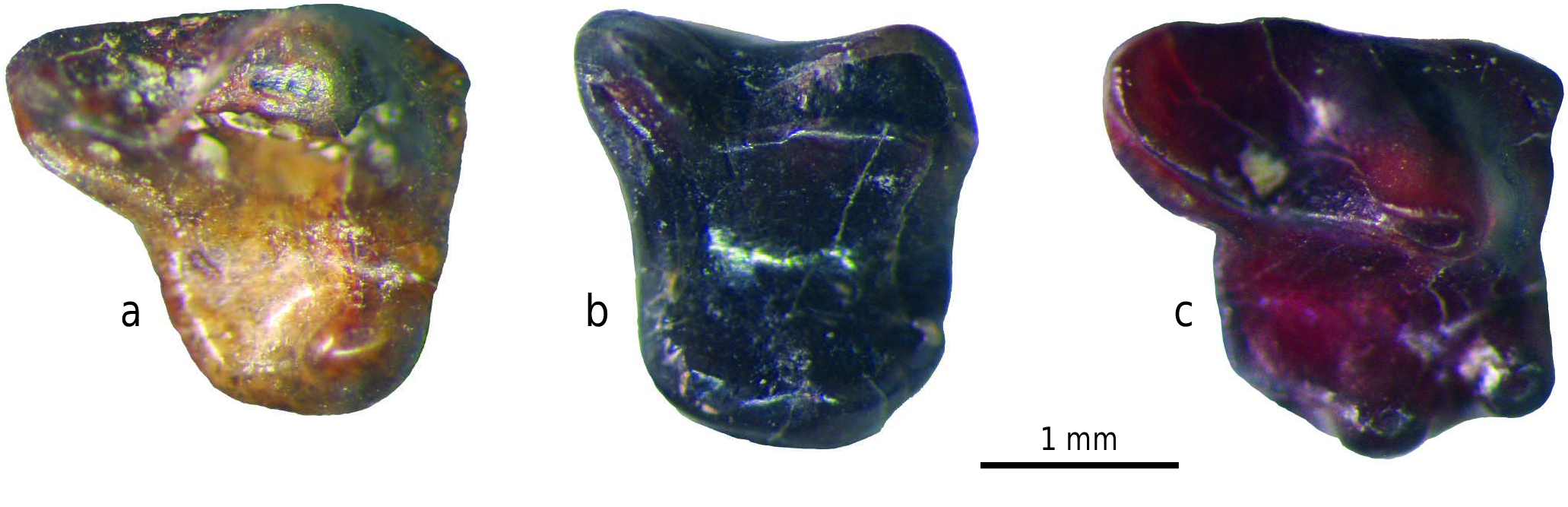
Text-fig. 2 View Text-fig , Pls 1–3
Synonymy for fissure BRS 25 material:
1986 Echinosoricinae cf. Galerix ; Costa et al., pp. 221–235.
1988 Galerix sp. aff. depereti ; De Giuli et al., pp. 65–67.
1989 Galerix sp. aff. depereti ; De Giuli, p. 198.
1989 Galerix sp. ; De Giuli, p. 199, tab. 1.
1989 Galerix sp. ; Masini, p. 296, tab. 1.
1989 Galerix sp. ; Masini and Thomas, p. 308, tab. 1.
1989 Galerix sp. ; Torre, p. 326, tab. 2.
1993 Galerix sp. aff. depereti ; Masini and Rook, p. 80, tab. 1.
1999 Galerix depereti ; Fanfani, pp. 28–35, tabs I.4, IX.4.
2013 Parasorex depereti ; Masini and Fanfani, pp. 101–102, tab. 2, fig. 9B.
H o l o t y p e. MTH 1, right M1.
T y p e l o c a l i t y. Mt. Hélène, Pyrénées-Orientales,
France.
O c c u r r e n c e s. Italy: Brisighella, Borro Strolla ( Abbazzi et al 2008), Sardinia (Capo Mannu D1 Mandriola) ( Furió and Angelone 2010); France: Celleneuve, Vendargues, Terrats, Mt. Hélène, Nimes ( Crochet 1986); Portugal: Esbarrondadoiro (Alvalade basin) ( Antunes and Mein 1989).
S t r a t i g r a p h i c r a n g e. Late Miocene (MN 13) –
Early Pliocene (MN 15).
S t u d i e d m a t e r i a l. Material included in this study originates from fissure BRS 25 (see Material and methods for details).
R e p o s i t o r y. Civic Museum of Natural History
“Malmerendi” of Faenza (for fissure BRS 25 material).
O r i g i n a l d i a g n o s i s. This is the largest species of the genus. Morphologically, it differs from G. exilis based on the same characters that discriminate the latter from G. socialis, i.e., the presence of a bi-cusped labial lobe on P3 and of a posterior crest on the metaconule of the upper molars extended to the postero-labial corner of the crown ( Crochet 1986, translated from French).
E m e n d e d d i a g n o s i s. Large-sized Parasorex . P3–P4 with a large, distally-expanded lingual lobe; P3 with a significant proportion of single cusped lingual lobe; large, prominent hypocone on P4; small protoconule on upper molars; more squarish M1–M2s respect to most of the other Parasorex species , with centrocrista but without mesostyles; with hypocone and protocone separated by deep notch and with low prehypocrista; p2 smaller than p3 and frequently with anterior cusp, with roots that tend to fuse; p4 with a high metaconid inclined lingually and with high paraconid; lower molars with low and short distal cingulid, never connected with the postentocristid (labial arm of entoconid).
D e s c r i p t i o n. Dental formula. I 3/?3 C 1/1 P 4/4 M 3/3.
Detailed description of the teeth. Fossil anterior premolars, canines and incisors of Galericini are poorly known, with a few remarkable exceptions (e.g. Galerix exilis; Ziegler 1983). This could result in some uncertainty in the identification of some of these teeth if they are isolated.
The canines, and the first lower and upper incisors can be easily distinguished from the other unicuspid teeth. More difficult are the second and third upper and lower incisors. In the case of the examined material, it is still difficult to assess if whether the BRS hedgehog had three or two lower incisors. In the upper anterior tooth row, I2 is usually larger than I3, P1 has a simple, single-cusped crown and two fused roots, between which lies a shallow vertical groove that separates them. P2 is much larger, has a talon-shaped posterior border, rarely an anterior bulge close to base of the crown and two widely separated roots.
I1. The first incisor is the largest of the anterior tooth row (Pl. 1, Fig. 1, Pl. 2, Fig. 1). It is a robust “canine-like” tooth, probably vertically implanted. The root is massive, longer than the crown, bent with posterior concavity and moderately flattened mesio-distally. The crown is pointed and sub-triangular in cross-section, with a somewhat flattened internal side faced to the contralateral I1 and a convex external (lateral) side. The crown is uniformly very worn, especially on its rear side, often all the way to the base of the crown.
I2 and I3. Many unicuspid teeth, smaller than I1, with sub-conical crown and bent root can be referred to I2s and/or I3s (Pl. 1, Figs 2, 3, Pl. 2, Figs 2, 3). The crown is subconical, with convex antero-labial side and flattened posterolingual side; in the larger ones the posterior side has a weak posterior bulge at the base and is generally worn in a way to generate a sharp rear edge. The root is proportionally shorter than in I1 and slightly curved with posterior concavity and moderately flattened labio-lingually. The smallest of these incisors are very slender and have a short, subconical, slightly procumbent crown. The posterior bulging is always faint. These teeth appear constantly unworn or only moderately worn. With respect to the crown, the root is proportionally longer than in the larger incisors and sub-oval in cross-section. Because in several Galericini species the incisors become smaller from I1 to I3, the smallest incisors from BRS 25 can be interpreted as I3s and the medium-sized ones as I2s. However, in Parasorex ibericus, which has a shorter muzzle than other gymnures, I2 is the smallest of the three incisors ( Mein and Martín-Suárez 1993: figs 1, 2).
C1. The upper canine is a two-rooted tooth, triangular in side view, somewhat similar to P2 (Pl. 1, Fig. 4, Pl. 2, Figs 4, 5). The crown consists of a single, high, pointed, slender cusp, compressed linguo-buccally, with a concave, sharp, rear side. It is higher than the second premolar and has a small basal bulge at the distal end of the crown. The roots are long, compressed labio-lingually, sub-triangular in cross-section, and strongly divergent antero-posteriorly. The anterior root is curved and the posterior one is slightly larger and straight.
P1. The single root of this tooth is often divided by a deep, vertical furrow (Pl. 1, Fig. 5, Pl. 2, Fig. 7). The crown consists of a single, slender, pointed cusp, whose tip bends slightly backwards. It is slightly procumbent, with a smooth weak anterior bulge and a low, variably-developed posterior one.
P2. This tooth resembles the upper canine (Pl. 1, Fig. 6, Pl. 2, Figs 6, 8). It has a single, high cusp, somewhat mesially situated, which is extended posteriorly by a low talon with a distinct accessory cusp on the labial corner. The main cusp is lower, less pointed and less flattened labio-lingually than that of the canine and its posterior edge is smooth. The crown is sub-triangular in side view and sub-oval in occlusal view. It has two sub-equal roots, the anterior one somewhat arched with posterior concavity and the posterior one straighter and slightly larger.
P3. This tooth is a sort of miniature P4 (Pl. 1, Fig. 7, Pl. 2, Figs 9–12). Occlusally, the outline of the tooth ranges from squarish to trapezoidal: the labial border is weakly oblique to the sagittal axis of the tooth, the posterior profile is moderately to very emarginated, the labial side is occupied by the large, high paracone whose posterior ridge (postparacrista) extends into a low, but well-developed crest (metastylar crest), often forming a carnassial notch; the lingual side consists of a low and wide lobe on which the protocone and hypocone are situated. The protocone is placed in the mesio-lingual corner of the tooth, and sometimes protrudes mesially. The part of the lingual lobe extending behind the hypocone forms a rather wide and flattened basin delimited lingually and posteriorly by a cingulum that becomes thicker at the postero-labial corner of the crown. The relative size, position and shape of the two lingual cusps, protocone and hypocone, vary considerably ( Text-fig. 2 View Text-fig , Tab. 2). In general, they are located rather anteriorly on the lingual lobe, with the hypocone that protrudes more lingually than the protocone. Morphologically, the protocone and hypocone sometimes form a ridge-like structure, whereas in one case only the hypocone is ridge-shaped. Six specimens have only one lingual cusp, which can be interpreted as a hypocone migrated mesially. Such inference is supported by the observed mesial displacement of both proto- and hypocone in the teeth with bicuspid inner lobes, and more so, by three single-cusped P3s, which have a rudimentary protocone placed mesially just against the base of the paracone. In seven other specimens the protocone is smaller than the hypocone and is situated more labially; in one case, the hypocone and protocone tend to merge; rare specimens (two) show an additional cusp behind the hypocone (Textfig. 2). The preprotocrista is absent; sometimes, a faint ridge-like parastyle occurs at the mesio-labial corner of the paracone. P3s have three roots; the lingual root is large and not subdivided, but shows a marked vertical furrow on its lingual side.
P4. The tooth is larger and less variable morphologically than P3 (Pl. 1, Fig. 8, Pl. 2, Figs 13–16, 20). Behind the high, robust paracone is situated a sharp postparacrista, which connects with the metastylar crest forming a carnassial notch in between. The metastylar crest extends to the postero-labial corner of the tooth. The tooth has a less emarginated posterior profile than P3. The lingual lobe is wide labio-lingually and extends posteriorly; it is delimited by a strong, posterolingual cingulum, which reaches the postero-labial corner of the crown and usually merges with the base of the hypocone, enclosing a wide basin. The protocone is the higher of the two lingual cusps, but the hypocone is still well-developed; in a few cases, the two cusps may be of similar size. Both are situated somewhat anteriorly. Sometimes a posterior bulge forms a sort of third lingual cusp on the posterior cingulum, close to the hypocone.
A preprotocrista is always present; sometimes it is directly connected with the parastyle, which occupies the anterolabial corner of the tooth. The latter is a lingually extended ridge, which can reach the preprotocrista without merging with it. The roots are like those of P3 but the vertical furrow on the inner side of the lingual root is somewhat deeper.
M1. The first upper molar is the largest tooth of the upper row (Pl. 1, Fig. 9, Pl. 2, Figs 17–21). Occlusally, it has a sub-rectangular (trapezoidal) outline, with a protruding postero-labial corner. The labial border is gently concave, due to the labio-distal projection of the mesostylar ridge. A fairly continuous narrow labial cingulum is always present. The parastyle is well-developed and connected with the low anterior cingulum. The mesostyle (centrocrista sensu Lopatin 2006) is practically absent; it consists of a very low and thin crest connecting the posterior arm of the paracone with the anterior arm of the metacone, and cut by a deep notch. The metaconule is large and crescent-shaped, with a posterior arm extended to the postero-lingual corner of the crown (a taxonomically significant character for taxa with Parasorex-Schizogalerix affinities).
The metacone is higher than the protocone. The latter is connected labially, through the preprotocrista, with a small, narrow cusped paraconule with no posterior arm. The mesial arm of the protoconule usually merges with the lingual side of the paracone, or rarely, dips and extends anteriorly along the base of the paracone. In rare specimens a low, additional tubercle may occur lingually at the base of the protocone. The high, crest-like posterior arm of the protocone (postprotocrista) joins the anterior arm of the hypocone, but in 17 % of the specimens, it branches also towards the metaconule and joins it (triple connection in Borrani et al. 2017). The valley between the metaconule and the posterior arm of the protocone is very shallow. The connection between the hypocone and the posterior arm of the protocone is always in the form of a low crest, which makes the tooth appear somewhat “primitive”. The posthypocrista is rarely visible, however, in worn teeth, the postero-labial side of the hypocone forms a sort of crista that merges with a weak posterior cingulum, which is interrupted by the posterior arm of the metaconule. In the sample from BRS 25 a continuous labial cingulum occurs, which does not extend to the postero-lingual corner. M1 has three roots: the lingual root is largest and seems to result from the fusion of two roots of different size, aligned antero-posteriorly to one another.
M2. The tooth is smaller than M1, from which it differs in having: a sub-rectangular occlusal outline, with convex anterior edge, shorter and non-prominent postero-labial corner and shorter metastylar ridge (metacone posterior arm) and posterior arm of the metaconule (Pl. 1, Fig. 10, Pl. 2, Figs 20, 22–25). The labial border is concave, with a narrow, variably-developed labial cingulum. A weak mesostylar crest rarely forms a bulge at the end of the anterior arm of the metacone. Unlike M1, in M2 the protocone is the highest cusp. Similarly to M1, in M2 the protocone has a high posterior arm directed towards the metaconule, and reaches it more frequently (26 %) than in M1. In some of these specimens the prehypocrista connecting the hypocone and the posterior arm of the protocone is very low and weak (Pl. 2, Fig. 24). In the M2, a low anterior arm of the hypocone meets the posterior arm of the protocone at open angles; rarely the two arms form a continuous, arched ridge. Like in M1, but more frequently and more markedly than in the latter, the labial arm of the paraconule dips and extends anteriorly along the base of the paracone. The roots are morphologically like in M1.
M3. The tooth is triangular-shaped in occlusal view, but can be variably extended antero-posteriorly (Pl. 1, Fig. 11, Pl. 2, Figs 26–28). It is much smaller than M2. The crown includes a conical paracone, higher than the metacone and slightly lower than the protocone. The crescent-shaped protocone, placed lingually to the paracone, is the largest cusp. The metaconule is absent. The anterior arm of the protocone runs towards the paracone, dipping and finally merging with its base; a small bulge-shaped paraconule may occur. A well-developed parastyle is always present; it extends lingually, fusing with the anterior cingulum. The labial and posterior cingulum are missing or extremely reduced. Protocone and metacone are always connected by a continuous crest. The mesostyle is absent and a straight, low crest connects the paracone with the metacone. The tooth has three roots, the lingual one being the largest.
i1. This is the largest lower incisor; it is stouter than i2 and i3 and slightly larger, on average, than the canine (Pl. 1, Fig. 12, Pl. 3, Fig. 1). The crown is spatulate and oblique labially. It is convex ventro-laterally (labially) and concave dorsal-lingually. A marked cingulid is present close to the collar, on the dorso-labial side of the tooth. The root is long, somewhat flattened labio-lingually and bent with dorsal (occlusal) concavity. The root ends in a flattened hook tip in some specimens.
i2. The second lower incisor (Pl. 1, Fig. 13, Pl. 3, Fig. 2) is a spatulate, procumbent tooth, smaller than both i1 and canine; it resembles the canine in many respects, having a less flattened crown than i1. The mesial side is rounder and the disto-dorsal side more concave than i1; the tooth bears a small lateral bulge on its dorso-labial side. Anteriorly, at the dental collar, crown and root are arranged in a weak arched pattern with ventro-lingual concavity. The root may present a longitudinal and shallow furrow separating a ventral and a dorsal portion. The distal end of the root is often fashioned as a flat hook.
i3?. A few incisors differ from i2 by being smaller, more concave ventro-lingually and by having shorter root. These teeth are tentatively identified as i3 (Pl. 1, Fig. 14, Pl. 3, Fig. 3).
c1. The incisor-like canine differs from the first lower incisor by being slightly smaller and by having a relatively shorter, straight root. It is implanted obliquely on the mandible (Pl. 1, Fig. 15, Pl. 3, Fig. 4). The crown is short, aligned with the root, with which, observed occlusally, it is arranged in an arched pattern labially. The root is flattened latero-medially and often furrowed longitudinally, with a larger dorsal portion and a smaller ventral one. The crown is blade-like, with a sharp postero-dorsal edge and blunt and worn tip, and is somewhat procumbent on the second/third incisor. The labial side is convex and the lingual one slightly concave. Wear affects also the lingual side. A small bulge is present close to the collar on the rear side of the dorsal edge.
p1. p1 and p2 are simple-crowned teeth with one or two roots, usually fused; p1 is smaller than p2 and has a simpler crown (Pl. 1, Fig. 16, Pl. 3, Fig. 5); morphologically, it is a simplified version of p2, with lower main cuspid and the accessory anterior labial cuspid fused to it in such a way as to form a procumbent anterior bulge in the crown. The posterior lingual cuspule is smaller than in p2. The tooth has one root; in some specimens a shallow vertical furrow may occur.
p2. Small tooth with a sub-elliptical occlusal profile (Pl. 1, Fig. 17, Pl. 3, Figs 6–8). The crown is sub-triangular in side view, with slender main cuspid. A smoothed accessory cuspid is present antero-labially, placed at about half the height of the crown; the posterior side prolongs distally and bears a low basal accessory cuspid placed slightly lingually. The height of the crown is variable; the mesial wall of the anterior cuspid is procumbent. The tooth usually has two fused roots, often distinguished by a furrow on each side, being deeper labially than lingually. There are specimens with single roots (3 out of 31), double roots (7 out of 31) and with fused roots separated by a vertical furrow (21).
p3. This premolar is much larger than p2 but somewhat smaller and narrower than p4 (Pl. 1, Fig. 18, Pl. 3, Figs 9–11). It appears sub-triangular in side view. The crown is dominated almost exclusively by a high, slender, sub-conical cuspid (protoconid), a low cuspid situated on the anterior side (paraconid) and a strong and low rear cingulum, whose postero-lingual corner protrudes distally and may carry a small cuspule. The posterior basin is divided in two by a weak, blunt longitudinal crest, but less markedly than in p4. The rear face of the protoconid is flat and its tip is blunted by wear. The metaconid is absent. The occlusal profile of the tooth is sub-oval, with the posterior edge straight and postero-lingually oblique to the sagittal axis of the tooth. The tooth has two roots, well-separated from one another and with the posterior one slightly larger than the mesial one.
p4. The fourth lower premolar is larger and wider than p3 (Pl. 1, Fig. 19, Pl. 3, Figs 12–13) and has a welldeveloped trigonid; the tooth is therefore molarized. The central part of the tooth is dominated by a sharp and high protoconid, with a sub-conical metaconid which tends to be situated mesio-lingually to it. These two cuspids are joined by a crest (protocristid sensu Lopatin 2006). The anterior part of the tooth consists of a high paraconid that elongates posteriorly into a sharp paralophid; the latter is connected at an obtuse angle with the sharp anterior arm of the protoconid (preprotocristid) and with a carnassial notch in between. The tip of the well-defined metaconid is slightly inclined lingually. The three cuspids enclose a narrow trigonid basin, open lingually. Posteriorly, the tooth shows a short and shallow basin delimited by a strong cingulum, which first rises and then levels off sub-horizontally towards the distally-protruding postero-lingual corner; the latter sometimes carries a small accessory cuspid. As in p3, the posterior basin is divided into two unequal parts by a weak and blunt longitudinal crest. The tooth has two unequallysized roots, the posterior one being the larger.
m1. The first lower molar is the largest of the lower cheek tooth row. (Pl. 1, Fig. 20, Pl. 3, Fig. 14) The protoconid is the highest cuspid; a paralophid issues from it anteriorly and bends lingually, ending into a swollen paraconid. The metaconid is conical and pointed, slightly lower than the protoconid from which it is separated by a notch. It is placed somewhat more anteriorly than the protoconid. A crescentshaped hypoconid is the lowest cuspid. The entoconid is large, sub-conical and somewhat lower than the metaconid. The posterior arms of the entoconid and hypoconid are fused, forming a sinuous crest (postcristid) that bounds, posteriorly, a deep talonid basin. The anterior arm of the hypoconid (crista obliqua) is low and ends against the posterior wall of the protoconid. The entocristid runs straight anteriorly reaching the distal wall of the metaconid, or joining a weak and low metacristid. The distal cingulid is weak, short and not fused with the postcristid. The antero-labial cingulid is narrow and bounds the base of the paralophid-paraconid complex; it disappears at the base of the protoconid.
m2. The second lower molar is smaller than m1 and less elongated mesio-distally. (Pl. 1, Fig. 21, Pl. 3, Fig. 15). It has no paraconid. The paralophid is low and curves lingually and distally, almost closing the lingual opening of the trigonid basin. The crest is oriented more vertically, and the metaconid is placed in a more anterior position than in m1. Such an arrangement produces a comparatively shorter trigonid: in fact in m2, the trigonid is around 47 % of the total length of the tooth, whereas it is 52 % in m1. The antero-labial cingulid is stronger than in m1. A weak labial cingulid is often present in the valley between protoconid and hypoconid. The tip of the entoconid is placed slightly more mesially than the hypoconid. The posterior cingulid is similar to that of m1.
m3. This tooth is smaller than m2, but the two teeth share trigonids morphologically similar (Pl. 1, Fig. 22, Pl. 3, Fig. 16). The third lower molar differs from m2 by having shorter and narrower trigonid and talonid, with the talonid narrower than the trigonid. The crest-shaped paraconid is similar to that of m2. The entoconid is a strong cusp that protrudes distally more than in m1 and m2. Compared to m2, m3 has a weaker hypoconid, situated more lingually and not protruding labially. The posterior cingulid is lacking.
Mandibles. The sample from BRS 25 includes a few fragmental horizontal rami without teeth. The specimens worth notice are:
– A fragmental right horizontal ramus with p1–m3 alveoli and the basal, anterior portion of the ascending ramus (MSF 3050). The mental foramen opens between the roots of p4.
– A fragment of a left horizontal ramus with p1–p4 alveoli and a broken mesial portion (MSF 3051). Based on the shape of the latter, p1 is single-rooted, p2 is single-rooted with root carved by a vertical groove. A large alveolus, presumably of the canine, occurs mesially to p1. Two cavities, possibly the alveoli of i1 and i2, are stretched under it. Based on comparisons with Parasorex socialis, the alveolus of i3 would be smaller and situated more dorsally than that of i2; hence, it cannot be excluded that it is not preserved. The mental foramen opens under the anterior root of p4.
– A fragmental left mandible with p4–m2, still included in the matrix, broken in front of p4 and with only part of the ascending ramus preserved (MSF 3052). The anterior margin of the ascending ramus is fairly vertical. The mental foramen opens between the mesial and distal roots of p4.
– A fragment of right mandible still embedded in the matrix, with p3, p4 and a broken m2 (MSF 3053).
– A fragment of left mandible with the alveoli of p3 (still preserving the anterior root of the tooth), of a single-rooted p2 and of p1. The mental foramen opens between the mesial and distal roots of p4 (MSF 3054).
Other specimens are listed in Table 3.
Maxillary. Left maxillary fragment with P4–M2 (Pl. 2, Fig. 20). The morphologies of the preserved teeth are included in the variability reported in the descriptions above.
a
No known copyright restrictions apply. See Agosti, D., Egloff, W., 2009. Taxonomic information exchange and copyright: the Plazi approach. BMC Research Notes 2009, 2:53 for further explanation.