
Talpacoxa brandini, Corgosinho, 2012
|
publication ID |
https://doi.org/ 10.1080/00222933.2012.725138 |
|
publication LSID |
lsid:zoobank.org:pub:E25DDEDF-2E30-4281-A54B-CC5826BD3913 |
|
persistent identifier |
https://treatment.plazi.org/id/7C118736-FF84-FFD7-B7F3-A7ADE961FD00 |
|
treatment provided by |
Felipe |
|
scientific name |
Talpacoxa brandini |
| status |
gen. et sp. nov. |
Talpacoxa brandini gen. et sp. nov.
Type locality
A small cove on the southern region of the Ilha do Mel (Honey Island; coordinates of the sampling site 25 ◦ 24 ′ 89 ′′ S, 48 ◦ 42 ′ 71 ′′ W), near the mouth of the Paranaguá Bay estuary.
Material examined
Holotype; one dissected male mounted on six slides ( INPA1374 View Materials a–f) . Paratypes: one dissected female mounted on four slides ( INPA1375 View Materials a–d), one undissected male ( INPA1697 View Materials ; size 370 µm) and one undissected female from Paranaguá Bay ( INPA1698 View Materials ; size 380 µm), one undissected female from the area of ECOPLAN / CEM / UFPR artificial reefs project ( INPA1699 View Materials ; size 350 µm) .
Description of male
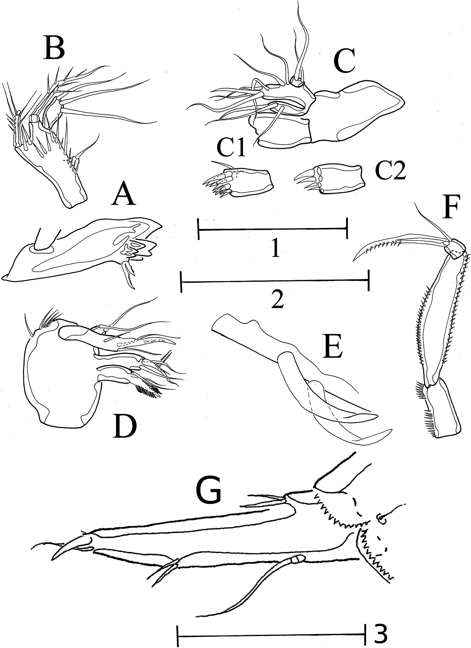
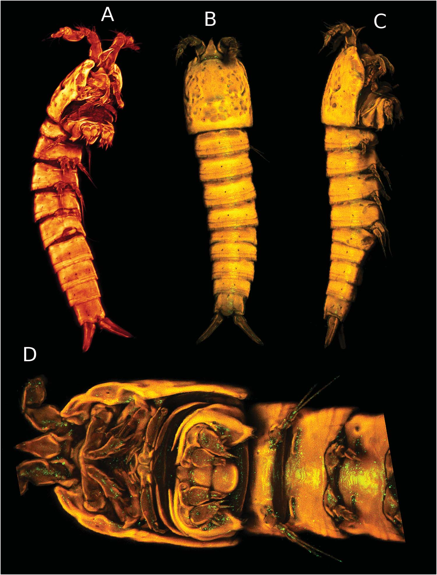
Habitus ( Figure 1A,B View Figure 1 and 8A View Figure 8 ). Length 385 µm (measured from tip of rostrum, to distal rim of anal operculum).
Body cylindrical, surface of cephalothorax irregular, with many rounded pits ( Figure 8A View Figure 8 ). For sensilla, pores and frill patterns see Figures 1A,B View Figure 1 and 8A View Figure 8 . Rostrum ( Figures 1A,B View Figure 1 and 8A View Figure 8 ) not fused to cephalothorax, long and triangular, with two sensilla near tip.
Furca conical ( Figures 1A,B View Figure 1 , 3G View Figure 3 and 8A View Figure 8 ), relationship between length and width is 3.5 × on the proximal portion and 7 × distally; setae I and II almost equal in length, inserted proximally, on the same level as seta VII, seta III and VI occupy an inner position, approximately in midlength between seta VII and distal end of furca, setae IV is short, spiniform and inserted distally, seta V is distally inserted on the inner margin.
A1 seven-segmented ( Figure 2A–H View Figure 2 ); segment 1 without outer process; aesthetasc on fifth (posterior schematic view of fifth segment on Figure 2I View Figure 2 ) and seventh segment; armature beginning with the proximal segment I [1 pinnate], II [4 + 4 pinnate], III [4 + 2 modified + 2 small spines], IV [1 + 1 reduced], V [8 + 2 stout spines + (1 + Ae)], VI [7 + acrothek].
A2 ( Figure 2J,K View Figure 2 ) with unornamented coxa; allobasis with abexopodal seta and well developed one-segmented exp armed with three setae; enp one-segmented, with two subdistal setae and five distal setae (three outermost setae geniculated). Additionally, the outer margin is ornate with a proximal and a distal row of spinules; a row of spinules decorates the distal margin.
Md with a well-developed gnathobasis ( Figure 3A View Figure 3 ); Md palp well developed ( Figure 3B View Figure 3 ) and well ornamented with many spinules of different sizes along its ventral margin; basis with two bipinnate setae, one bi-articulate at base; exp represented by a single seta at the tip of a small lobe; enp one-segmented, with three distal seta.
Mx1 ( Figure 3C, C View Figure 3 1,2) with a short praecoxal arthrite ( Figure 3C View Figure 3 1,2) armed with one single dorsal seta and eight distal spines arranged in two parallel rows of four spines each; coxa with three setae; basis with one endite armed with five distal setae; enp represented by two setae; exp one-segmented, with two setae.
Mx2 ( Figure 3D View Figure 3 ) syncoxa fused to the basis; basis with two endites; distal endite with one fused spine, strong and bipinnate, with upwardly curved spinules ( Figure 3E View Figure 3 ) and two bare setae; proximal endite with one spinulose seta and two bare setae; enp- 1 with four elements represented by one claw, one inferodistal seta, one posterior seta and one subdistal anterior seta; enp-2 one-segmented with two bare setae.
Mxp ( Figure 3F View Figure 3 ) very long, prehensile; syncoxa rectangular, with two inner rows of spinules; basis long, with a row of spinules along the outer and inner margins; enp quadrate, short, with a strong, pinate claw and a superodistal seta.
P1 ( Figures 4A–F View Figure 4 and 8A View Figure 8 ) strongly modified, transformed into a burrowing structure; praecoxa well developed, without crista; coxa hypertrophied ( Figure 4C View Figure 4 ), with a well-developed crista, caudally extended, with bifid tip; outer margin with a row of small spinules on its midlength, inner margin serrated on its distal portion; basis short ( Figure 4D View Figure 4 ), rectangular, with outer seta and a stout inner spine inserted anteriorly; ornamentation of basis ( Figure 4A,B View Figure 4 ) composed of a row of strong spinules near insertion of enp, a row of strong spinules near insertion of inner spine, an inner row of strong spinules inserted on the posterior side ( Figure 4B View Figure 4 ), two outer pores and three outer spinules; exp one-segmented ( Figure 4E View Figure 4 ), strong, trapezoidal, ornamented with many conical cuticular processes and armed with four strong distal spines and one posterior spine; enp two-segmented ( Figure 4F View Figure 4 ); enp-1 strong and rectangular, almost as large as the exp, ornamented with many conical cuticular processes; enp- 2 short, quadrate, not ornamented, with a strong distal spine and a subdistal seta on the posterior margin; intercoxal plate well developed, almost of same size of coxa, with a transversal groove on its midlength, as in female ( Figure 8D View Figure 8 ), conferring to it an apparent articulated condition.
P2 ( Figure 4G View Figure 4 ) strongly reduced; coxa and basis separated as in female ( Figure 6C View Figure 6 ); limbs laterally displaced with no indication of intercoxal plate ( Figure 8D View Figure 8 ); basis with one outer seta; exp two-segmented, exp-1 and exp-2 with row of spinules on inner posterior margin, exp-1 with an outer spine; exp-2 with one inner bipinnate seta and an outer unipinnate seta; enp one-segmented (arrowed, lost on the left limb), with distal seta.
P3 ( Figure 4H View Figure 4 ) strongly reduced; coxa and basis fused, intercoxal plate indiscernible; basis with one outer seta and one outer pore; exp two-segmented; exp-1 with one outer spine; exp-2 with one inner bipinnate seta, one distal spine and one outer bare and small seta; enp one-segmented, slightly longer than the exp-1 and with a distal apophysis.
P4 ( Figure 4I View Figure 4 ) strongly reduced; coxa and basis fused, intercoxal plate indiscernible; basis with outer seta, outer pore and a row of spinules between insertion of enp and exp; exp two-segmented, exp-1 armed with an outer spine and ornamented with an inner posterior row of spinules; exp-2 with one bipinnate seta, one distal bipinnate spine, one outer bare and small seta and ornamented with two spinules inserted on the inner posterior margin; enp one-segmented, reduced and with a distal bare seta.
P5 ( Figure 4J, J′ View Figure 4 ) basoendopod with one outer basal seta and outer pore, inner endopodal lobe with one bipinnate spine and one inner short spine; exp onesegmented, with two outer bare setae, one distal bare seta and an inner short spine; intercoxal plate reduced (like in female).
P6 ( Figure 4K View Figure 4 ) asymmetrical, with outer seta, two distal small setae and a row of spinules along the inner margin; right limb fused to the somite, left limb is free.
Spermatophore ( Figures 1A,B View Figure 1 , 4L View Figure 4 ) small, a little longer than P4.
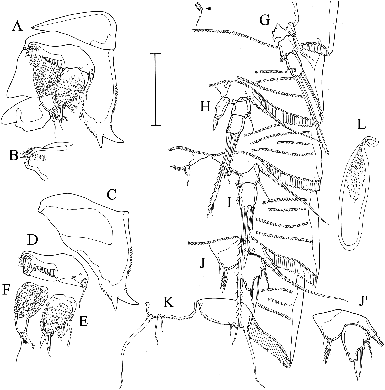
Description of female
Habitus ( Figures 5A View Figure 5 and 8B,C View Figure 8 ). Length 400 µm (measured from tip of rostrum, to distal rim of anal operculum). Sexual dimorphism in A1, P1, P3, P5, P6 and genital somite. For sensilla, pores and frill patterns see Figures 5A View Figure 5 and 8B,C View Figure 8 .
Cephalothorax ornamentation as in males, with many rounded pits ( Figure 8B,C View Figure 8 ). A1 ( Figure 5B View Figure 5 ) five-segmented, aesthetasc on the fourth and fifth segments; armature beginning from the first segment I [1], II [6 + 3 pinnate], III [4], IV [3 + (1 + Ae)], V [7 + acrothek].
P1 ( Figures 6A,B View Figure 6 and 8C,D View Figure 8 ) constructed as in male; remarkable differences can be seen in the coxa, that has a stronger inner margin, with three well-developed processes and the exp, with one seta on the place of the posterior spine.
P3 ( Figure 6D View Figure 6 ) basis and exp much alike as in male; enp short, with a distal bare and short seta.
P5 ( Figure 7B View Figure 7 ) basis with an outer seta, an outer pore and a well-developed endopodal lobe armed with an inner spine, a distal and an outer spine, inner margin ornamented with long setules; intercoxal plate is reduced; exp rectangular, onesegmented, with two outer setae, two distal setae and one inner seta subdistally inserted, inner margin ornamented with a row of spinules along its midlength, outer margin with a proximal row of spinules. Genital double somite incompletely fused ( Figures 5A View Figure 5 , 7C View Figure 7 ).
P6 represented by small, lateral and unarmed protuberances. Genital field with one copulatory pore located midventrally, just anterior to the inner cuticular bar ( Figure 7C View Figure 7 ); gonopore as a transversal slit located midventrally; seminal receptacles represented by a pair of midlateral vesicles; three to four large eggs are carried in a single midventral sack.
Urosomites 2 and 3 are dorsally and laterally separated, with a less developed hyaline frill in the joint of both segments (arrowed on Figure 5A View Figure 5 ); ventrally fused, but with a cuticular bar demarcating the fusion zone ( Figure 7C View Figure 7 ).
Etymology
The generic name Talpacoxa is built by combining the Latin lexemes “ Talpa ” meaning mole (a mammal with fossorial behaviour), and “ coxa ”, which means hip, and refers to the superficial resemblance between the burrowing forelegs of a mole and the modified P1, with a hypertrophied coxa, that can be observed on both sexes of the new species. The specific epithet “ brandini ” is given in honour of Dr Frederico Pereira Brandini, from the Centre of Marine Studies ( CEM / UFPR), Paraná, Brazil, who helped us in one of our fieldwork expeditions.


| VI |
Mykotektet, National Veterinary Institute |
| V |
Royal British Columbia Museum - Herbarium |
No known copyright restrictions apply. See Agosti, D., Egloff, W., 2009. Taxonomic information exchange and copyright: the Plazi approach. BMC Research Notes 2009, 2:53 for further explanation.