
Colobomatus, Hesse, 1873
|
publication ID |
https://doi.org/ 10.1080/00222933.2012.737483 |
|
DOI |
https://doi.org/10.5281/zenodo.4631776 |
|
persistent identifier |
https://treatment.plazi.org/id/813A87D1-FF80-3A1A-565F-FC964BB3FF6D |
|
treatment provided by |
Carolina |
|
scientific name |
Colobomatus |
| status |
|
Colobomatus pupa Izawa, 1974
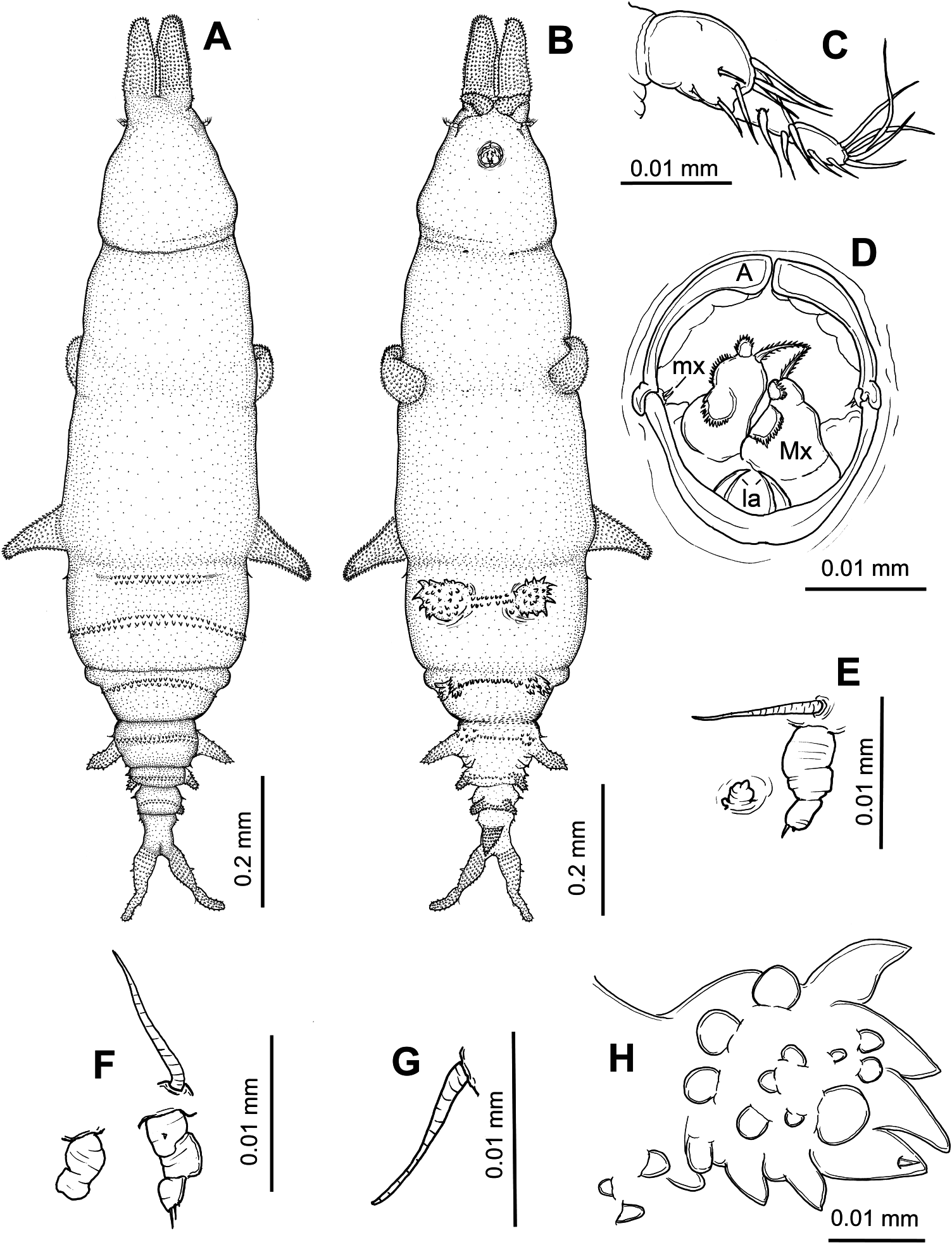
( Figure 2A – H View Figure 2 )
Material examined
Thirteen adult females (NSMT-Cr 21859), from lateral line scales of two Parupeneus ciliatus Lacepéde ( Perciformes : Mullidae ), and nine adult females, from lateral line scales of one Parupeneus multifasciatus (Quoy and Gaimard) , both hosts captured in the western North Pacific Ocean off Okinawa-jima Island and subsequently purchased at Hama Fisheries Cooperative (26 ◦ 34 ′ N, 127 ◦ 14 ′ E) in Nakagusuku, Okinawa-jima Island, 9 August 2009.
Description
Adult female. Body ( Figure 2A,B View Figure 2 ) 1.1 (0.9–1.3) mm long (excluding cephalic processes and caudal rami) (n = 4). Pre-oral area of cephalosome ( Figure 2B View Figure 2 ) with anterodorsal pair of blunt processes and anteroventral pair of anterolaterally directed processes; latter about one-third length of dorsal pair; all cephalic processes bearing spinulose ornamentation. Cephalosome widest posteriorly and demarcated from body by slight transverse constriction. First to fourth pedigerous somites fused to form cylindrical trunk, with slightly swollen lateral margins at posterior end of third pedigerous somite and two pairs of lateral processes; anterior pair of processes blunt, arising from ventral surface and anterolaterally directed; posterior pair slightly longer than anterior pair, posterolaterally directed and with blunt tips; both pairs of processes bearing spinulose ornamentation. Second and third pairs of legs occurring ventrally near base of anterolateral and posterolateral processes, respectively. Fourth pedigerous somite with one ventral pair of papillose lobes ( Figure 2H View Figure 2 ) intersected by two rows of spinules. Fifth pedigerous somite with one ventral pair of papillose lobes intersected by one row of spinules. Genital somite ( Figure 2B View Figure 2 ) with anteroventral pair of spinule clusters intersected by one row of spinules and lateral pair of processes; latter with pointed tip and spinulose ornamentation. Abdomen ( Figure 2B View Figure 2 ) composed of three somites, decreasing in width posteriorly; first two somites with small ventral pair of posterolaterally directed, spinulose processes; last abdominal somite bearing single ventromedian spinulose process, projecting posteriorly and one-third length of caudal rami. Third to fifth pedigerous somites, genital somite and first two abdominal somites each with two dorsal rows of denticles. Caudal ramus ( Figure 2B View Figure 2 ) fused to last abdominal somite, slender, tapering to simple blunt tip and bearing proximolateral spiniform seta and spinulose ornamentation.
Antennule ( Figure 2C View Figure 2 ) short, laterally directed, arising near base of cephalic processes and apparently three-segmented, with armature of 7, 3 and 7. Antenna ( Figure 2D View Figure 2 ) modified, forming longitudinally divided anterior margin of buccal capsule; latter ( Figure 2B,D View Figure 2 ) tube-like, projecting ventrally from conical base. Labrum and mandibles not seen. Maxillule ( Figure 2D View Figure 2 ) minute, situated mid-laterally in buccal capsule and bearing two short spines on distal margin. Maxilla ( Figure 2D View Figure 2 ) robust, two-segmented; basal segment with two semicircular rows of spinules and one apical spinulated element; distal segment with spinules along apical margin. Maxillipeds absent. Labium ( Figure 2D View Figure 2 ) divided, tapering into sharp tips. Posterior rim of buccal capsule longitudinally undivided.
Leg 1 ( Figure 2E View Figure 2 ) biramous; protopod completely fused to somite and represented by long, lateral surface seta; exopod indistinctly two-segmented, with unarmed basal segment and distal segment bearing two tiny spines on distal margin; endopod vestigial, unsegmented and unarmed. Leg 2 ( Figure 2F View Figure 2 ) similar to leg 1, except exopod indistinctly three-segmented, with basal segment carrying minute spinule on anterior surface and distal segment bearing three spines on distal margin and endopod indistinctly two-segmented. Leg 3 ( Figure 2G View Figure 2 ) vestigial, reduced to single surface seta.
Remarks
Colobomatus pupa was originally described by Izawa (1974) based on females and males collected from Parupeneus spilurus (Bleeker) captured in Tanabe Bay, Wakayama Prefecture, Japan. Slight differences in the body length, position of the antennule and infection site of C. pupa were evident between Izawa’s and our specimens. The body length of the specimens of C. pupa described in this study ranged from 1.1 to 1.3 mm (excluding the cephalic processes and caudal rami), while those of Izawa’s are 1.6 to 3.2 mm long (excluding the cephalic processes and caudal rami). Moreover, the antennules arise posterior to the ventral pair of cephalic processes in our specimens, while in Izawa’s specimens the antennules are anterior to the ventral cephalic processes. Finally, our specimens of C. pupa were found in the lateral line scales of P. ciliatus and P. multifasciatus , whereas Izawa collected his specimens from the supra-orbital, infra-orbital and pre-opercular canals of P. spilurus .
Colobomatus pupa resembles C. haeckeli , C. exilis , C. collettei , C. creeveyae and C. ornatus in the presence of a single, ventromedian process on the last abdominal somite. However, in C. ornatus the anterior pair of thoracic processes are bifurcated and the abdominal somites lack lateral processes. Colobomatus haeckeli , C. collettei and C. exilis can be distinguished from C. pupa in the length of the ventromedian process on the last abdominal somite, which is visible in dorsal view in the former three species. Colobomatus creeveyae can be distinguished from C. pupa by having only one pair of cephalic processes.
This study reports for the first time C. pupa parasitic in the lateral line scales of P. ciliatus and P. multifasciatus . Moreover, our finding represents a new locality record for this species in the subtropical waters off Okinawa-jima Island, Japan.
No known copyright restrictions apply. See Agosti, D., Egloff, W., 2009. Taxonomic information exchange and copyright: the Plazi approach. BMC Research Notes 2009, 2:53 for further explanation.
|
Kingdom |
|
|
Phylum |
|
|
Class |
|
|
Order |
|
|
Family |