
Colobomatus gymnocranii, Madinabeitia & Tang & Nagasawa, 2013
|
publication ID |
https://doi.org/10.1080/00222933.2012.737483 |
|
DOI |
https://doi.org/10.5281/zenodo.4631781 |
|
persistent identifier |
https://treatment.plazi.org/id/813A87D1-FF8B-3A1C-55EA-FAD54C0FF908 |
|
treatment provided by |
Carolina |
|
scientific name |
Colobomatus gymnocranii |
| status |
sp. nov. |
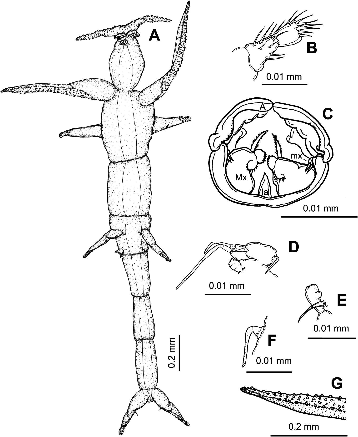
Colobomatus gymnocranii sp. nov.
( Figure 5 View Figure 5 A–G)
Material examined
Holotype female (NSMT-Cr 21862), from head canal of Gymnocranius griseus (Temminck and Schlegel) (Lethrinidae) , captured in the western North Pacific Ocean off Okinawa-jima Island and subsequently purchased at Yonashiro Fisheries Cooperative (26 ◦ 22 ′ N, 127 ◦ 58 ′ E) in Uruma, Okinawa-jima Island, 12 September 2009.
Description
Adult female. Body ( Figure 5A View Figure 5 ) 1.89 mm long (excluding cephalic processes and caudal rami). Pre-oral area of cephalosome ( Figure 5A View Figure 5 ) bearing one anterior pair of long, laterally directed processes ornamented with papillae on ventral surface and one short anteroventral pair of laterally directed processes ornamented with papillae at tip. Cephalosome ovate posterior to buccal capsule. First to third pedigerous somites fused to form cylindrical body and bearing two lateral pairs of laterally directed processes; anterior pair of processes elongated, tapering into blunt tips and with papillose ornamentation along posteroventral margin ( Figure 5G View Figure 5 ); posterior pair of processes dorsolaterally located, half as long as anterior pair and spinulose at rounded tip. Second and third pairs of legs occurring ventrally along same plane as anterolateral and posterolateral processes, respectively. Fourth and fifth pedigerous somites fused, about as long as preceding tagma. Genital somite ( Figure 5A View Figure 5 ) bearing one mid-lateral pair of slender posterolaterally directed processes and one posterolateral pair of naked, ventral swellings; each process as long as posterior pair of thoracic processes and with spinulose ornamentation posteroventrally. Abdomen ( Figure 5A View Figure 5 ) elongated, four-segmented and gradually tapering towards caudal rami. Caudal ramus ( Figure 5A View Figure 5 ) fused to anal somite, as long as posterior pair of thoracic processes, posterolaterally directed and bearing proximomedial seta and spinulose ornamentation along posteroventral surface toward blunt tip.
Antennule ( Figure 5B View Figure 5 ) apparently two-segmented, with armature of 9 and 8. Antenna ( Figure 5C View Figure 5 ) modified, forming longitudinally divided anterior margin of buccal capsule; latter ( Figure 5A,C View Figure 5 ) tube-like, projecting ventrally from conical base. Labrum and mandibles not seen. Maxillule ( Figure 5C View Figure 5 ) small, located mid-laterally in buccal capsule and bearing two apical spines. Maxilla ( Figure 5C View Figure 5 ) large, twosegmented; basal segment bearing one semicircular row of spinules and distolateral spinulated element; distal segment short, ornamented with spinules along distal margin. Maxillipeds absent. Labium ( Figure 5C View Figure 5 ) undivided. Posterior rim of buccal capsule undivided.
Leg 1 ( Figure 5D View Figure 5 ) biramous; protopod carrying lateral seta; exopod twosegmented, with basal segment bearing one distolateral spine and distal segment bearing two unequal setae apically; endopod vestigial, unsegmented and unarmed. Leg 2 ( Figure 5E View Figure 5 ) uniramous; protopod completely fused to somite, represented by long, lateral surface seta; exopod vestigial, apparently unsegmented and unarmed. Leg 3 ( Figure 5F View Figure 5 ) rudimentary, reduced to long surface seta.
Etymology
The species epithet, gymnocranii , refers to the generic name of the host.
Remarks
The female of C. gymnocranii sp. nov. resembles C. steenstrupi , C. sparsi and C. similis in having the anterior pair of thoracic processes longer than the posterior pair. Most Colobomatus species have the anterior and posterior thoracic processes of equal length or if unequal, the posterior processes are usually longer than the anterior ones. However, C. gymnocranii sp. nov. can be distinguished from those three congeners by having the anterior pair of thoracic processes twice as long as the posterior pair of thoracic processes and adorned with papillae.
This study represents the second report of a species of Colobomatus parasitic in finfishes of the family Lethrinidae , after West (1989) reported C. icopaius parasitic in the pre-opercular canals of Lethrinus miniatus (Foster) from Australian waters.
No known copyright restrictions apply. See Agosti, D., Egloff, W., 2009. Taxonomic information exchange and copyright: the Plazi approach. BMC Research Notes 2009, 2:53 for further explanation.
|
Kingdom |
|
|
Phylum |
|
|
Class |
|
|
Order |
|
|
Family |
|
|
Genus |