
Colobomatus pteroisi, Madinabeitia & Tang & Nagasawa, 2013
|
publication ID |
https://doi.org/ 10.1080/00222933.2012.737483 |
|
DOI |
https://doi.org/10.5281/zenodo.4631778 |
|
persistent identifier |
https://treatment.plazi.org/id/813A87D1-FF8F-3A18-5595-FE9C49CEFD14 |
|
treatment provided by |
Carolina |
|
scientific name |
Colobomatus pteroisi |
| status |
sp. nov. |
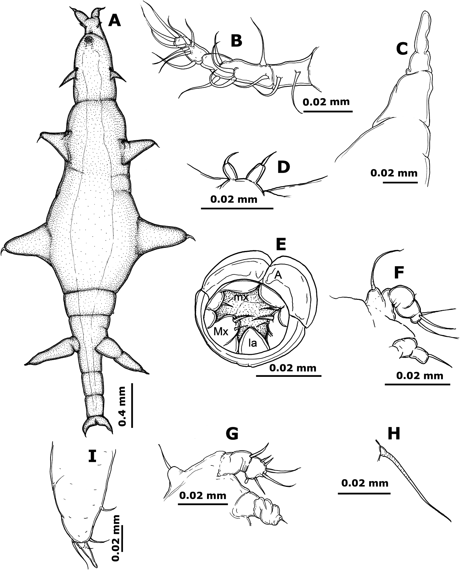
Colobomatus pteroisi sp. nov.
( Figure 3 View Figure 3 A–I)
Material examined
Holotype female (NSMT-Cr 21860), from head canal of Pterois volitans Linnaeus ( Scorpaeniformes : Scorpaenidae ), captured in the western North Pacific Ocean off Okinawa-jima Island and subsequently purchased at Hama Fisheries Cooperative (26 ◦ 34 ′ N, 127 ◦ 14 ′ E) in Nakagusuku, Okinawa-jima Island, 25 August 2009.
Description
Adult female. Body ( Figure 3A View Figure 3 ) 3.18 mm long (excluding anterior cephalic processes and caudal rami). Pre-oral area of cephalosome ( Figure 3A View Figure 3 ) slightly extended anteriorly and bearing one anterior pair of short, unadorned cephalic processes each with digitate tip ( Figure 3C View Figure 3 ). Minute pair of papillae ( Figure 3A,D View Figure 3 ), each bearing one sensillum, arising anterior to buccal capsule. Cephalosome swollen around oral area and bearing additional pair of processes arising anterolateral to posterior margin of cephalosome; processes posterolaterally directed, naked and pointed. First to fourth pedigerous somites fused to form pear-shaped body. Small first pair of legs situated on ventral surface near anterior margin of body. Body widest at fourth pedigerous somite and bearing two pairs of lateral processes; posterior pair of processes dorsolaterally located and larger than anterior pair; both pairs of processes bearing four sensilla and digitate structure on distal margin ( Figure 3I View Figure 3 ). Fifth pedigerous somite naked and demarcated from preceding somites by transverse constriction. Genital somite ( Figure 3A View Figure 3 ) bearing ventrolateral pair of slender, posterolaterally directed processes; latter as long as posterior pair of thoracic processes, naked and with digitate tip. Abdomen ( Figure 3A View Figure 3 ) indistinctly three-segmented and gradually tapering towards caudal rami. Caudal ramus ( Figure 3A View Figure 3 ) fused to anal somite, short, apically pointed.
Antennule ( Figure 3B View Figure 3 ) anterolaterally directed, arising near base of anterior cephalic processes and apparently three-segmented with armature of 2, 6 and 8. Antenna ( Figure 3E View Figure 3 ) modified, forming longitudinally divided and robust anterior margin of buccal capsule; latter tube-like, projecting ventrally from conical base. Labrum and mandibles not seen. Maxillule ( Figure 3E View Figure 3 ) located mid-laterally in buccal capsule, with one spine on distal margin. Maxilla ( Figure 3E View Figure 3 ) robust, apparently onesegmented and bearing two apical spines. Labium ( Figure 3E View Figure 3 ) undivided. Maxillipeds absent. Posterior rim of buccal capsule undivided.
Leg 1 ( Figure 3F View Figure 3 ) biramous; protopod carrying long, lateral seta arising from basal protrusion; exopod indistinctly two-segmented, with tiny lateral element on basal segment and three long apical spines on distal segment; endopod papillose, indistinctly two-segmented and bearing one spine on distal margin. Leg 2 ( Figure 3G View Figure 3 ) biramous; protopod bearing lateral seta arising from basal protrusion; exopod indistinctly two-segmented, with one outer spine on basal segment and two short outer spines, three long apical spines and one blunt inner spine on distal segment; endopod, papillose, indistinctly two-segmented and bearing minute apical spine on distal segment. Leg 3 ( Figure 3H View Figure 3 ) vestigial, reduced to long lateral seta.
Etymology
The species epithet, pteroisi , refers to the generic name of the host.
Remarks
The presence of two pairs of unbranched cephalic processes in the female of C. pteroisi sp. nov. separates it from all species of the genus except C. ornatus and C. pupa. However, C. pteroisi sp. nov. is unique in possessing the posterior pair of cephalic processes arising posterior to the buccal capsule and a digitate tip on all the processes except the posterior pair of cephalic processes.
This study reports for the first time a member of the genus Colobomatus from the family Scorpaenidae (Scorpaeniformes) . Previously, only C. rothae West, 1992 was reported from the lachrymal, infra-orbital and inter-orbital canals of the scorpaeniform fish, Platycephalus fuscus Cuvier (Platycephalidae) , collected from Deception Bay, Australia ( West 1992).
No known copyright restrictions apply. See Agosti, D., Egloff, W., 2009. Taxonomic information exchange and copyright: the Plazi approach. BMC Research Notes 2009, 2:53 for further explanation.
|
Kingdom |
|
|
Phylum |
|
|
Class |
|
|
Order |
|
|
Family |
|
|
Genus |