
Schizosairhynchus Gustafsson & Bush, 2017
|
publication ID |
https://doi.org/ 10.11646/zootaxa.4313.1.1 |
|
publication LSID |
lsid:zoobank.org:pub:A5Fdfba5-F992-44A8-84C2-1756C943C19B |
|
DOI |
https://doi.org/10.5281/zenodo.5297051 |
|
persistent identifier |
https://treatment.plazi.org/id/832187E9-FE9E-FED6-FF74-6024FB5BF8EF |
|
treatment provided by |
Plazi |
|
scientific name |
Schizosairhynchus Gustafsson & Bush |
| status |
gen. nov. |
Schizosairhynchus Gustafsson & Bush , new genus
Sturnidoecus Eichler, 1944: 81 (in partim).
Type species. Schizosairhynchus erysichthoni new species
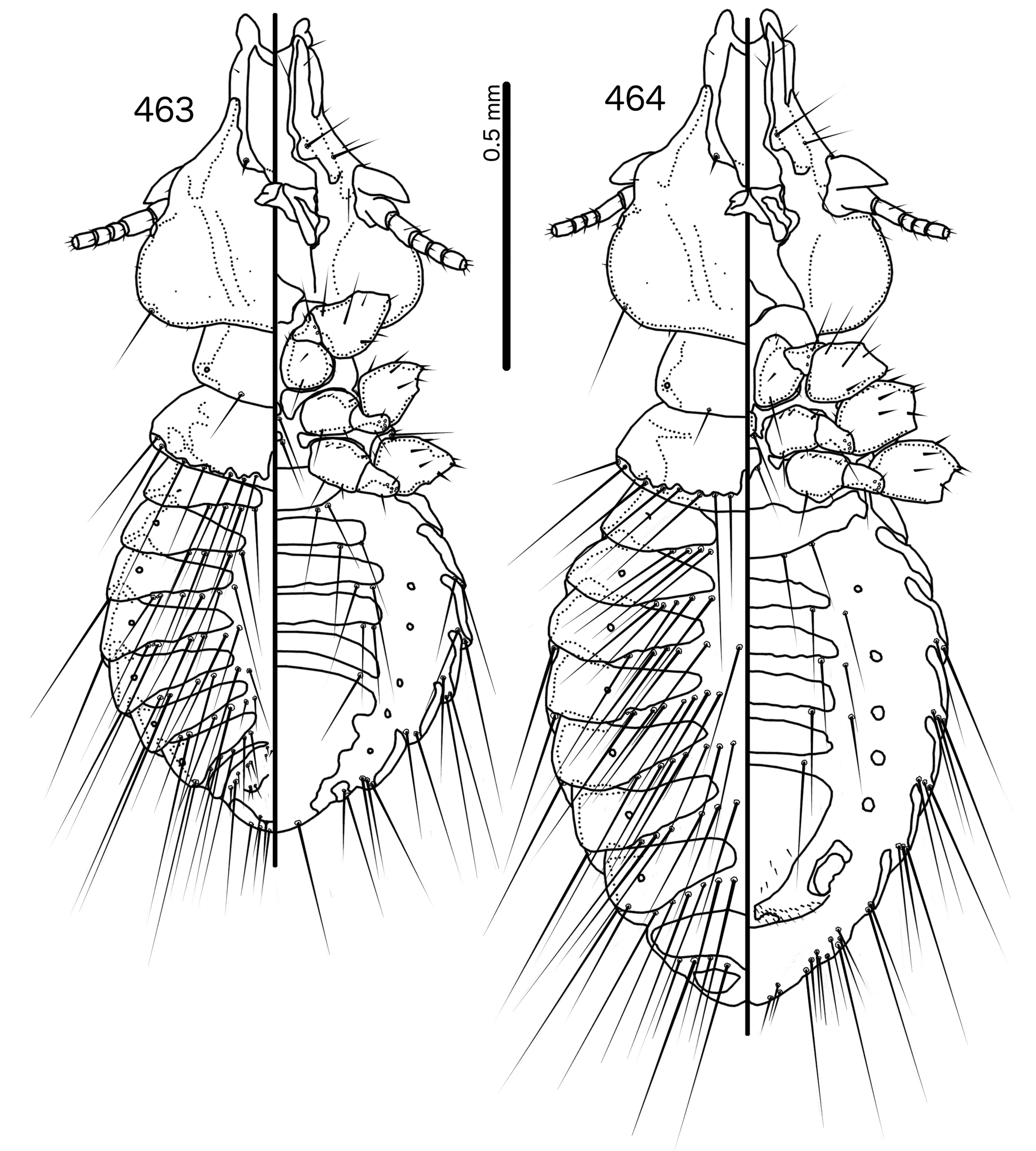
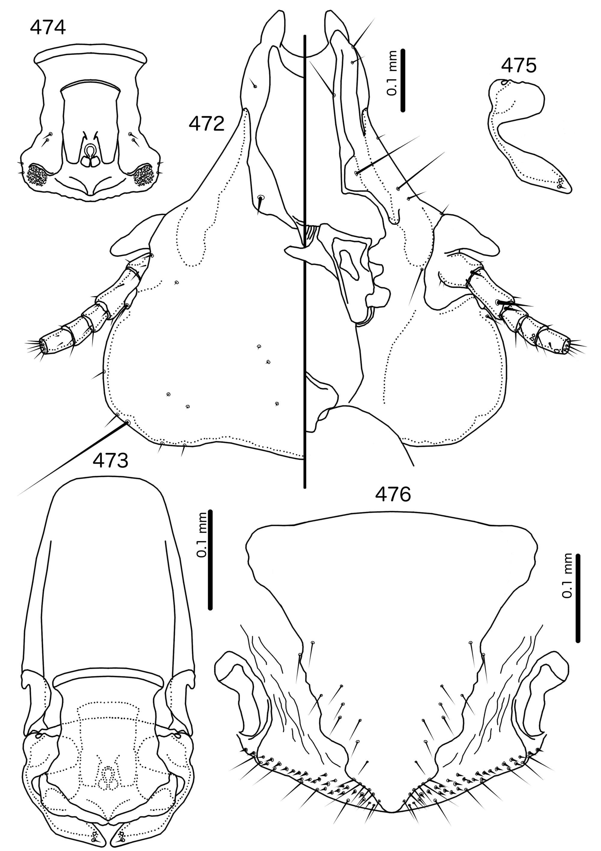
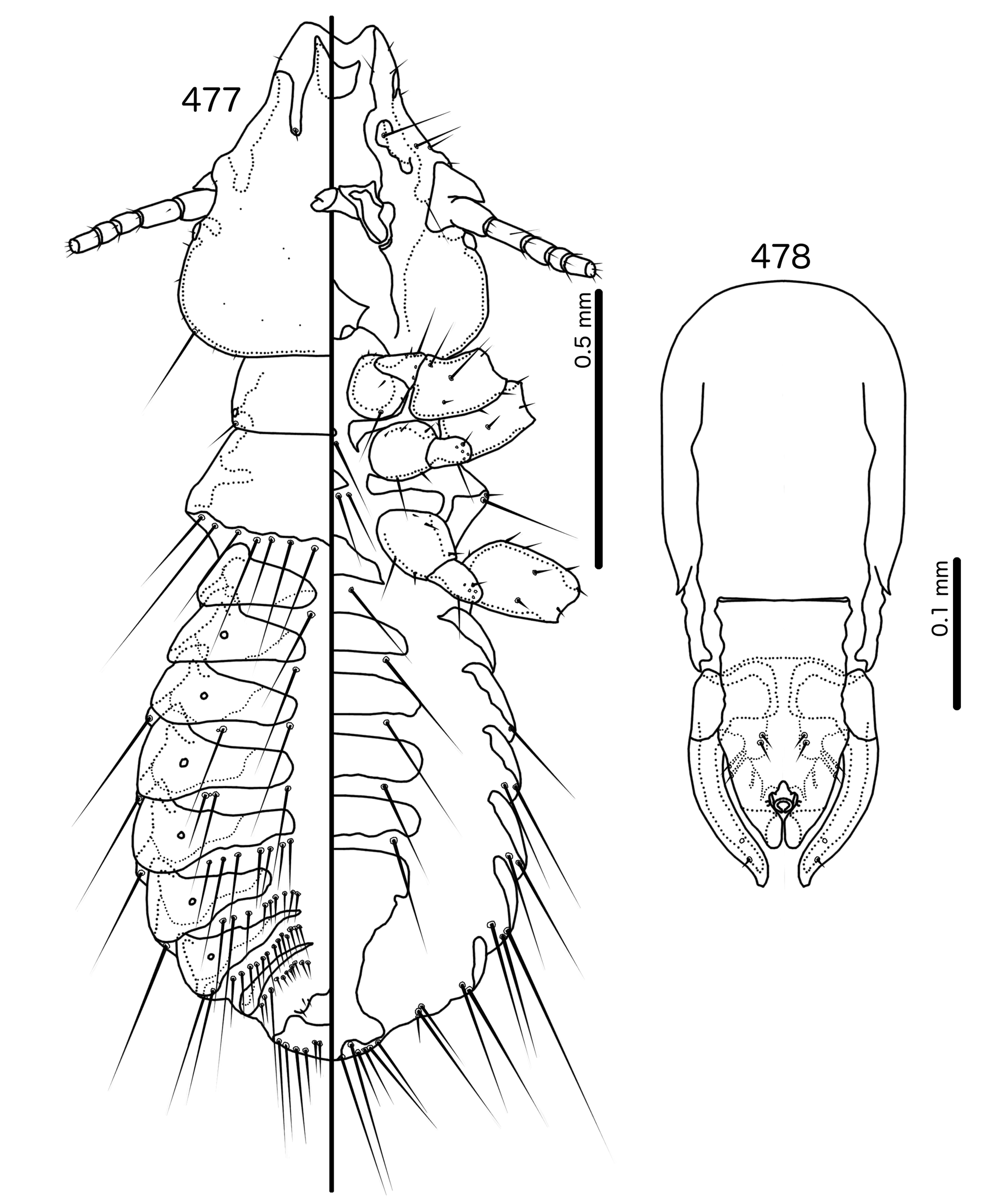
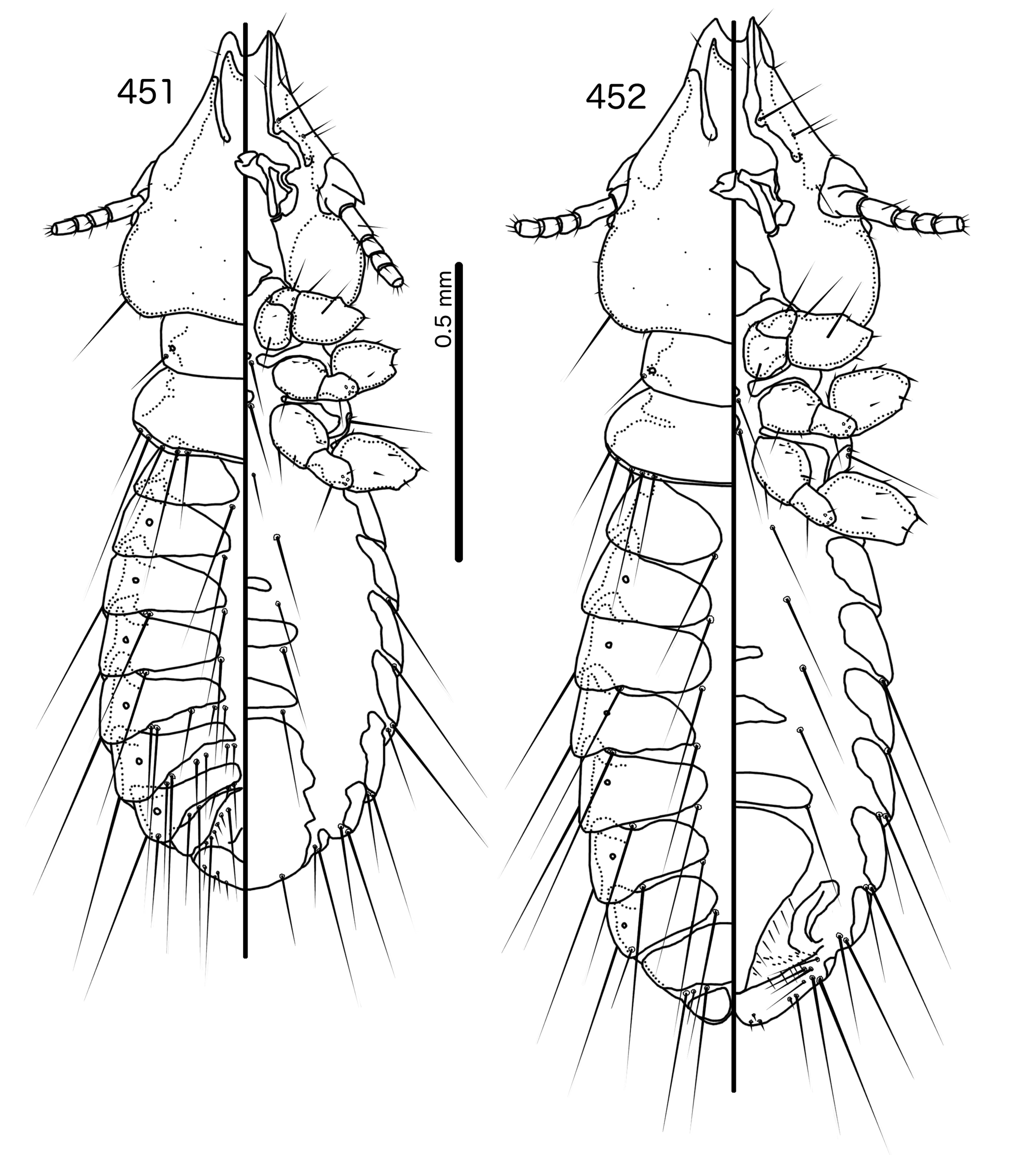
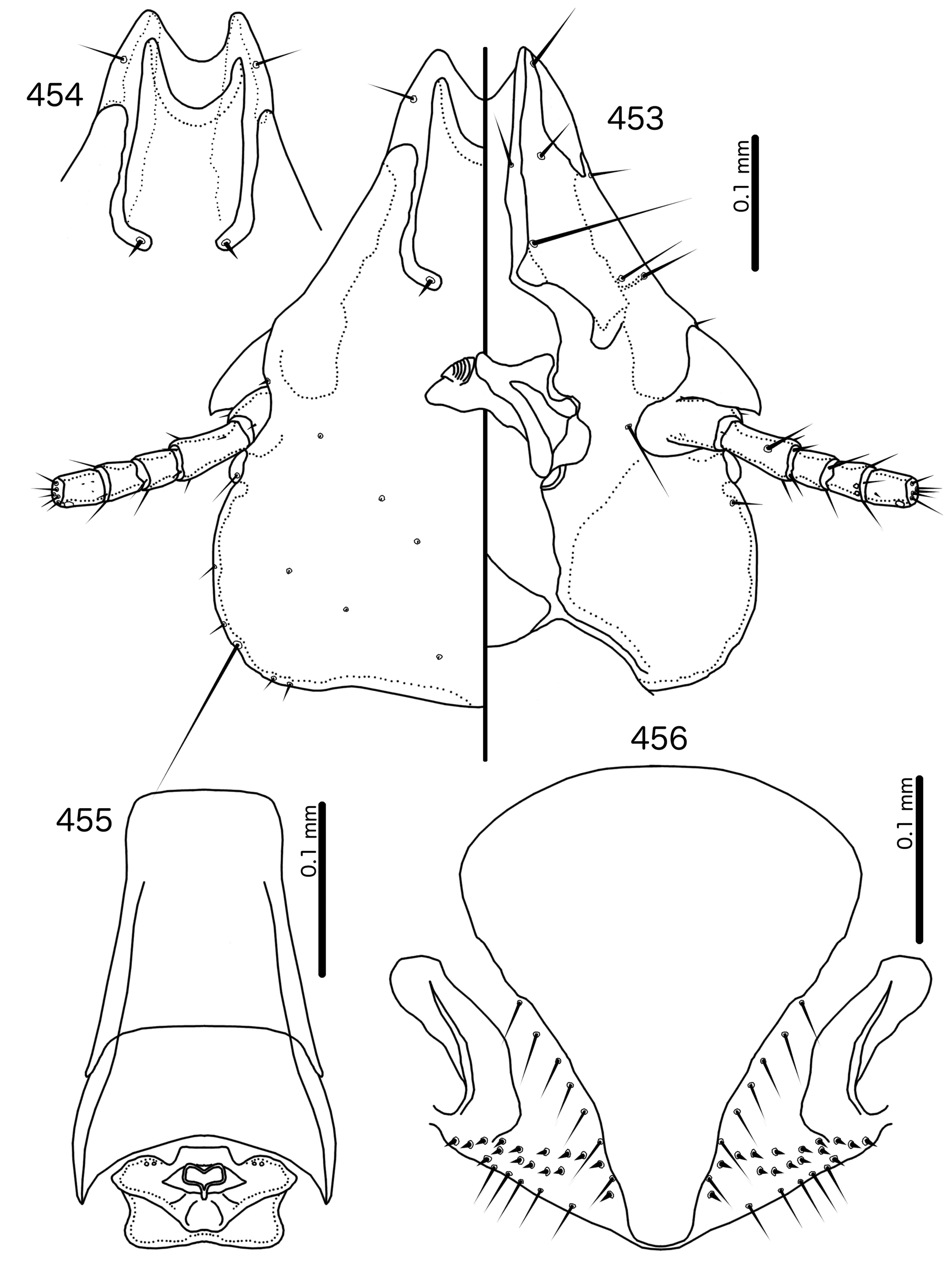
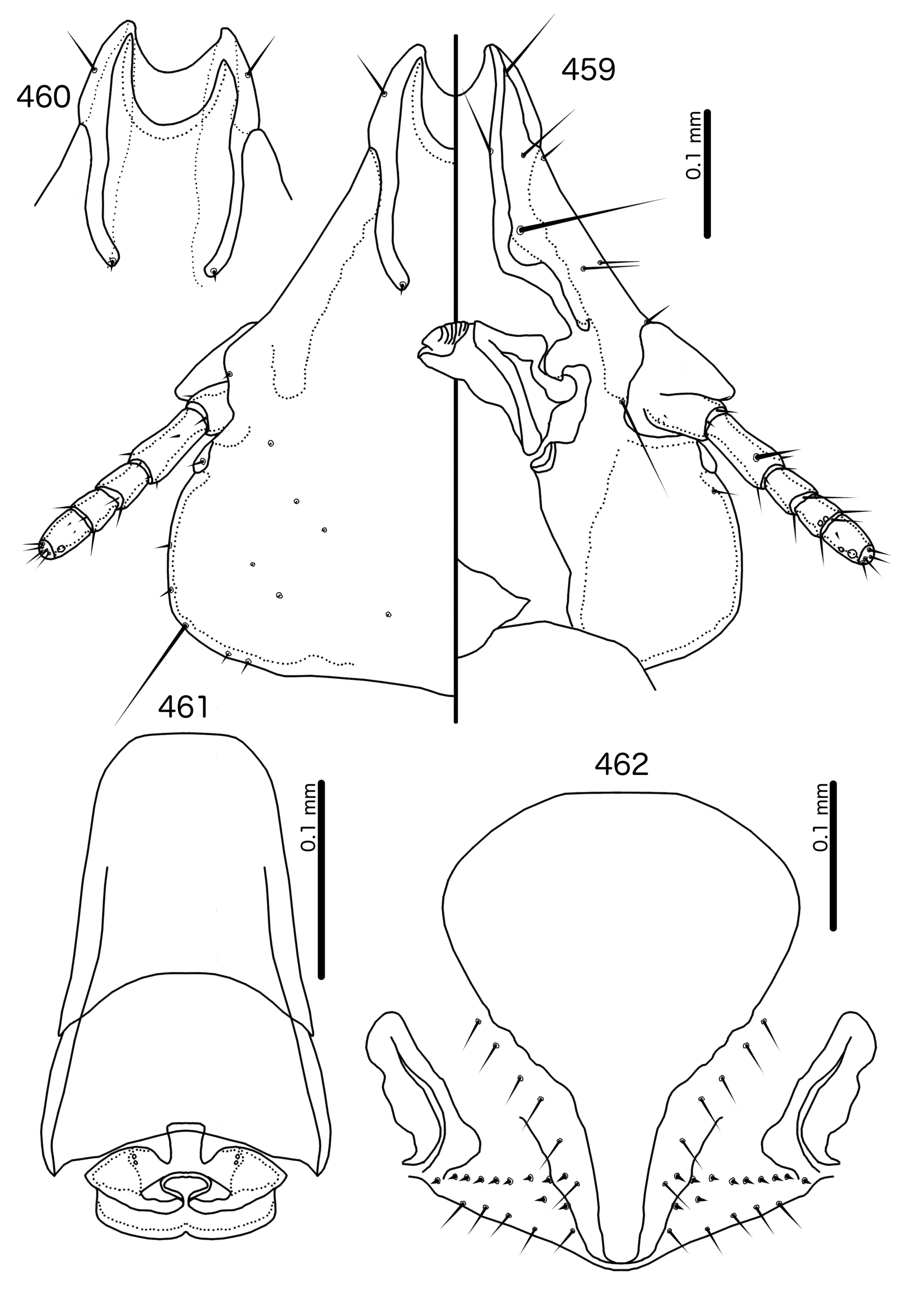
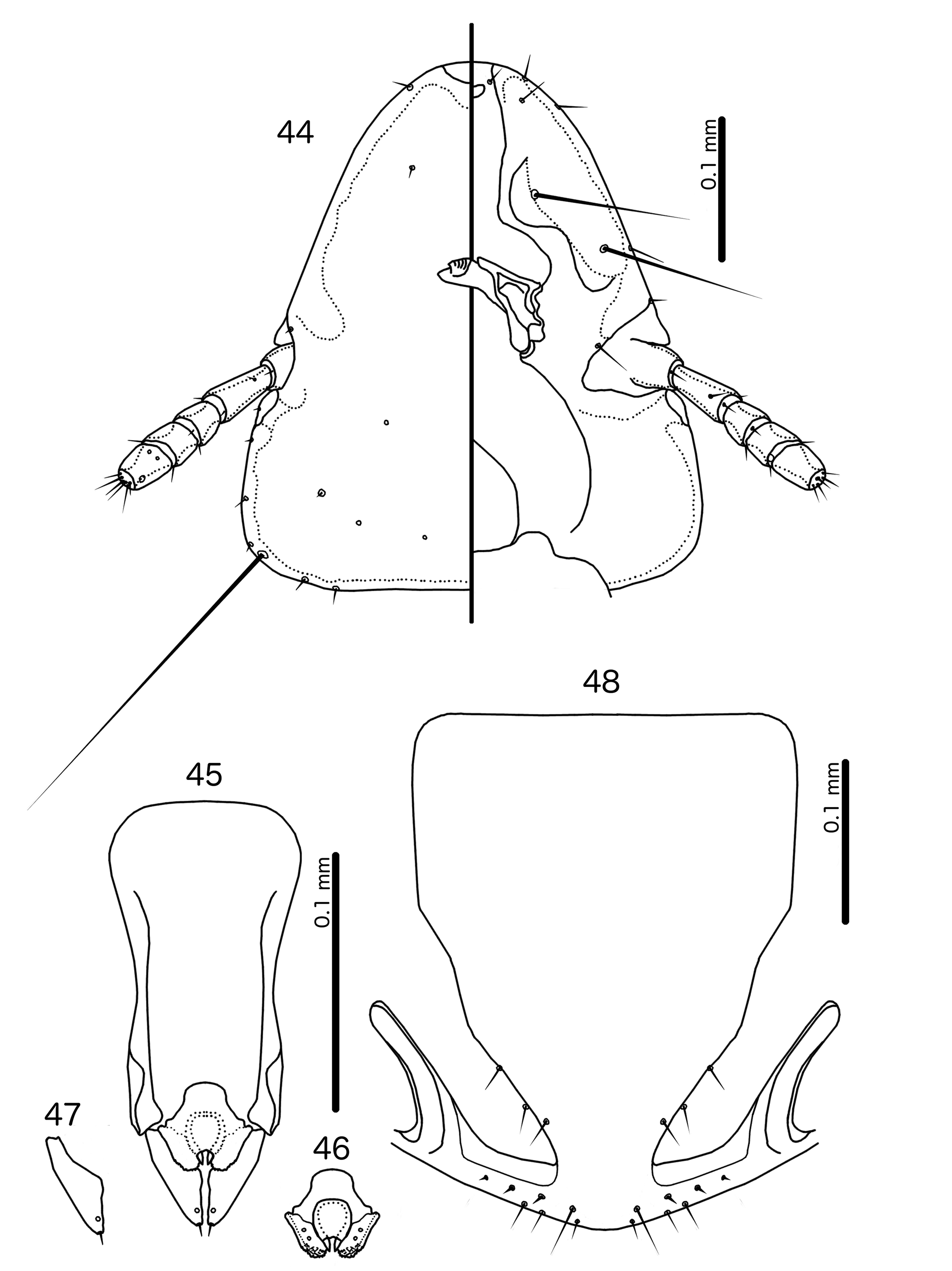
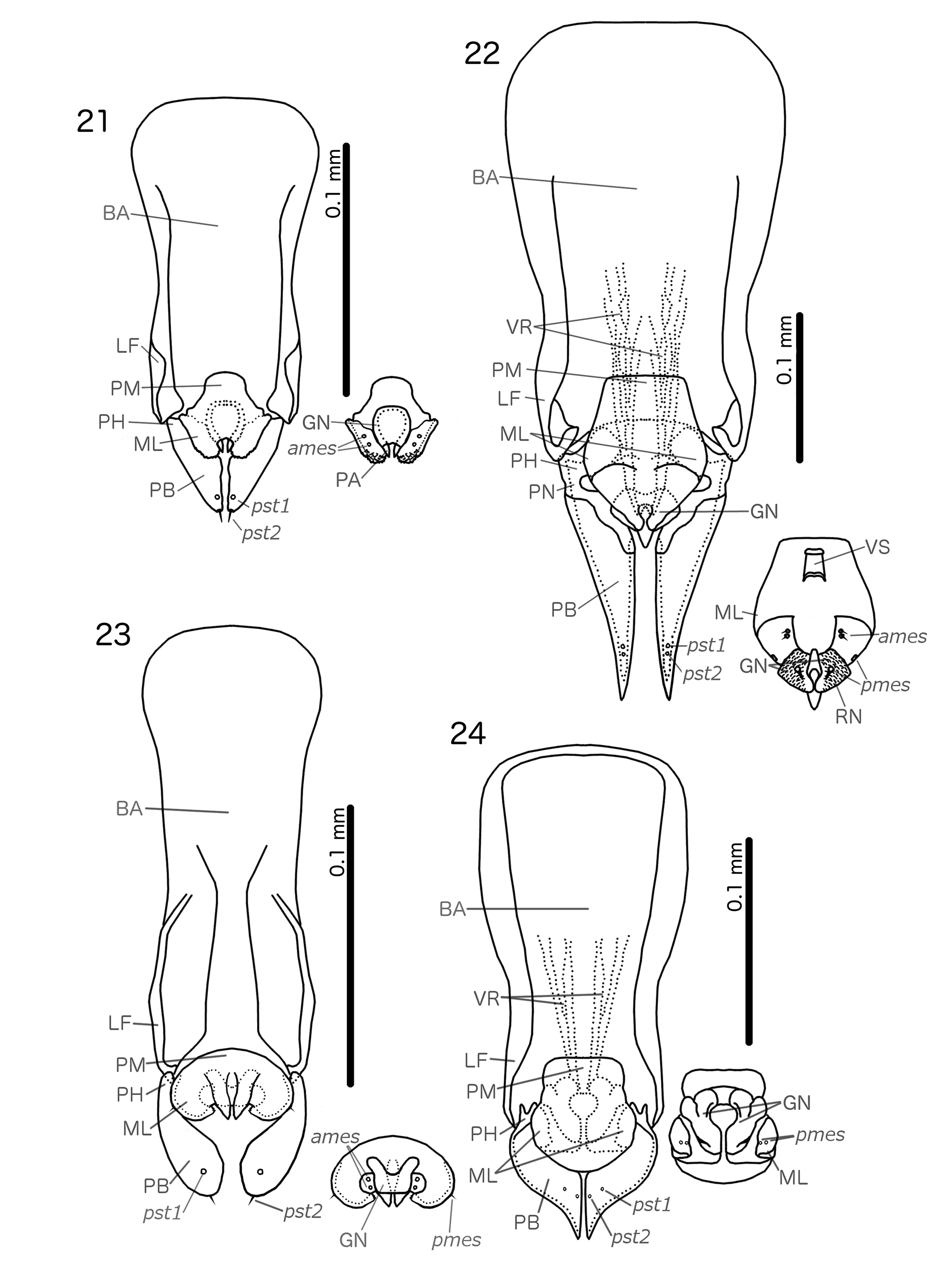
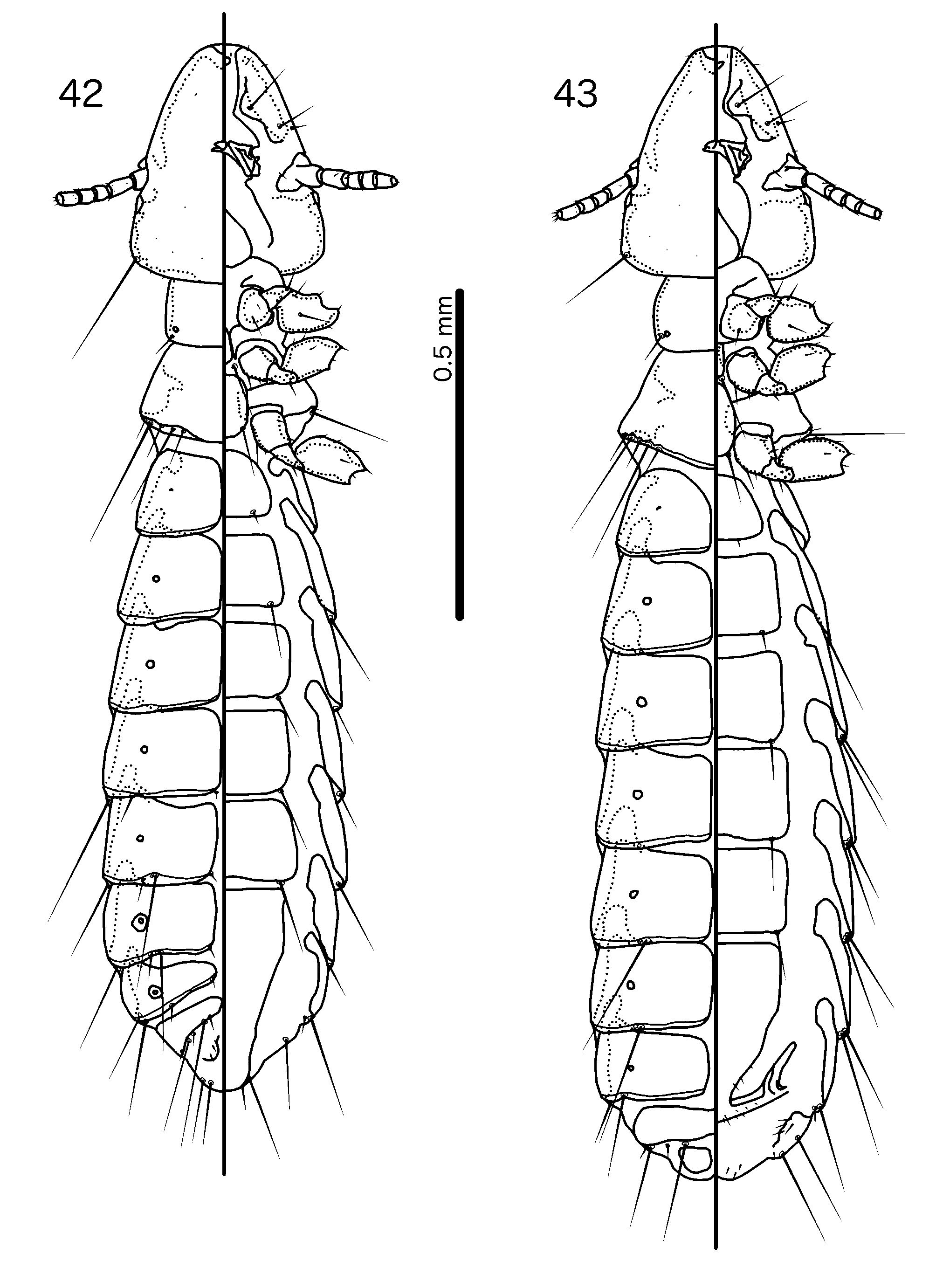
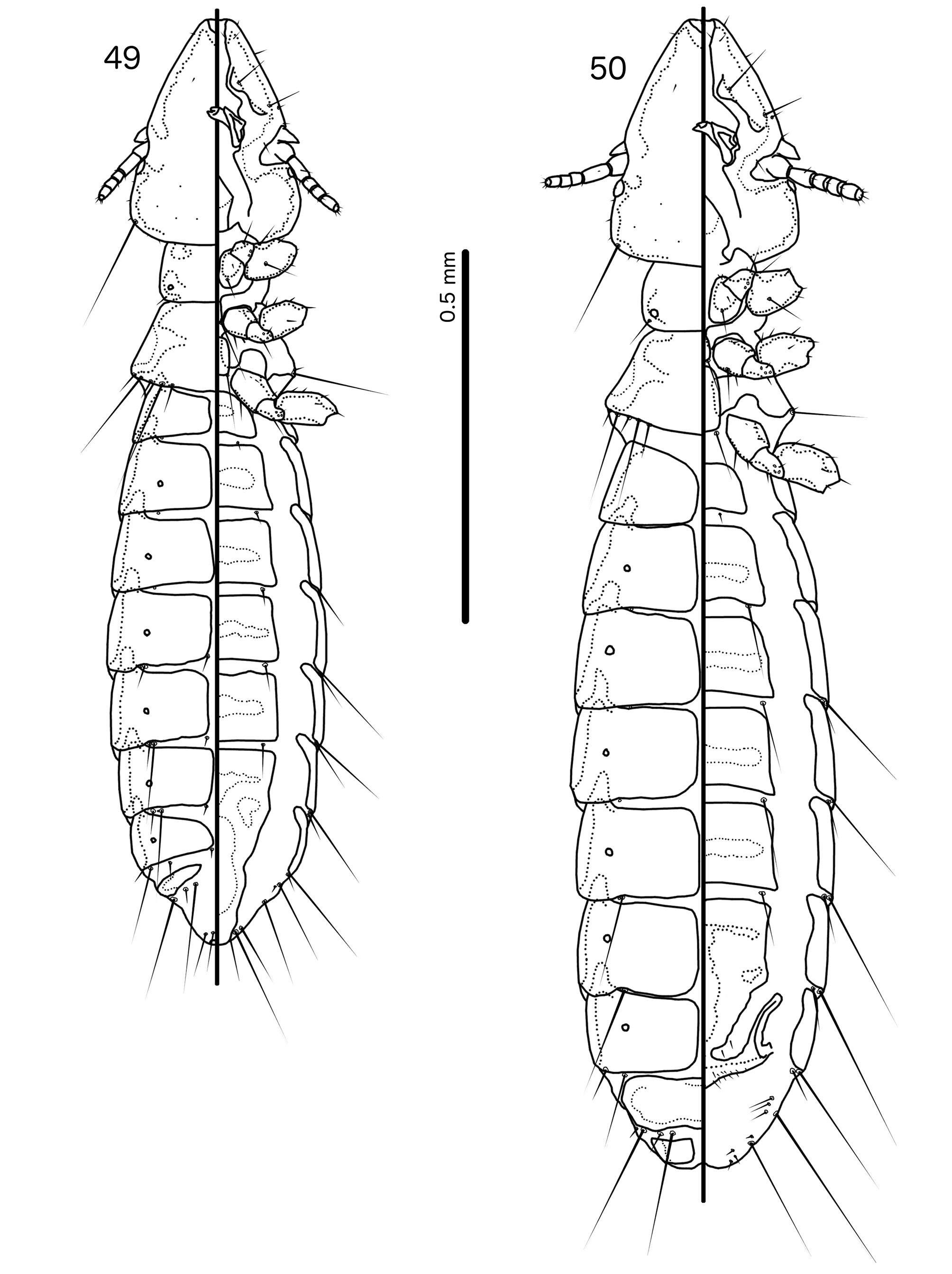
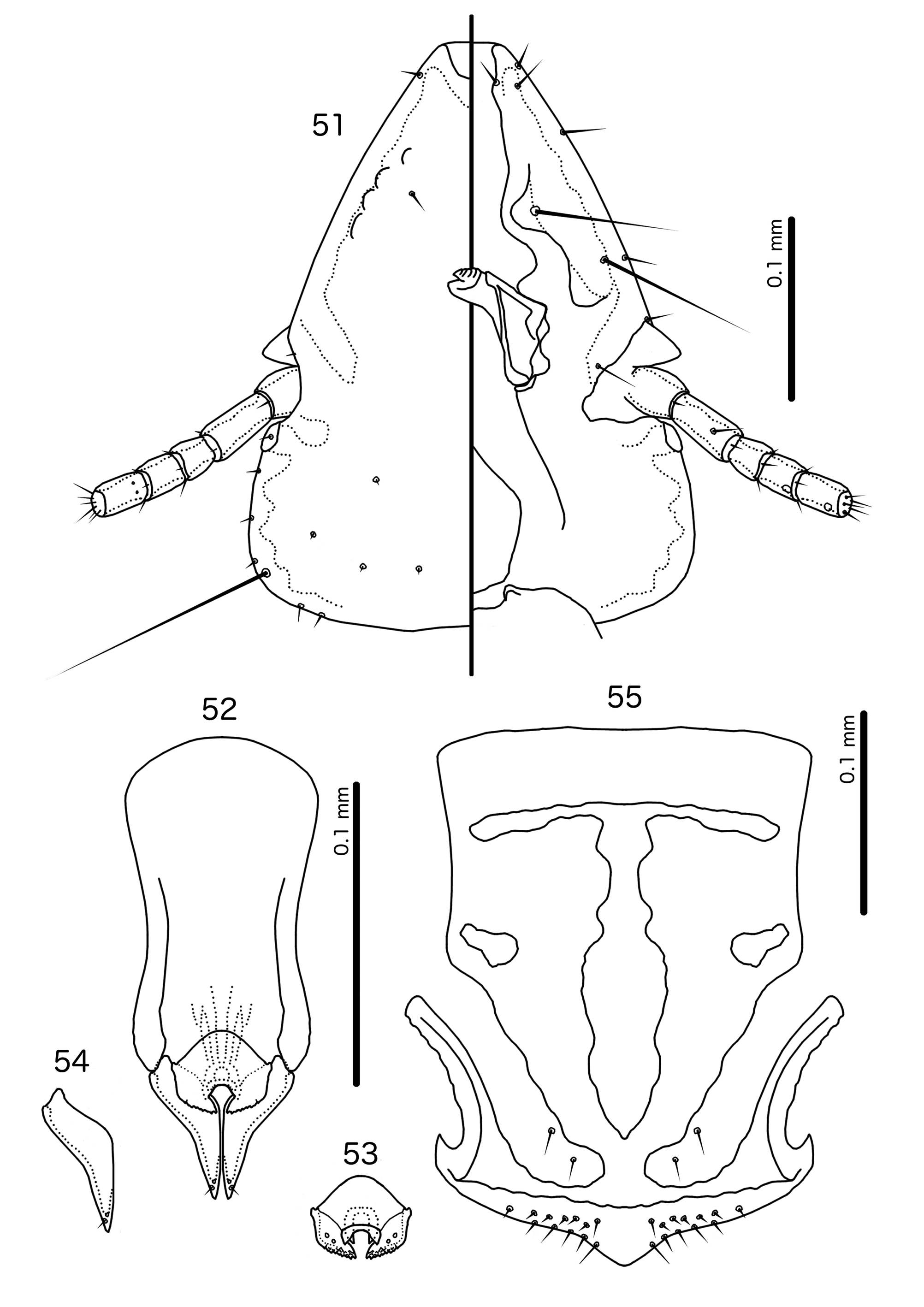
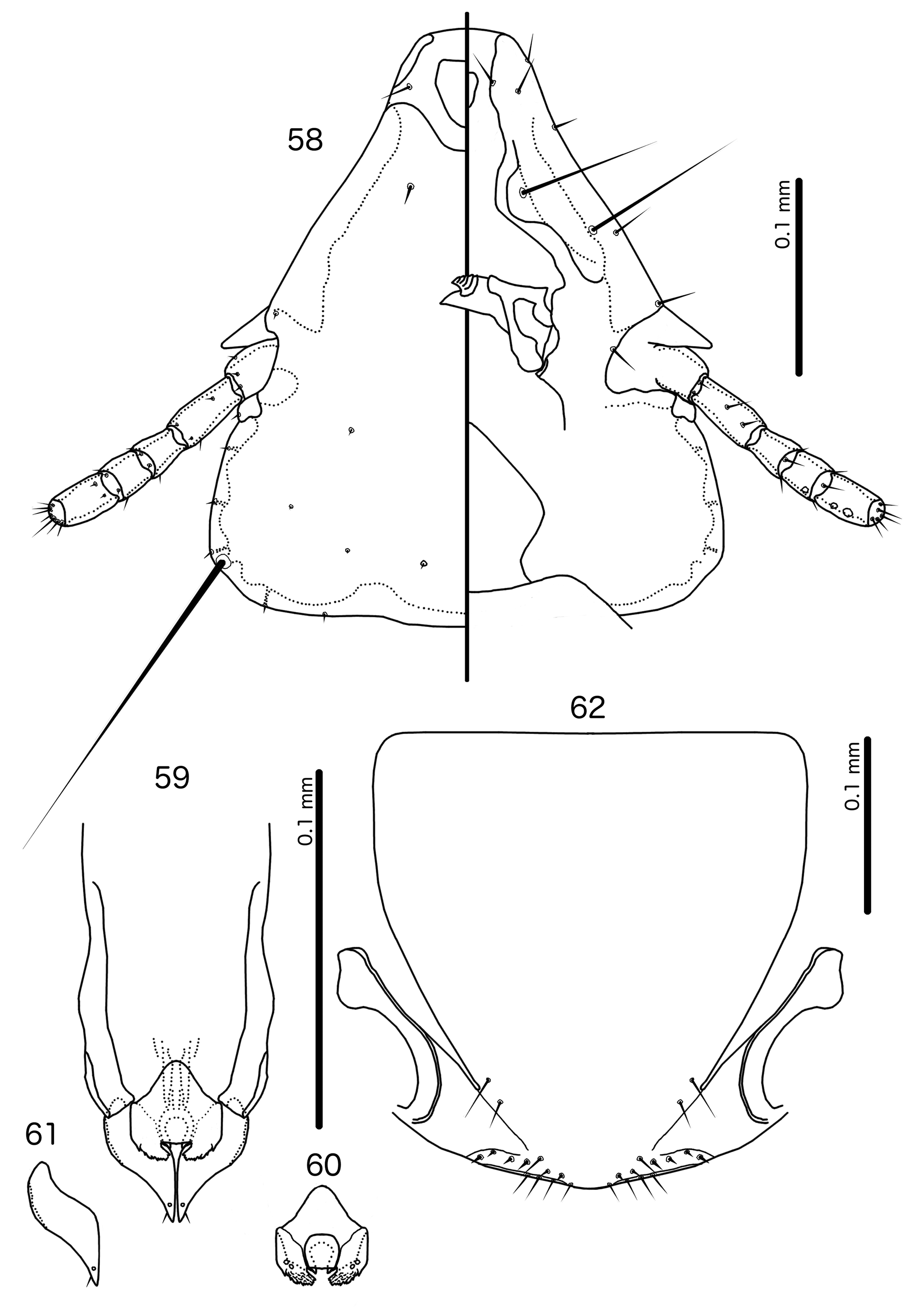
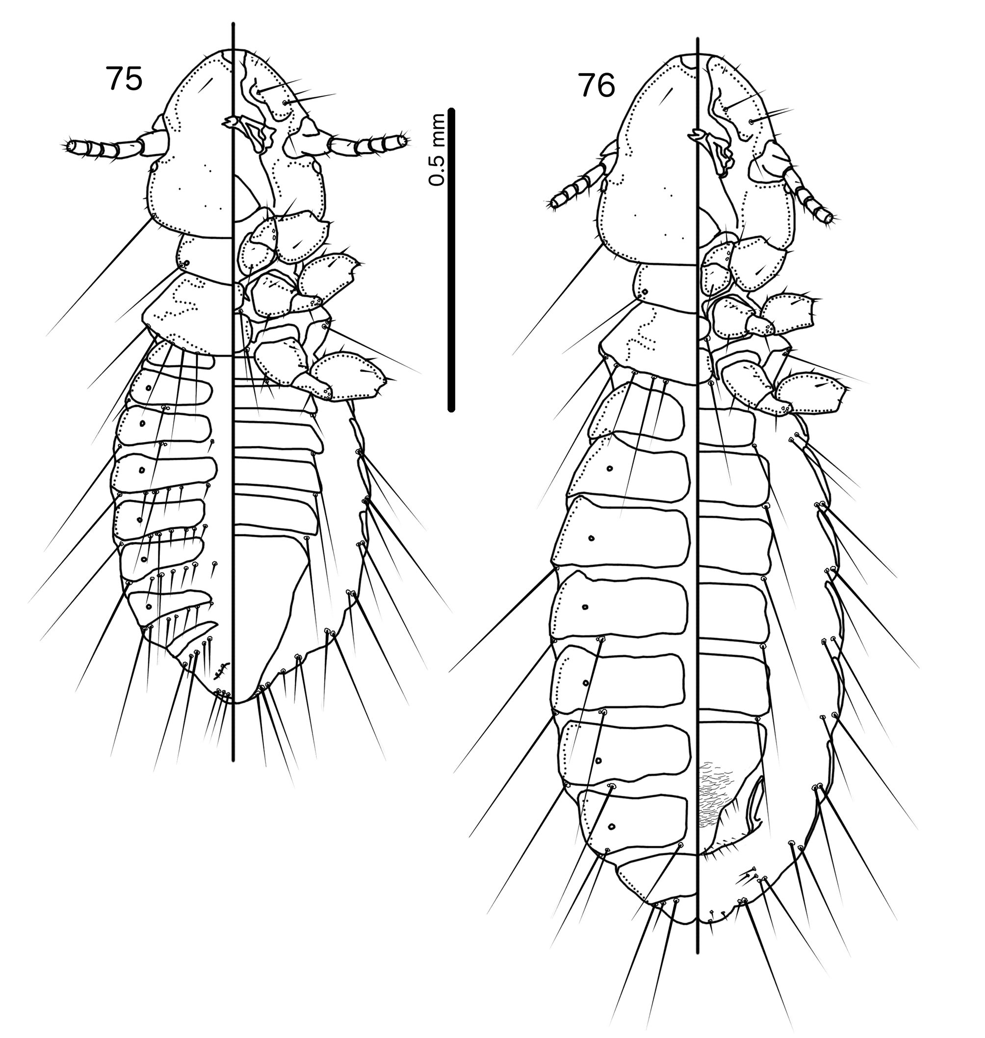
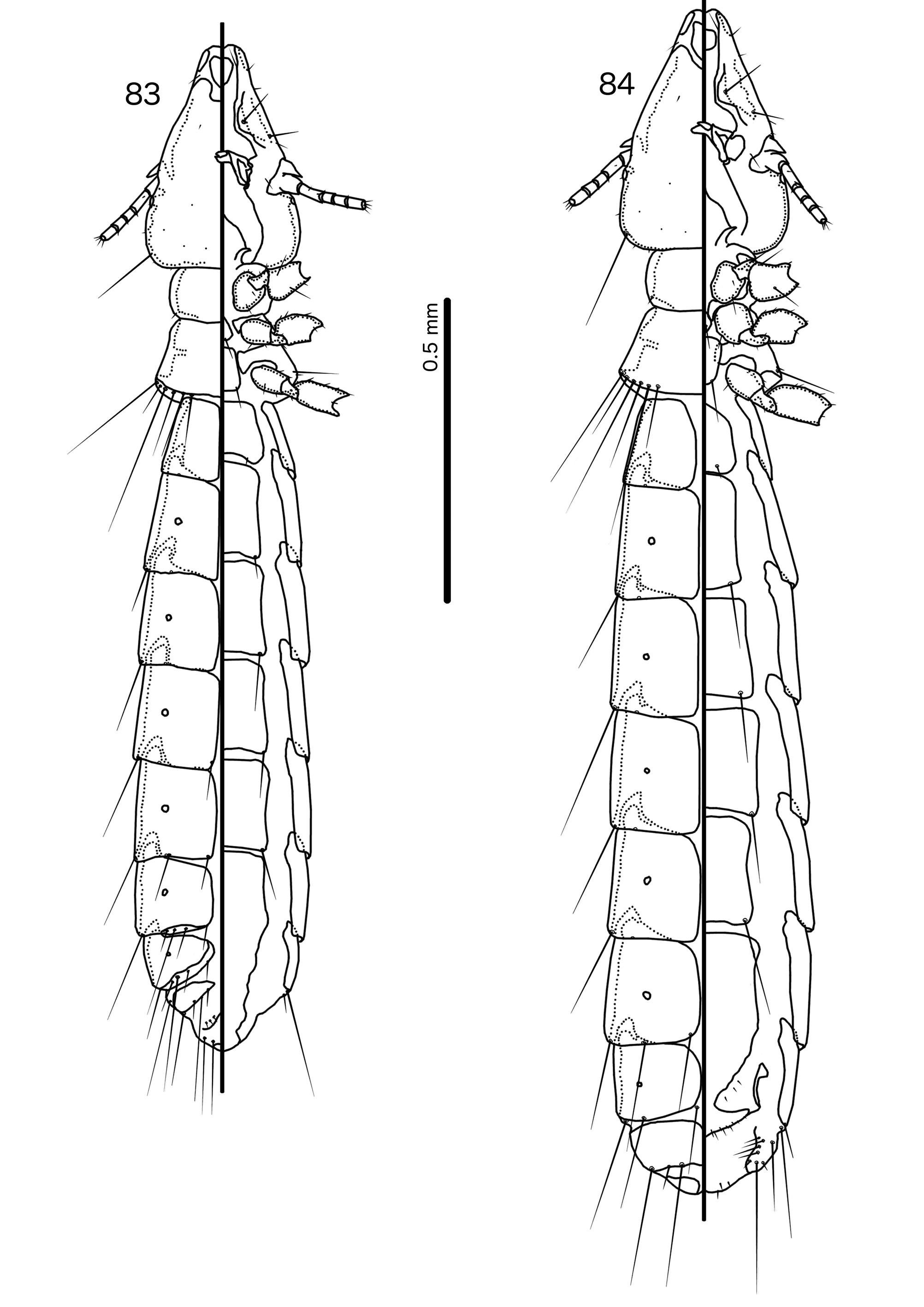
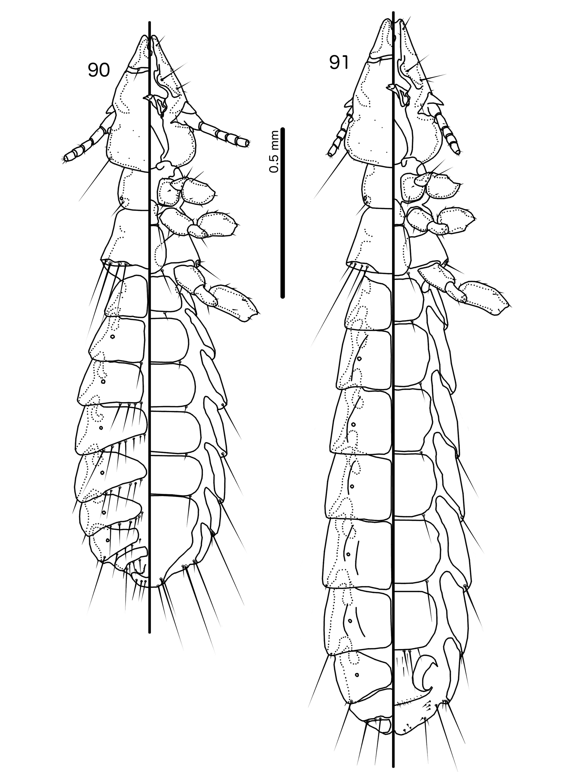
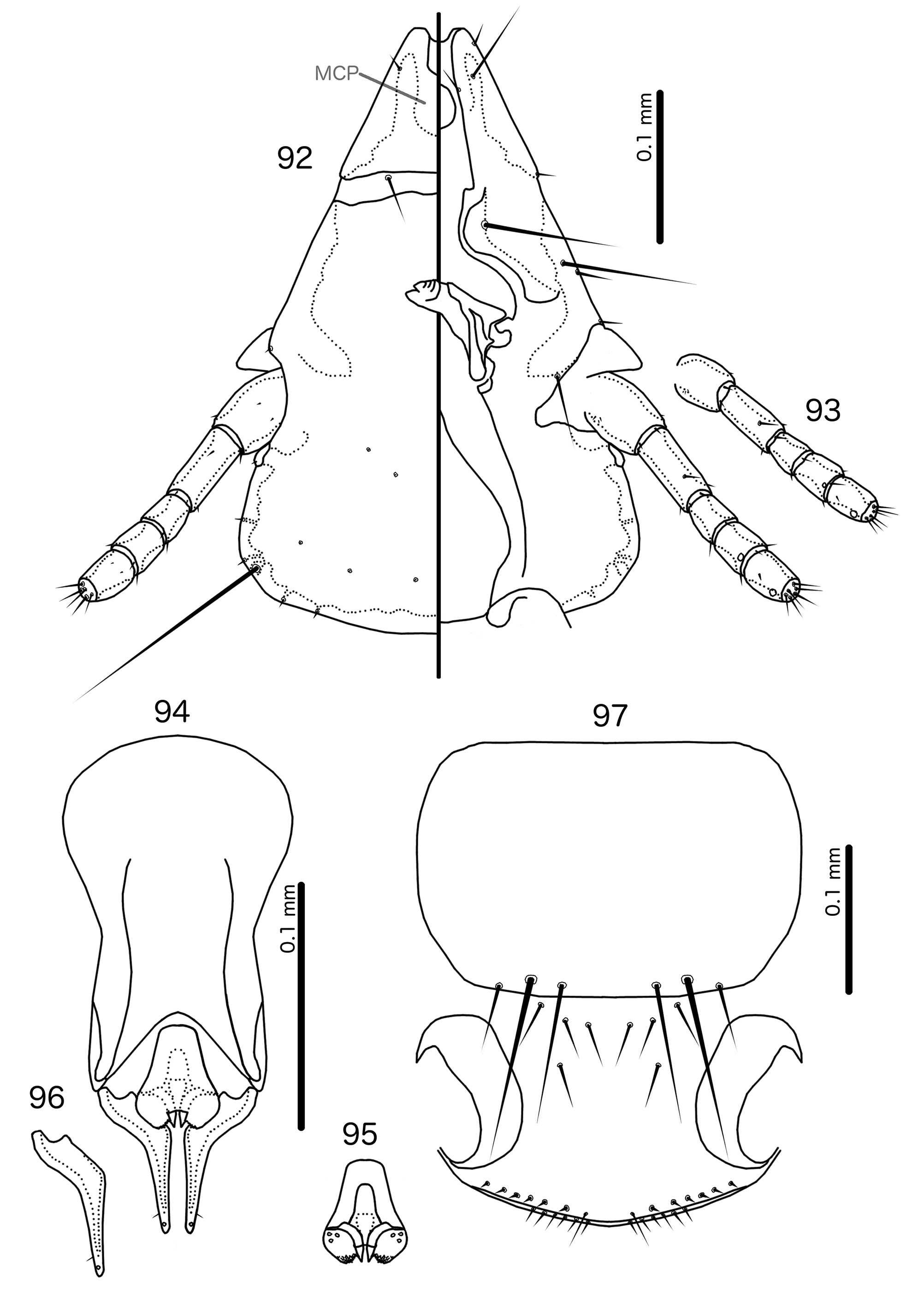
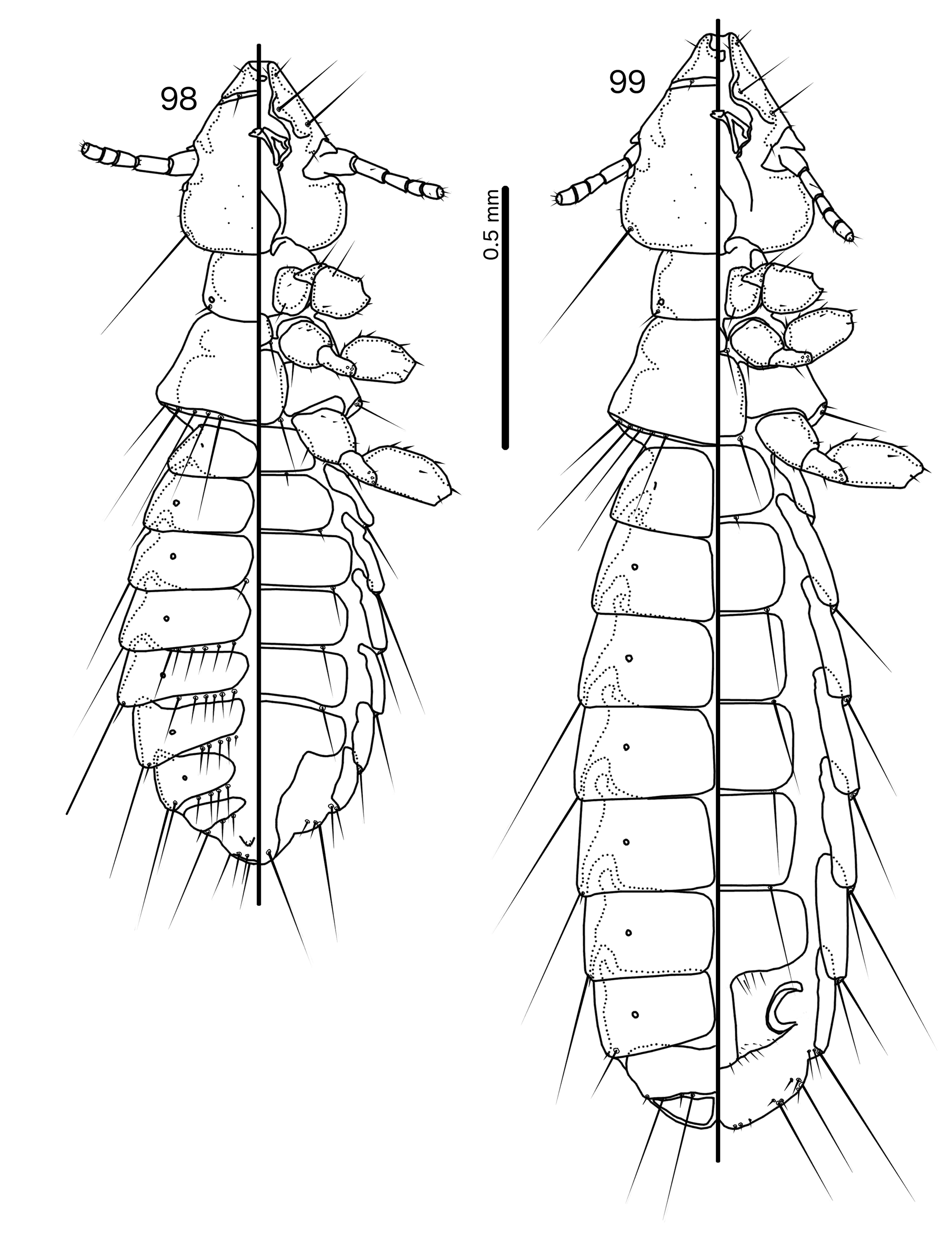
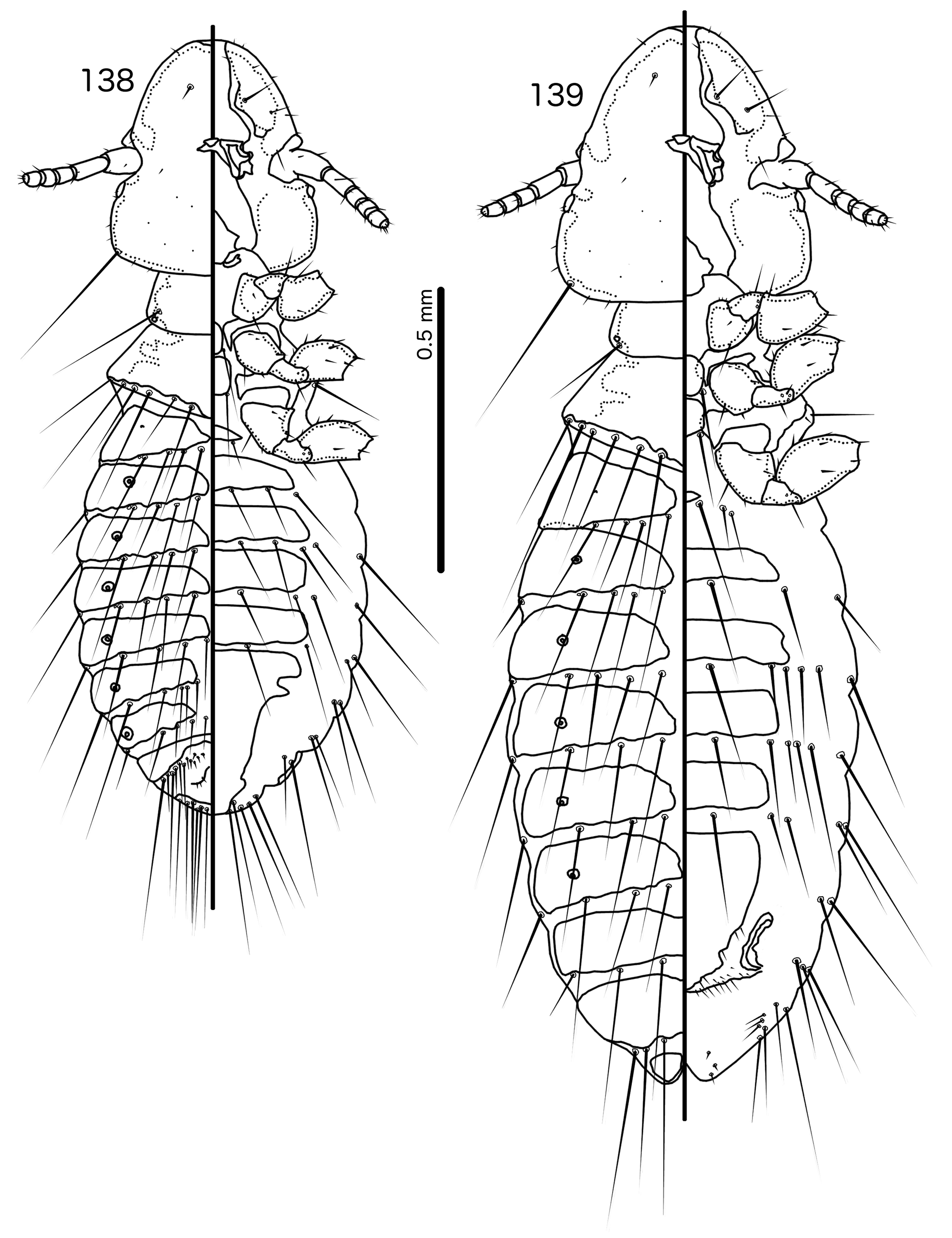
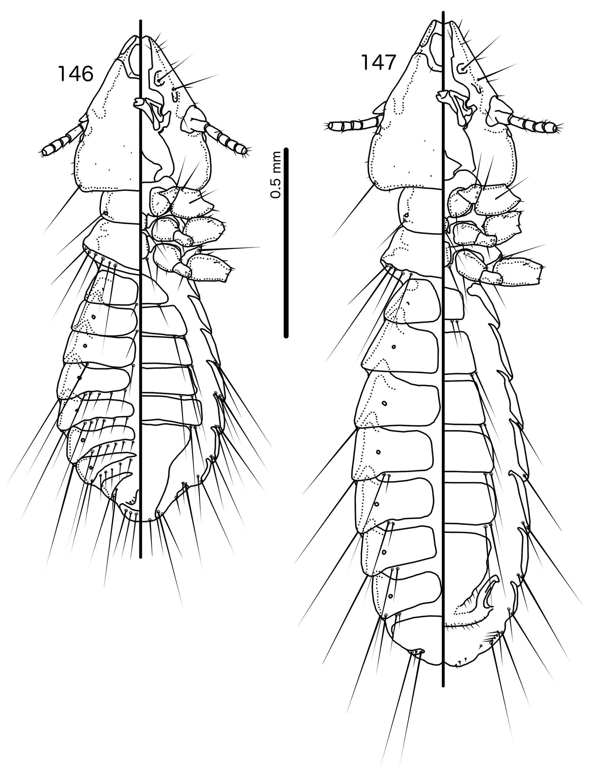
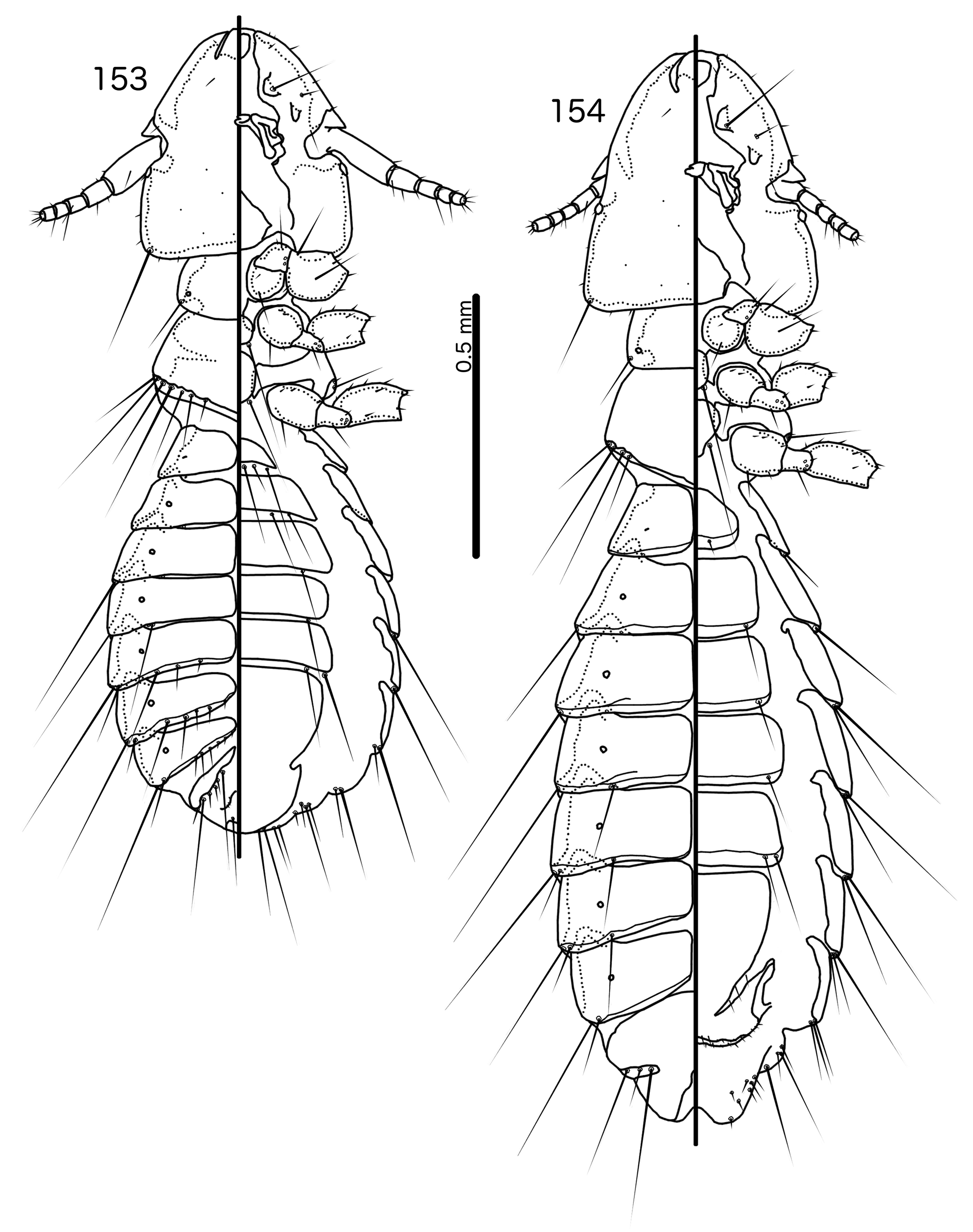
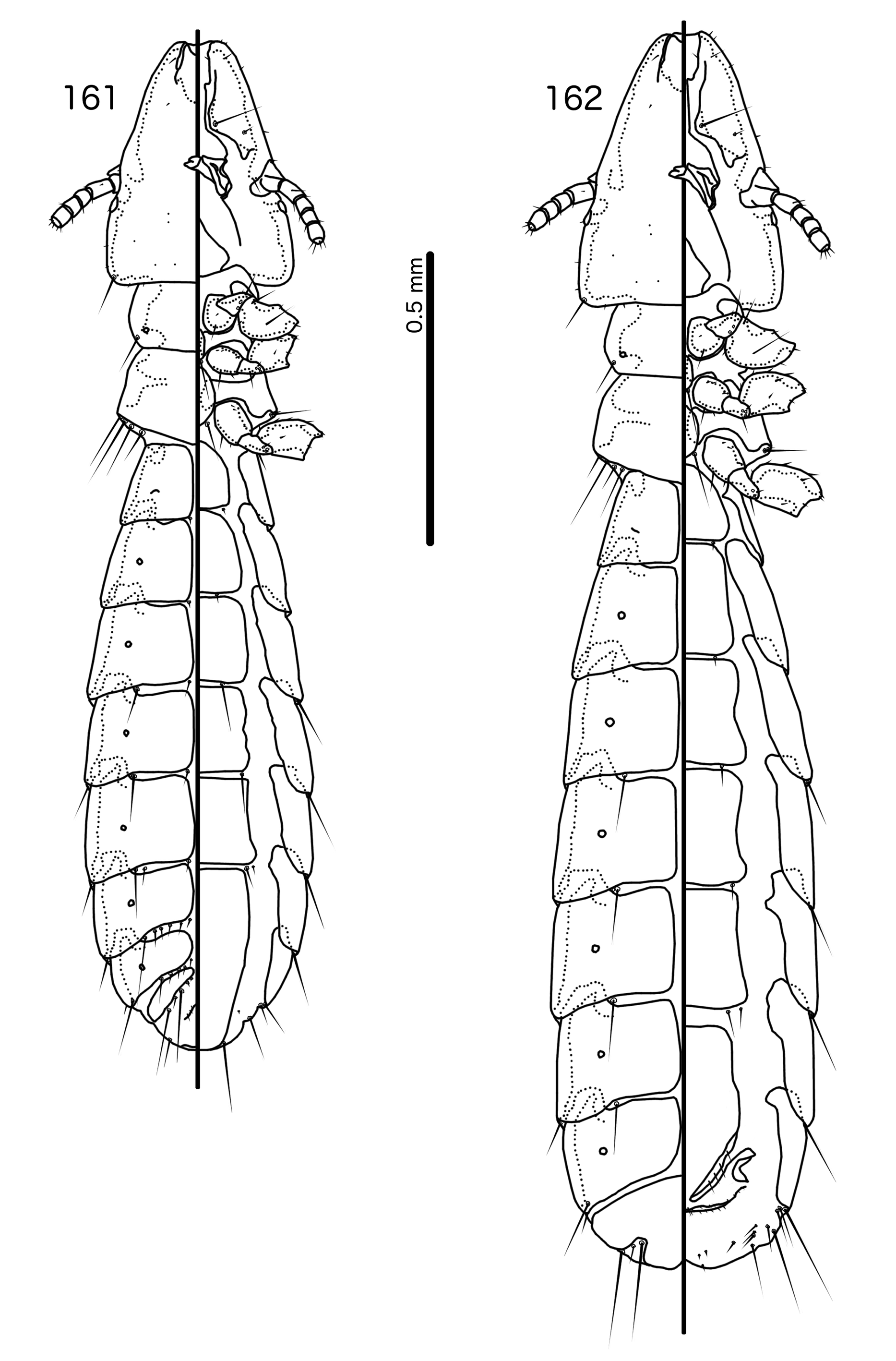
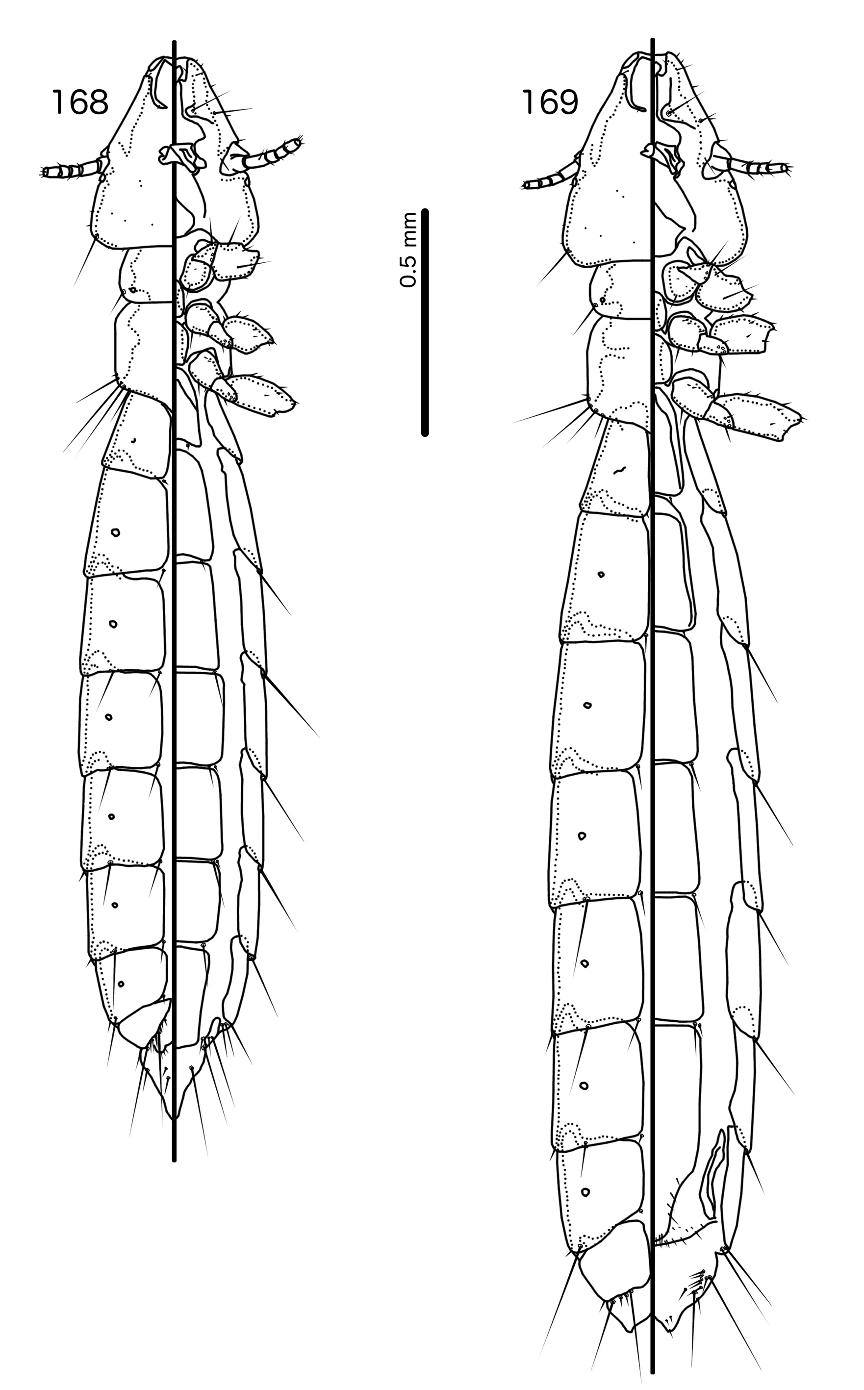
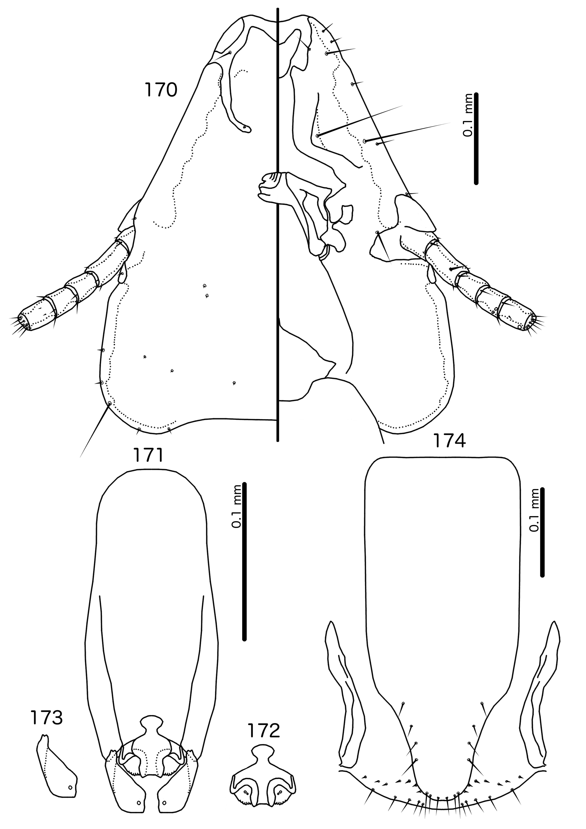
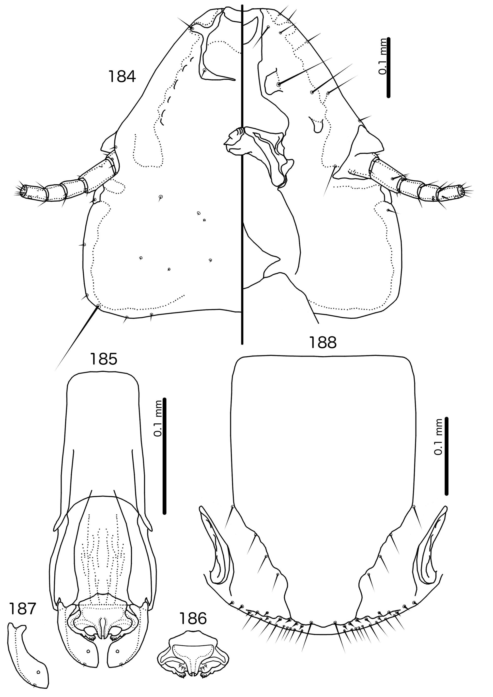
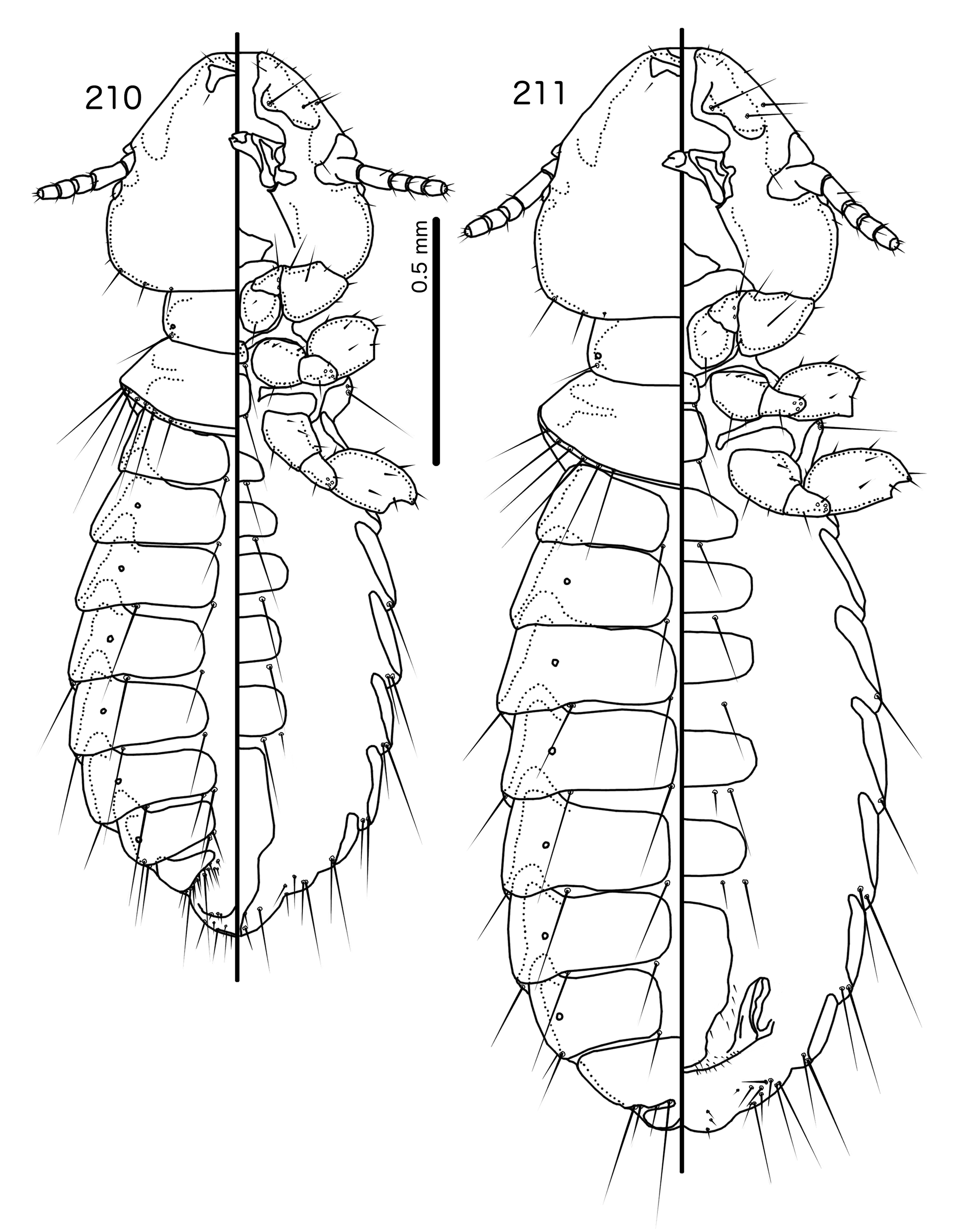
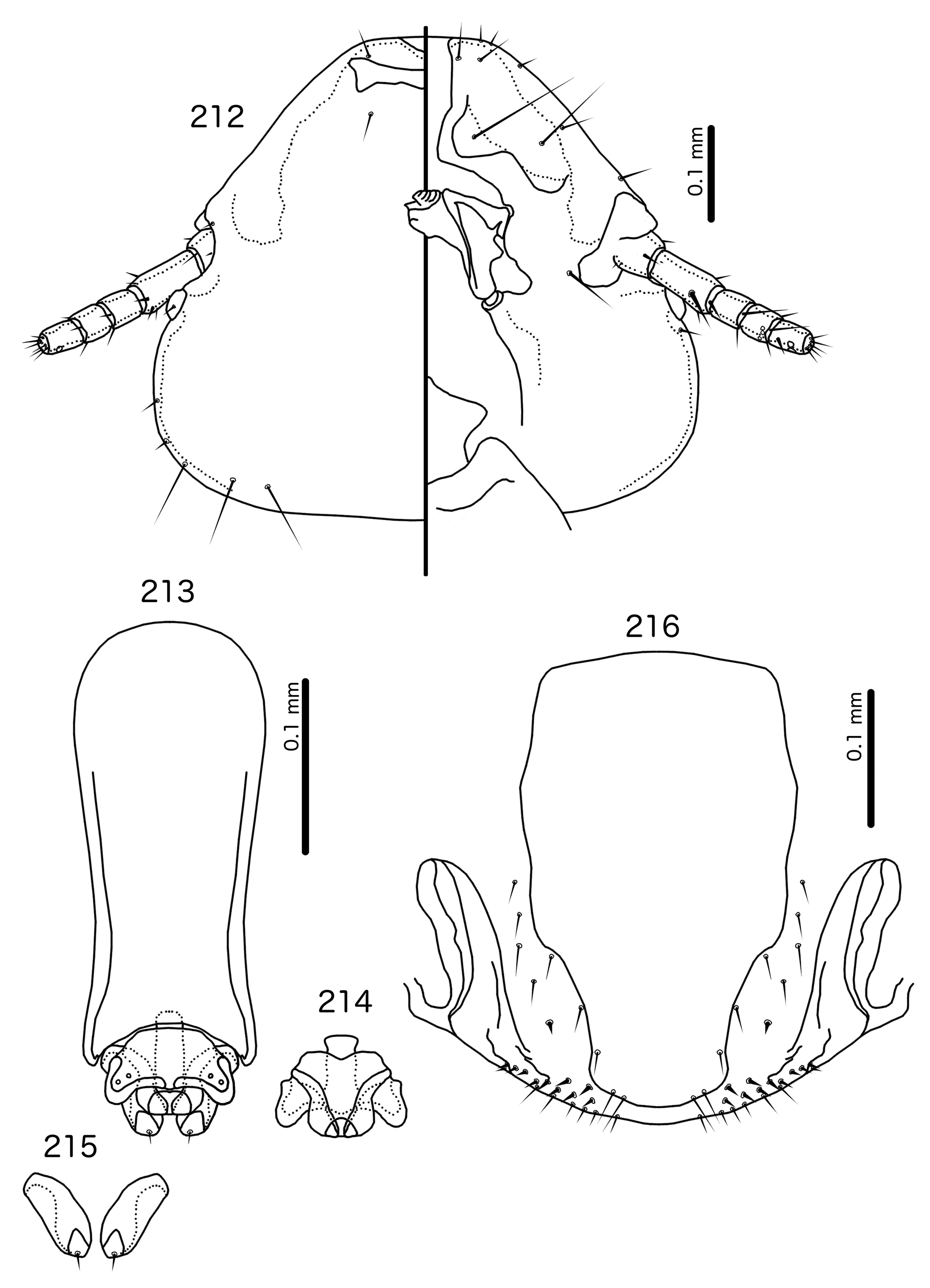
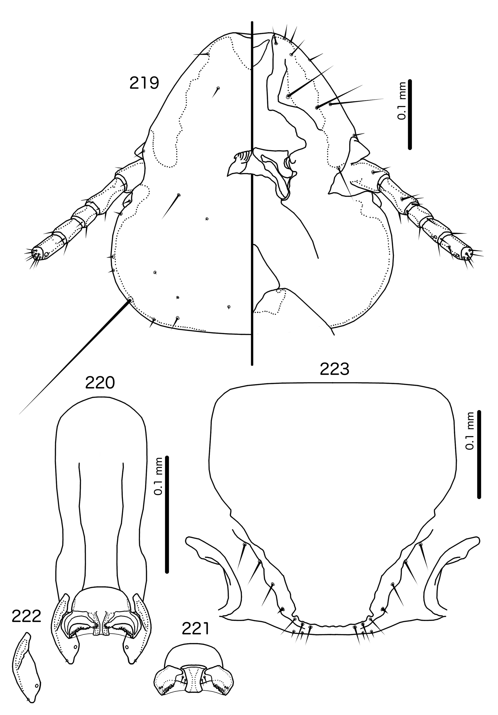
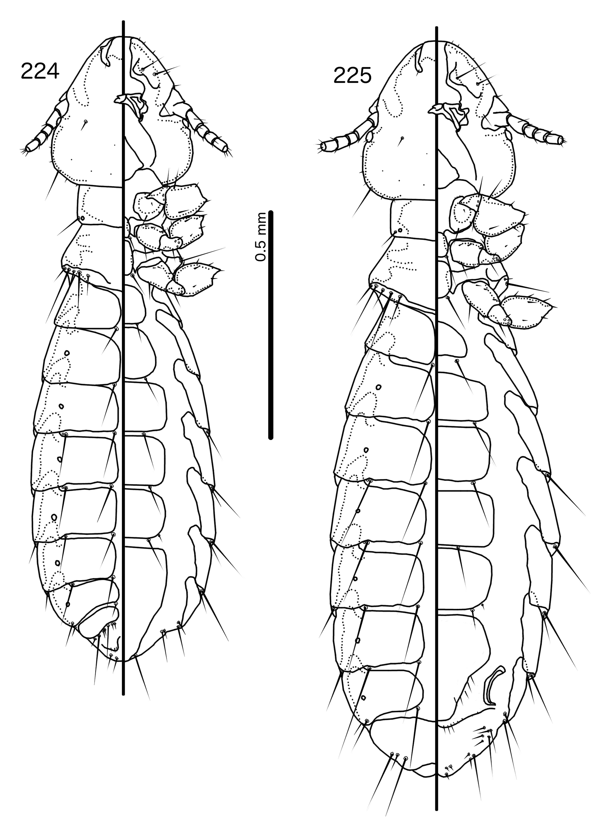
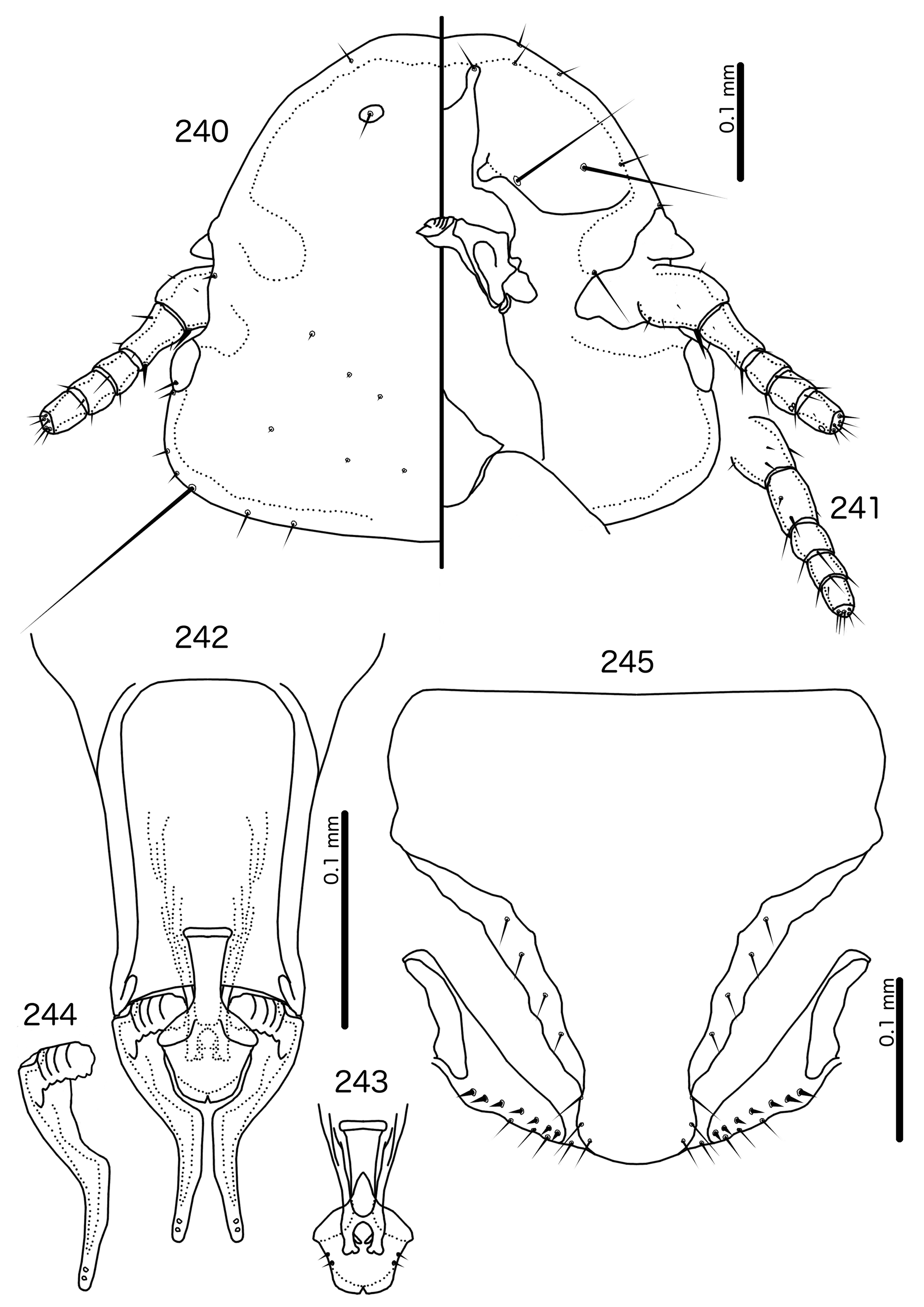
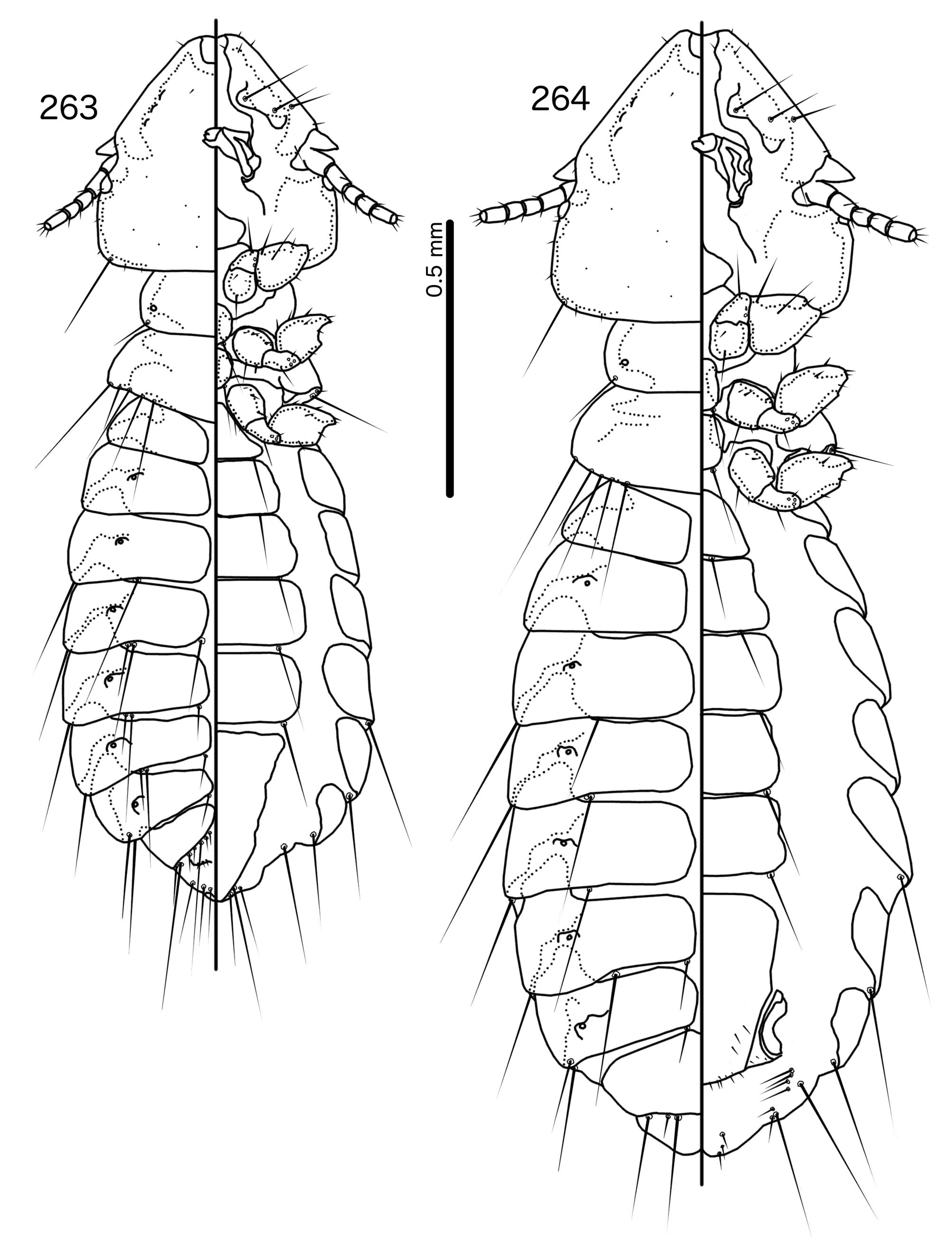
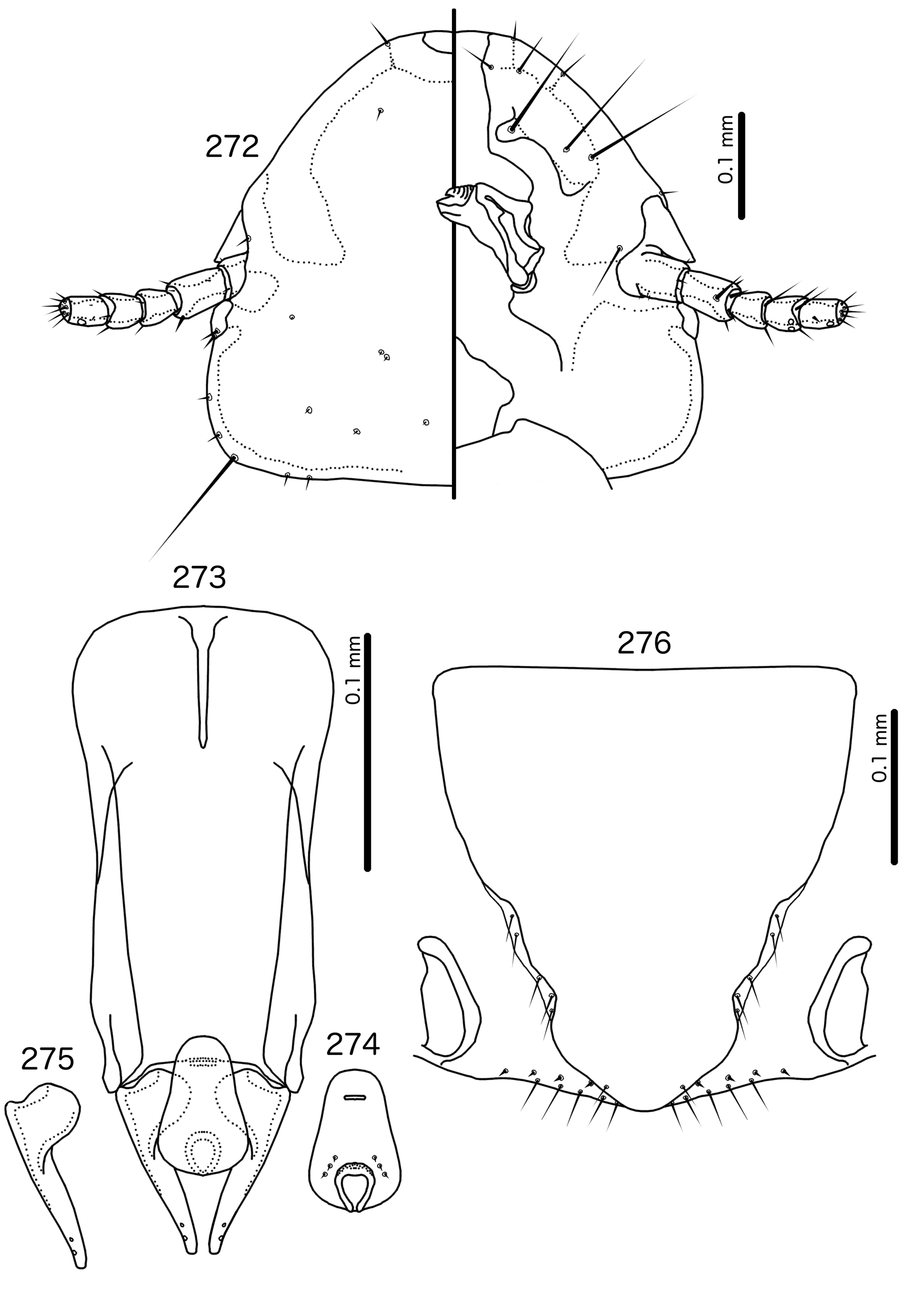
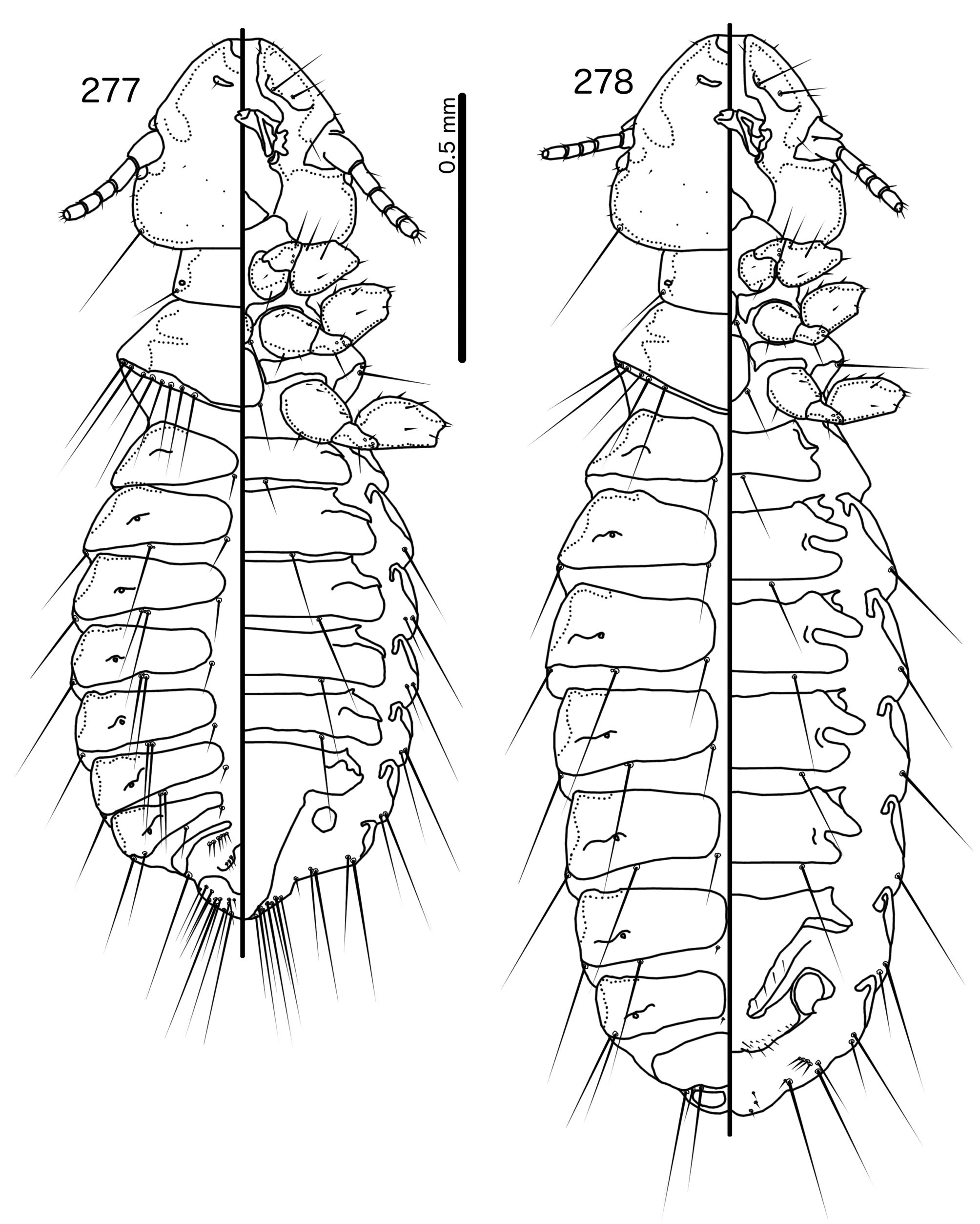
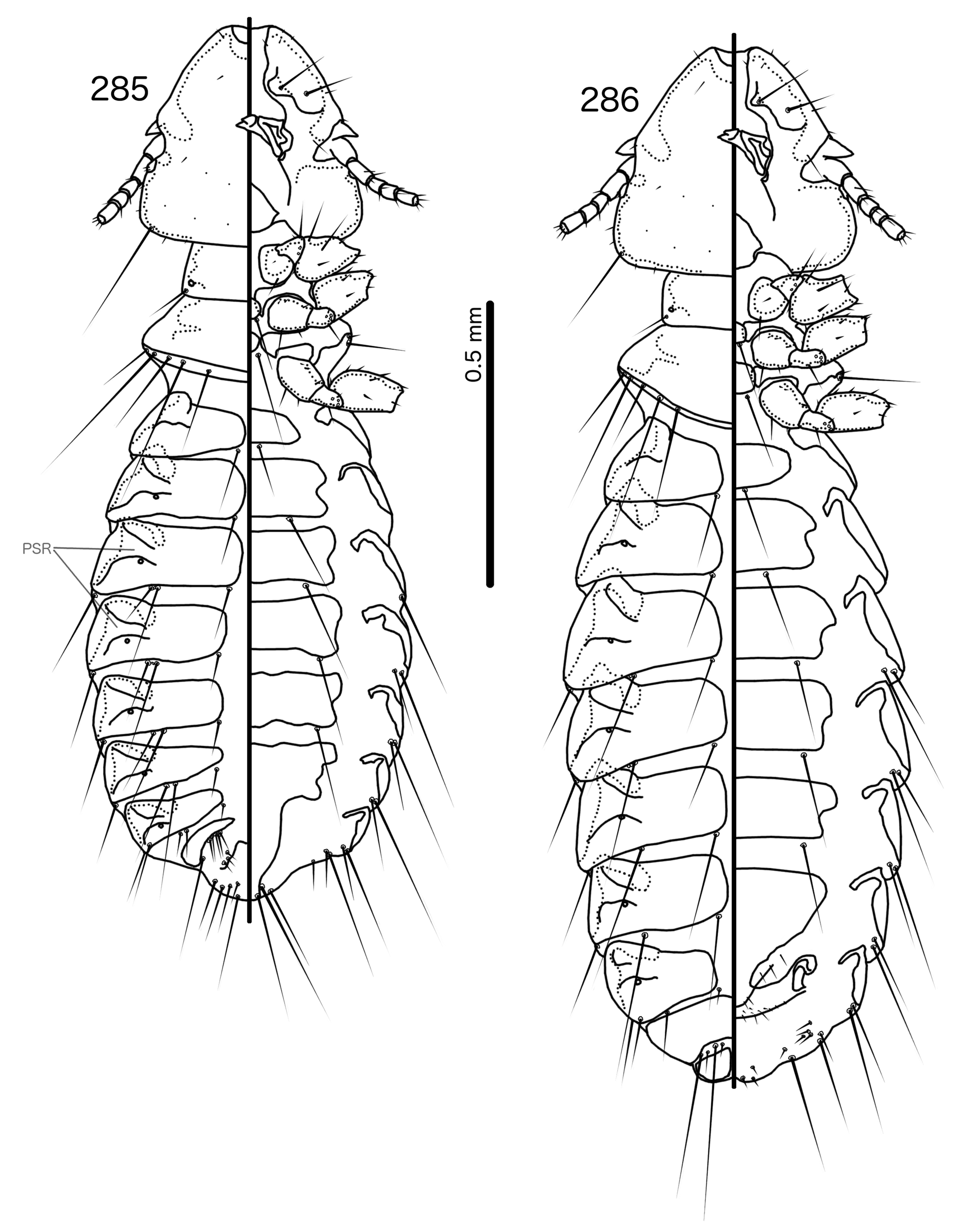
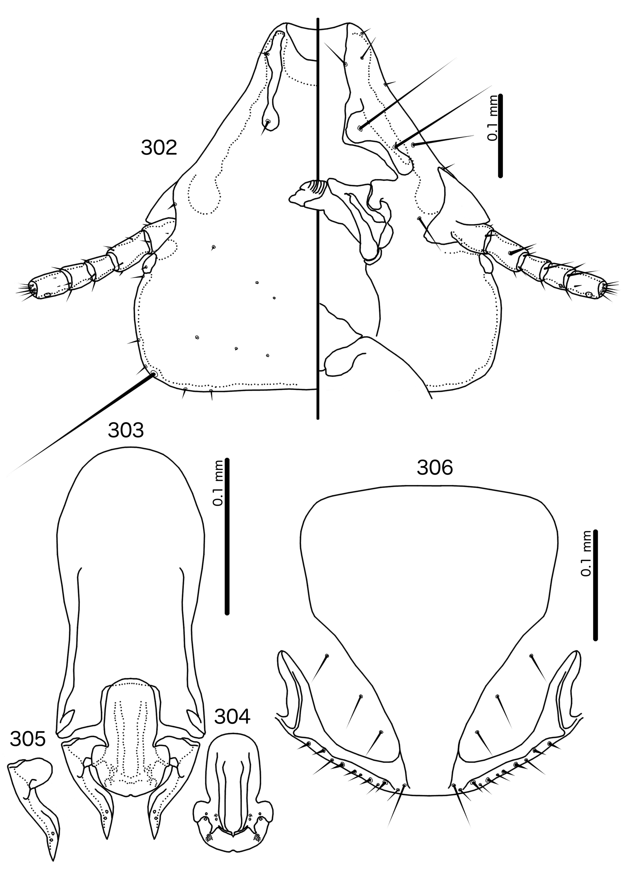
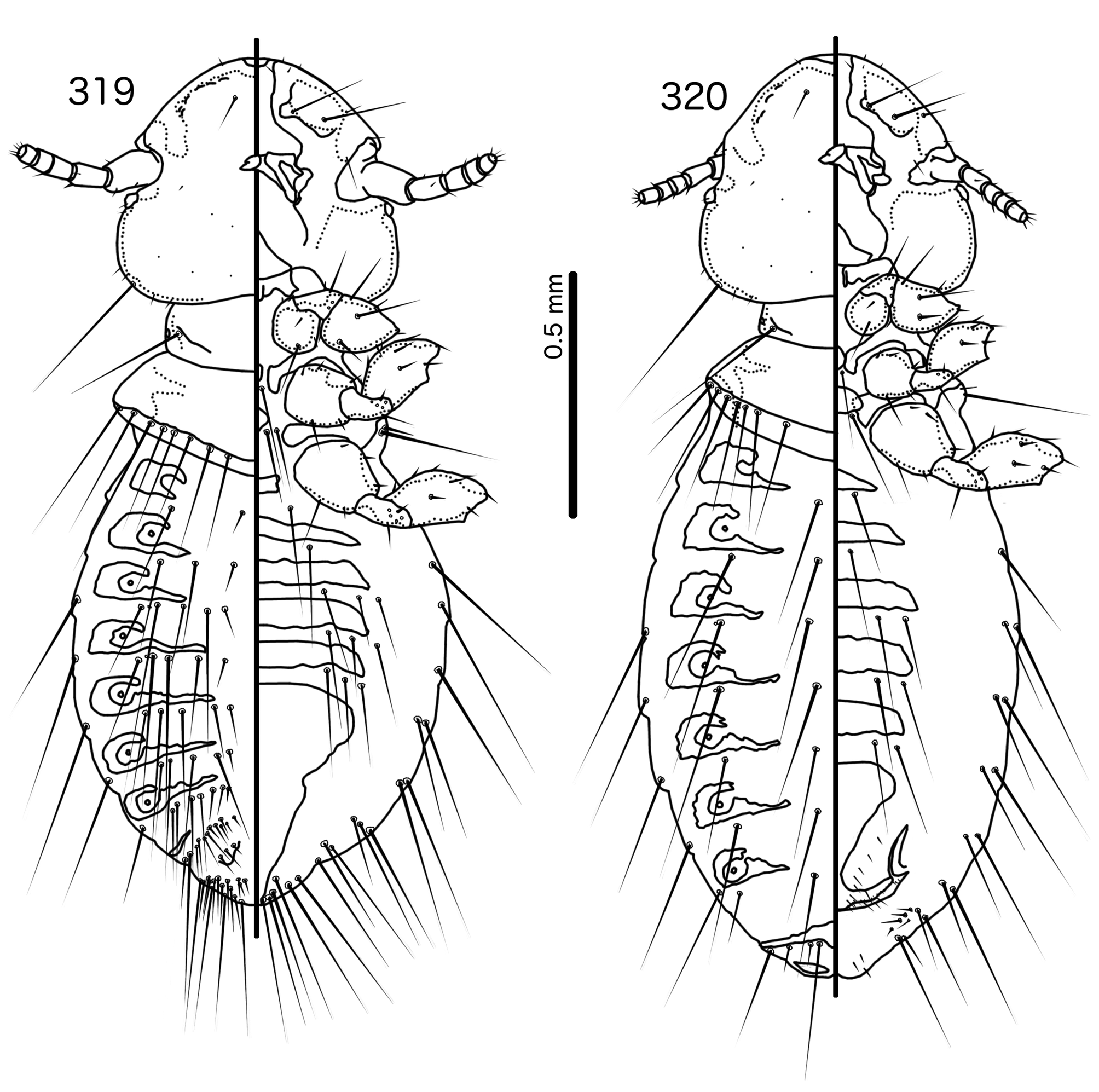
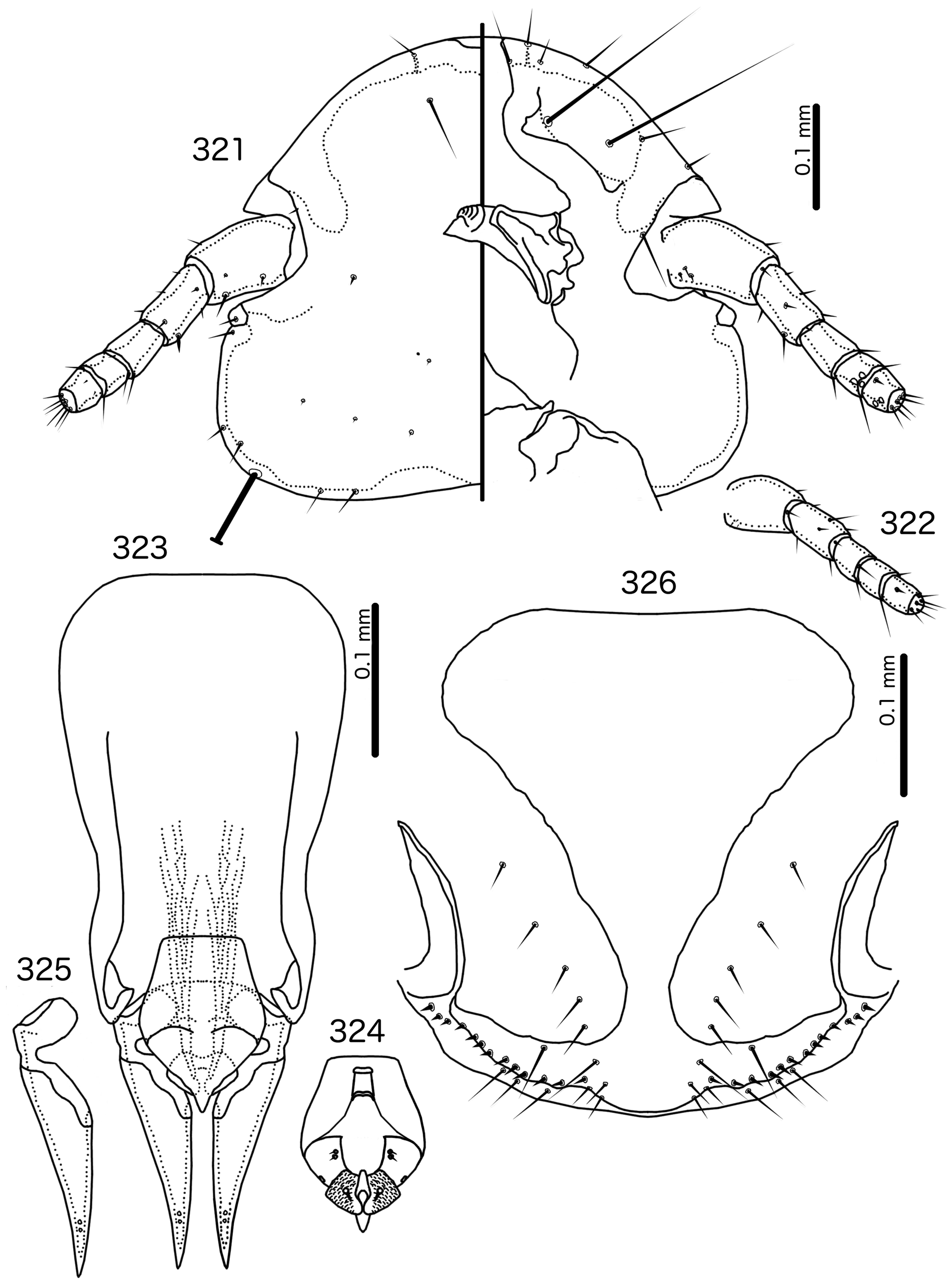
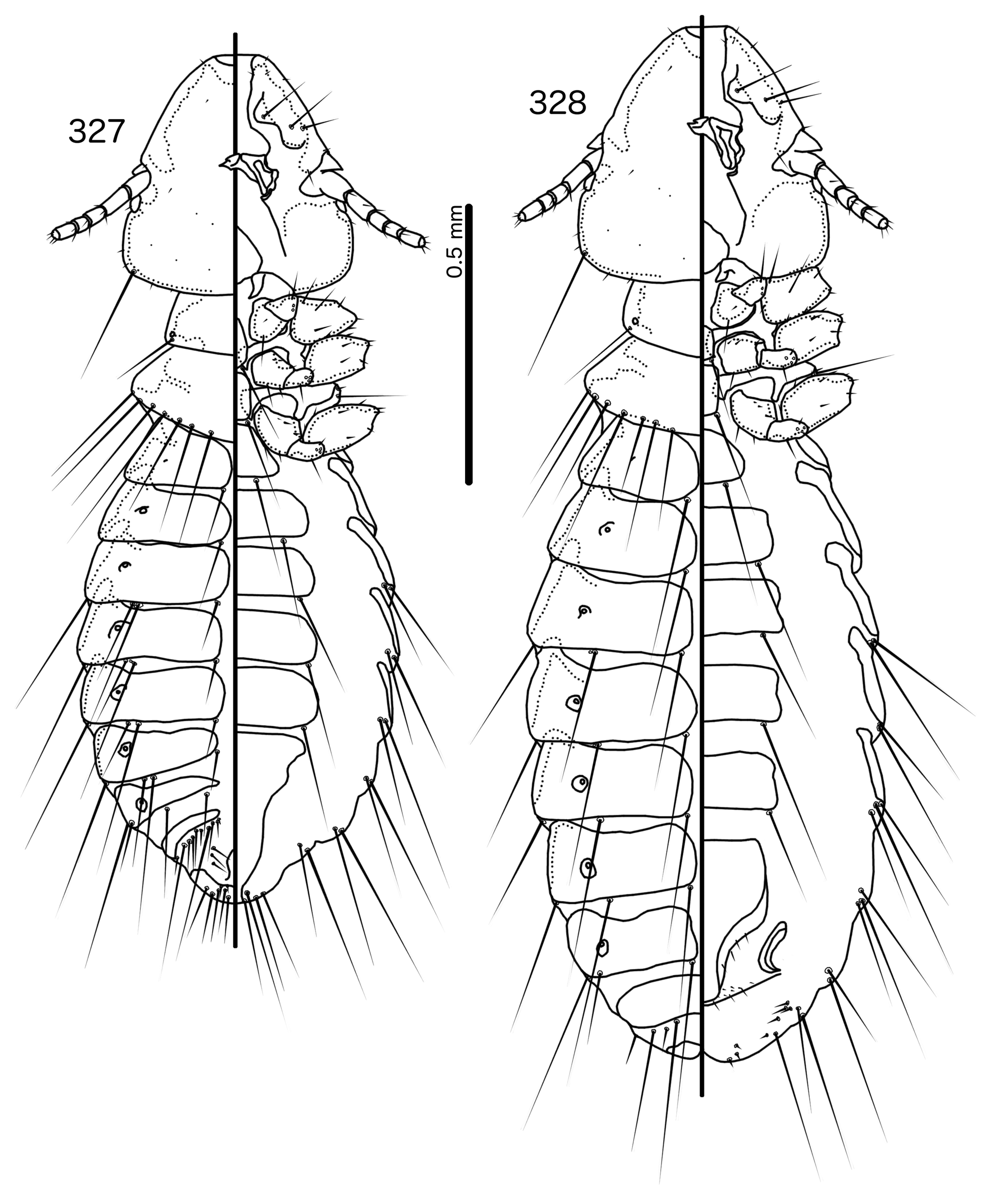
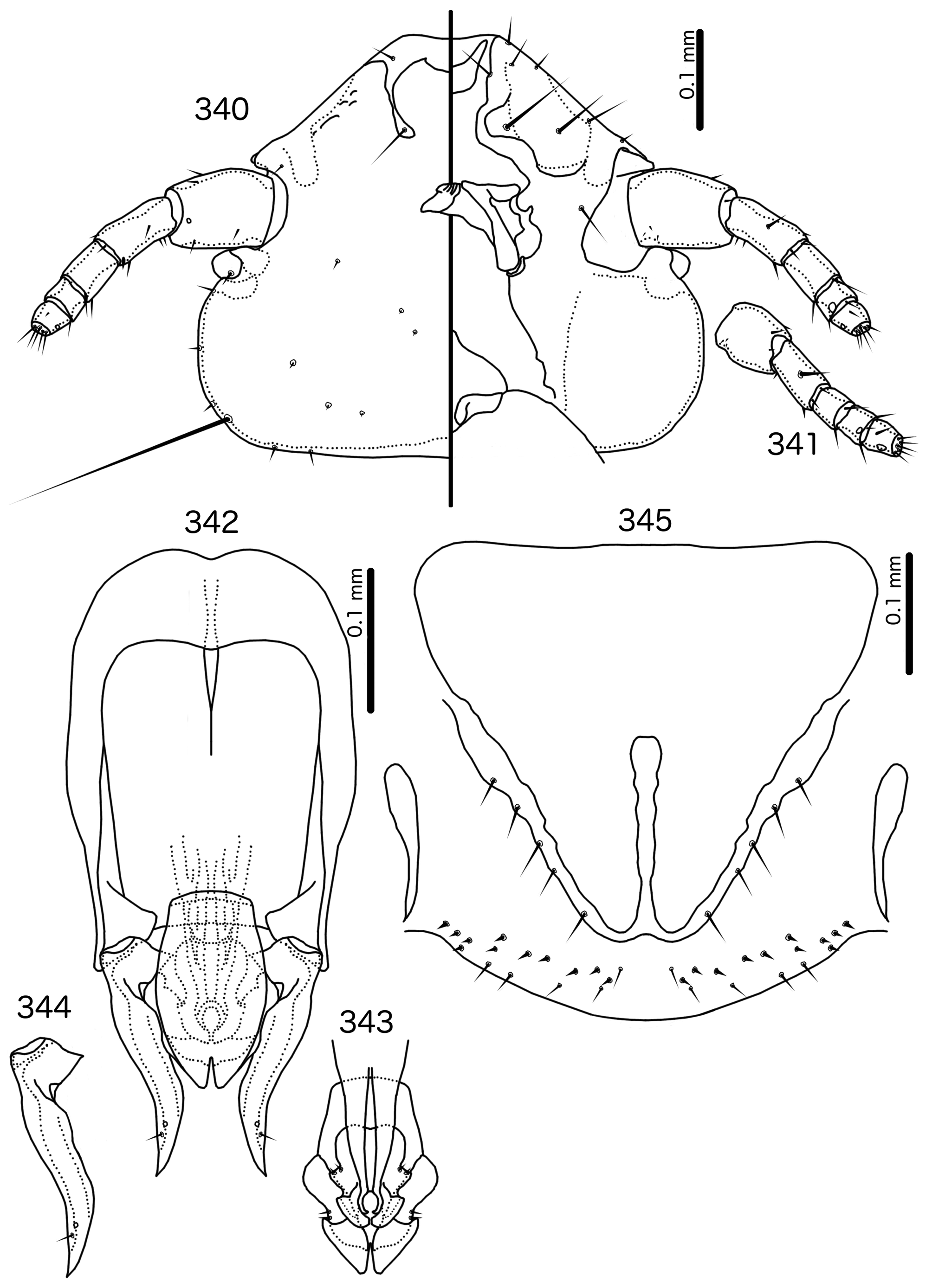
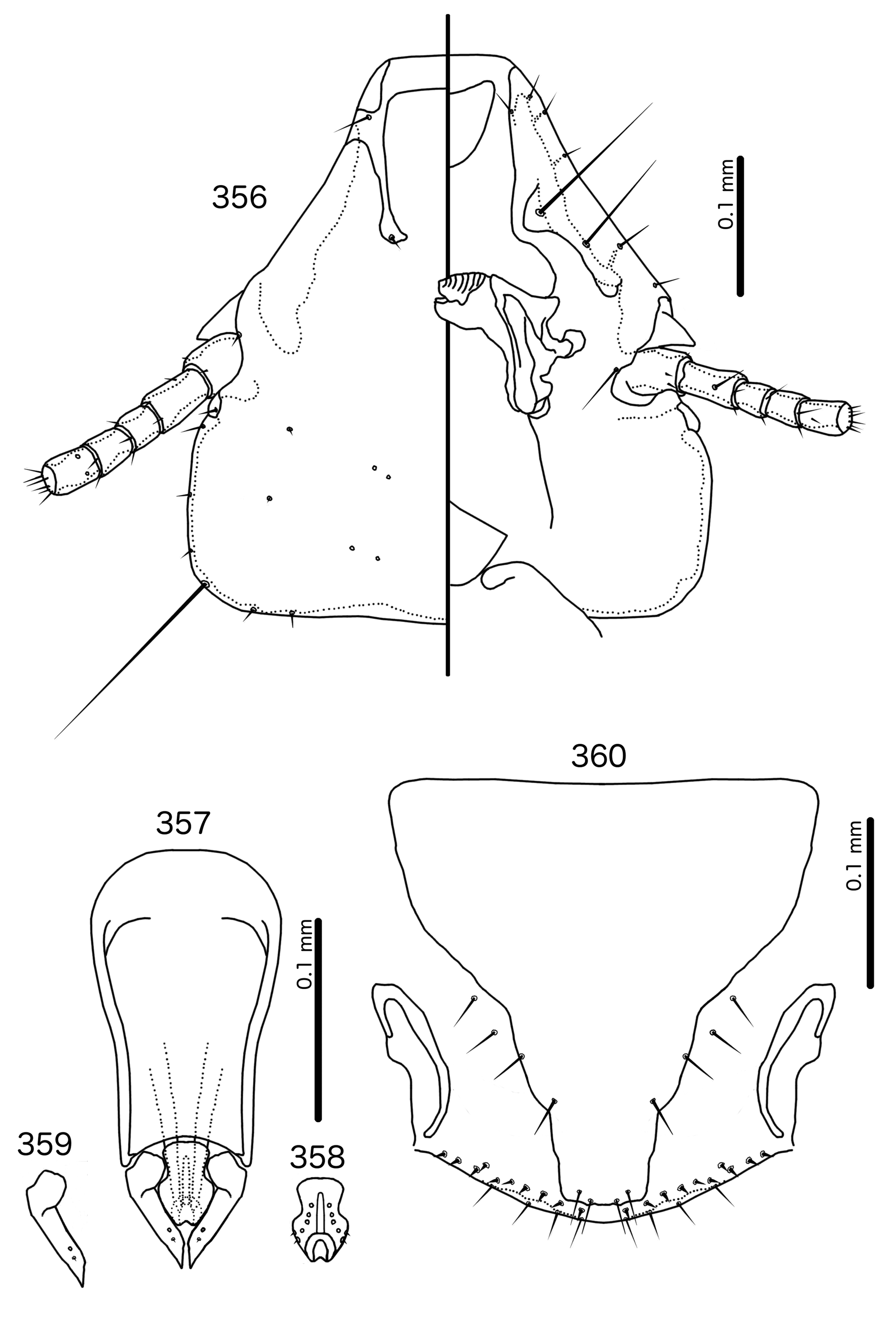
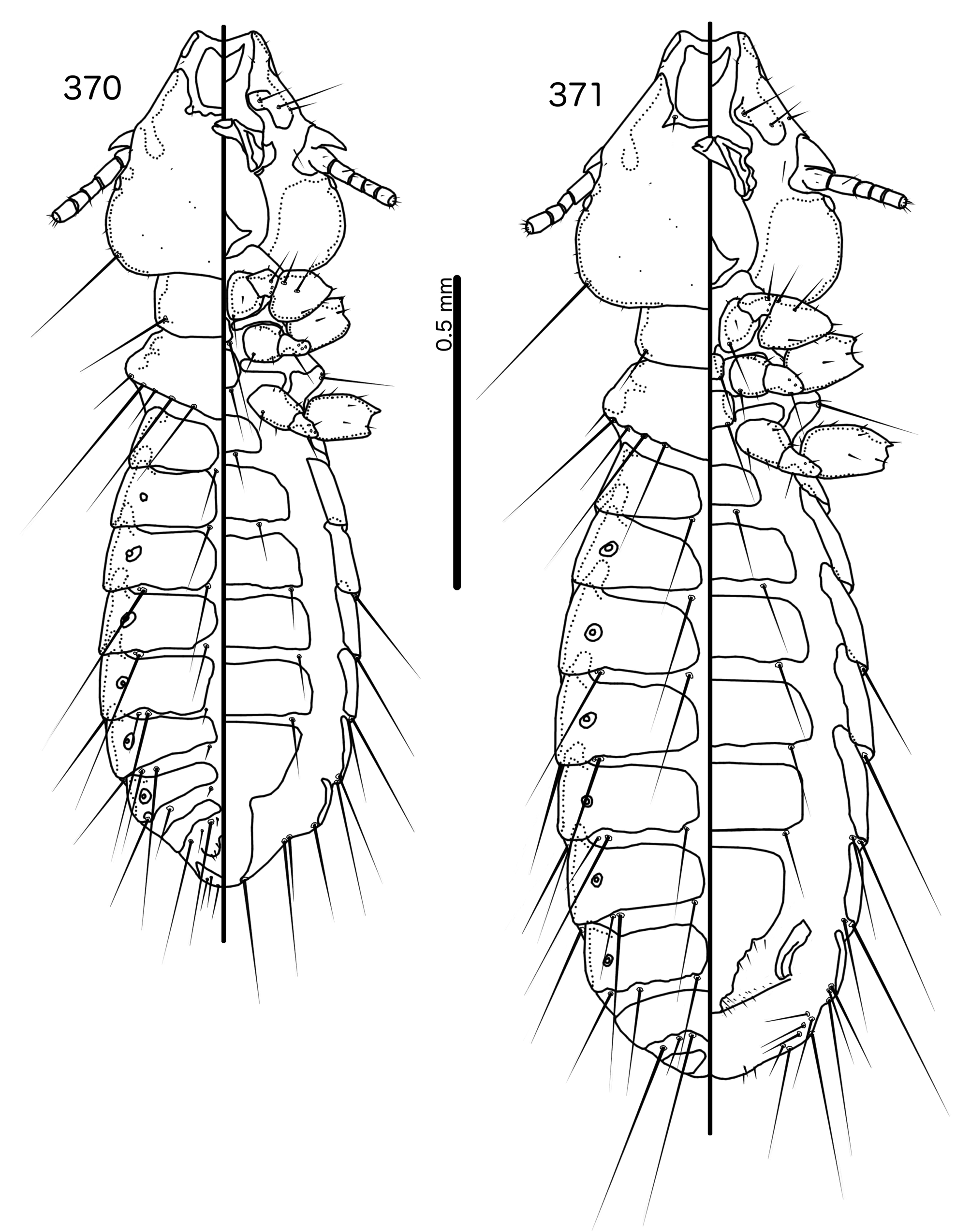
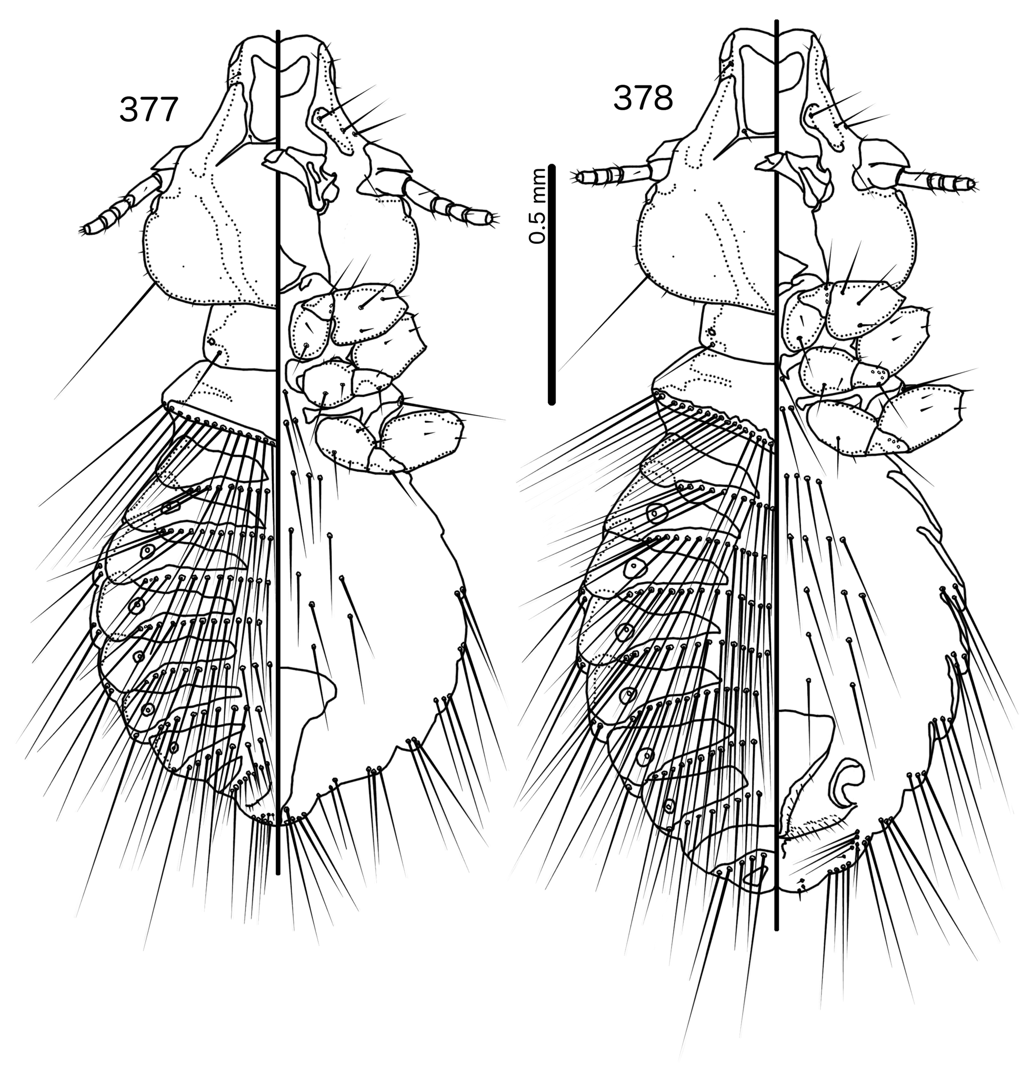
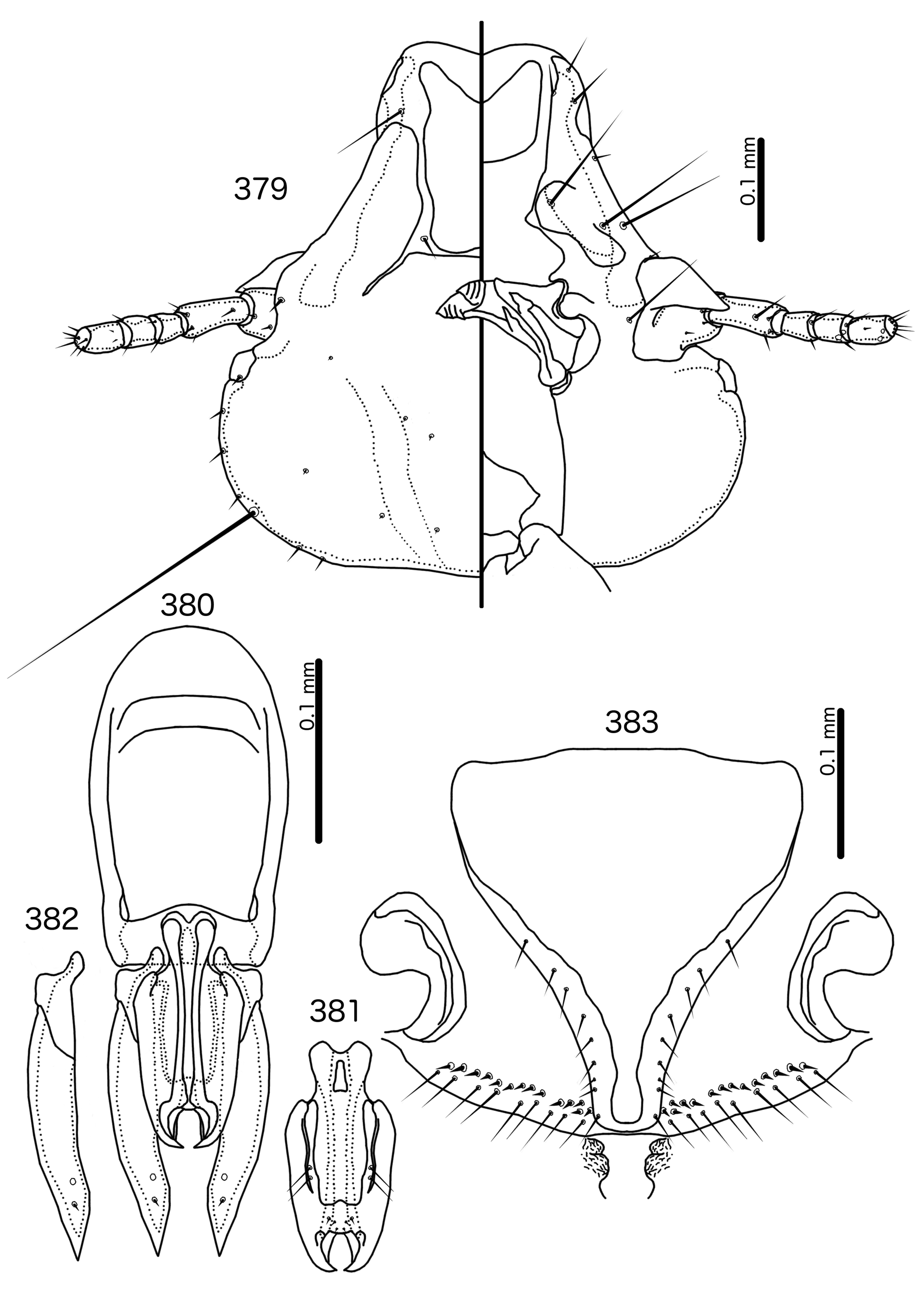
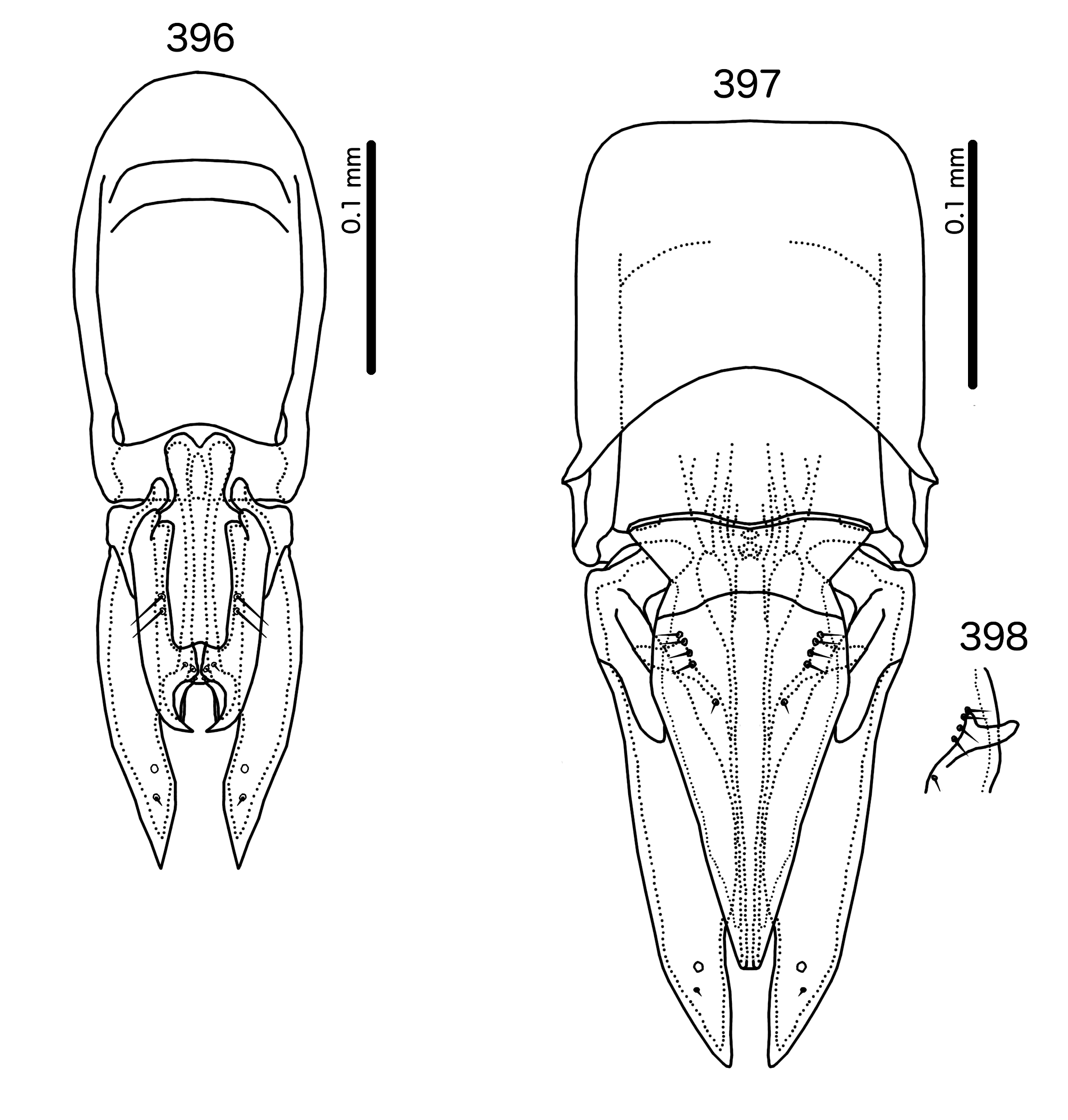
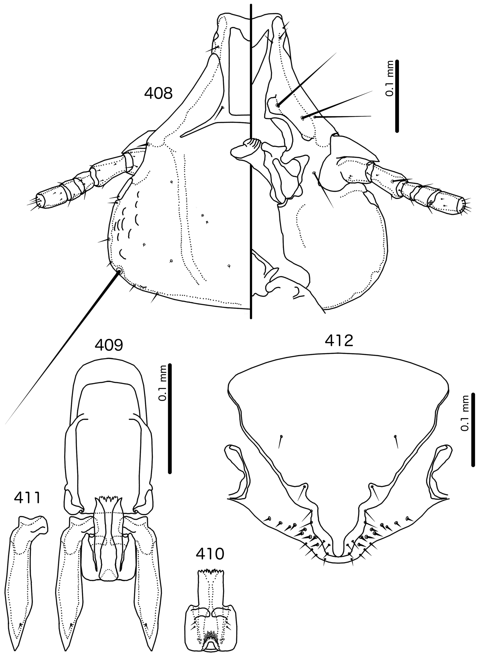
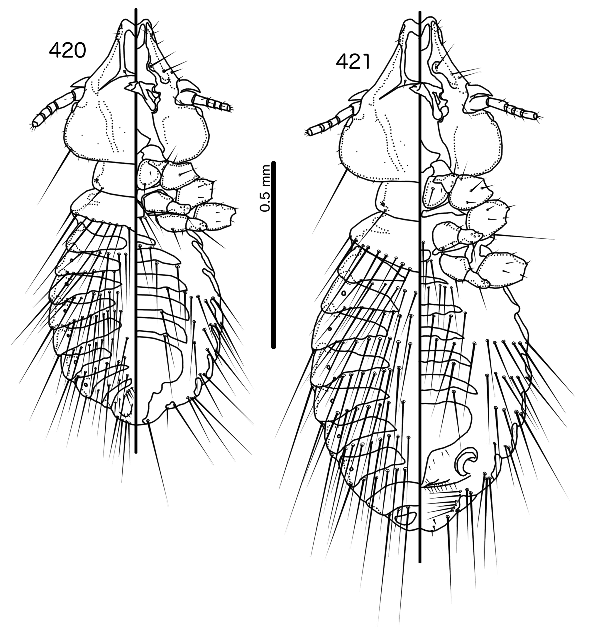
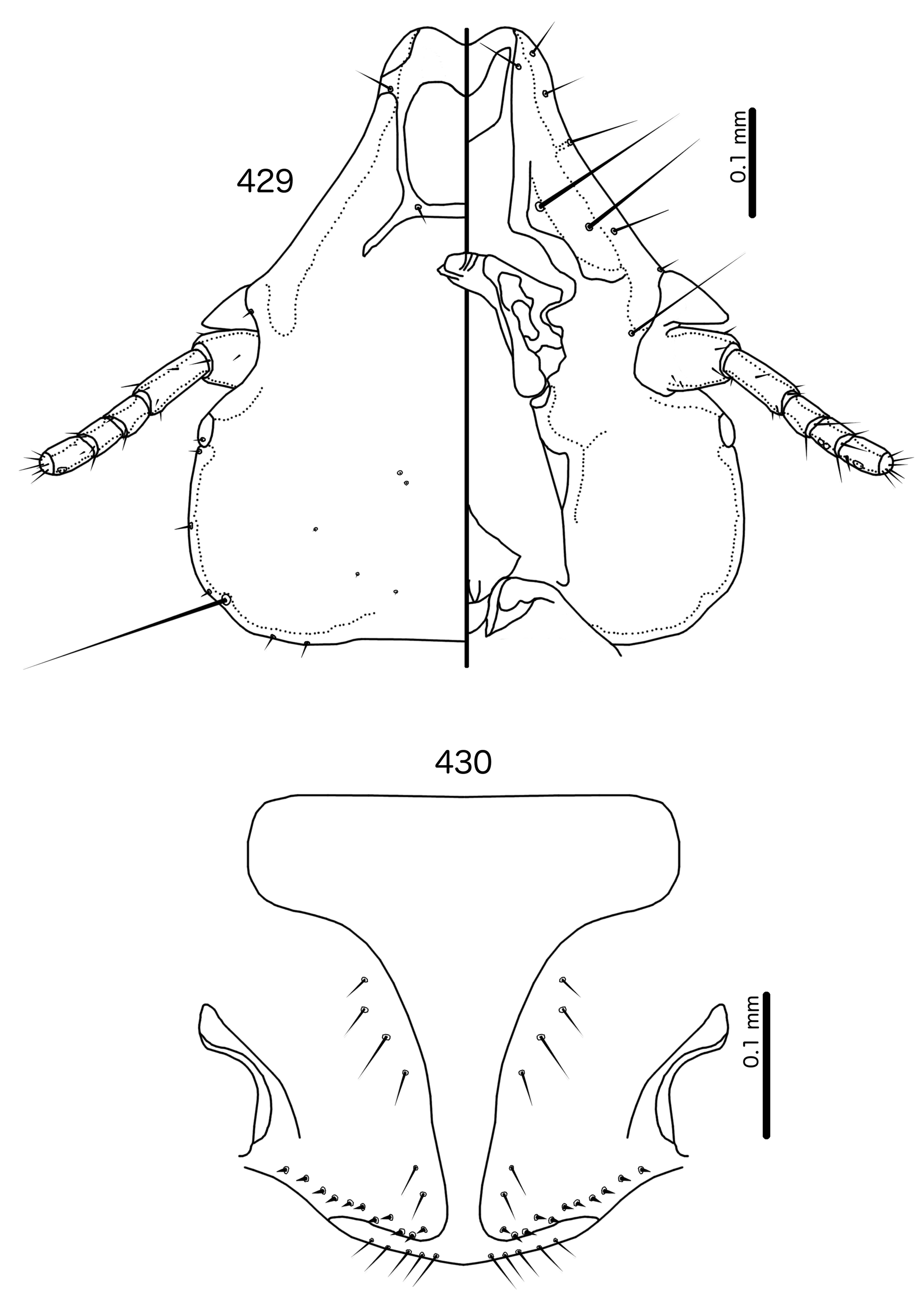
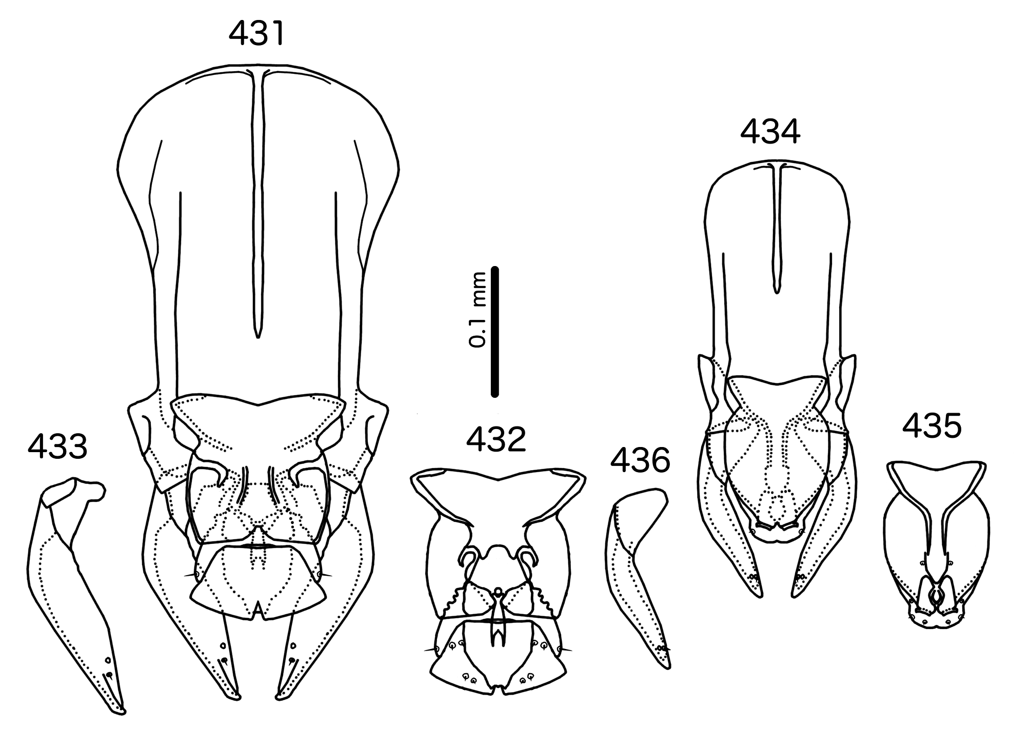
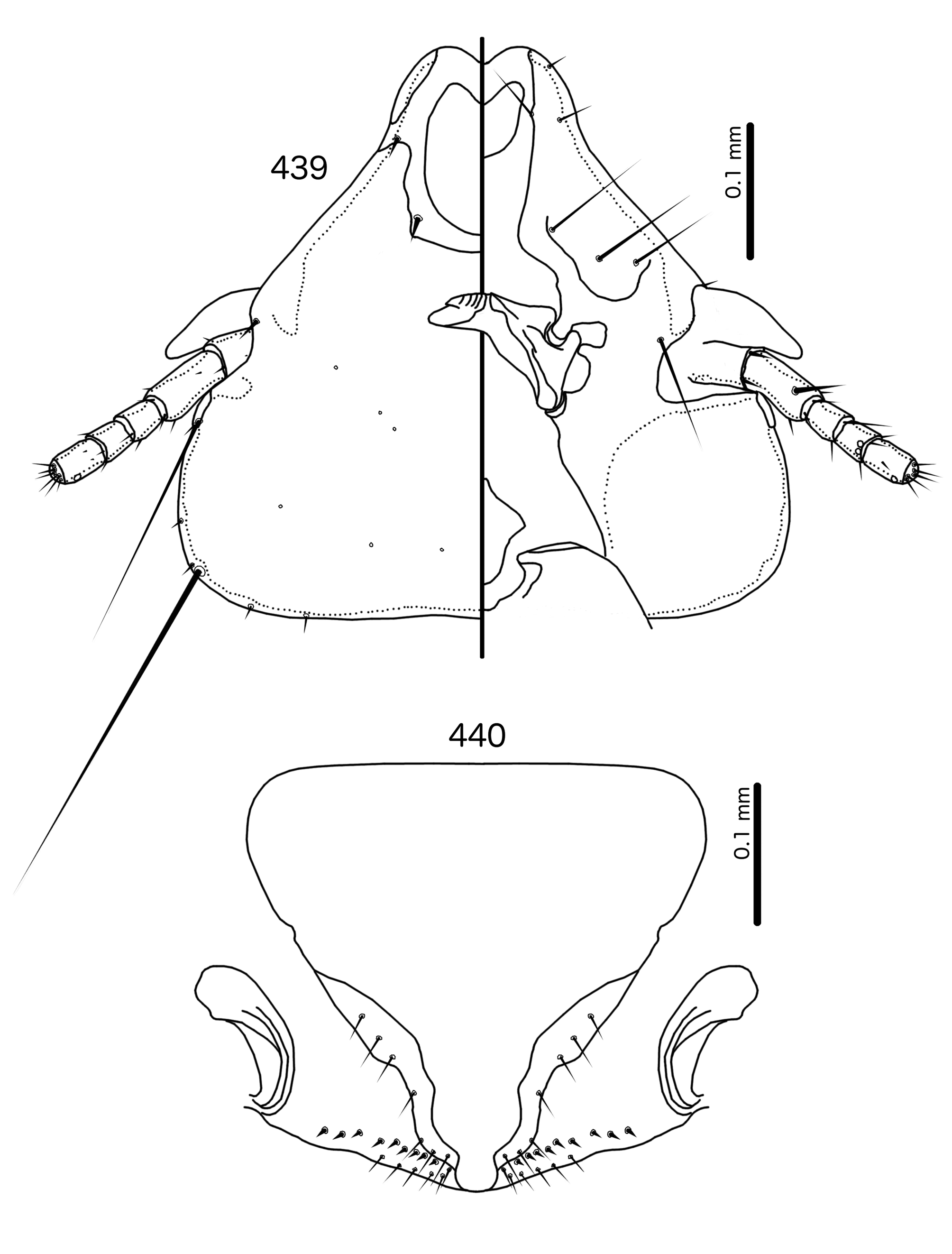
Diagnosis. Members of the genus Schizosairhynchus n. gen. ( Figs 463–476 View FIGURES 463 – 464 View FIGURES 465 – 469 View FIGURES 470 – 471 View FIGURES 472 – 476 ) share the following characters with Bizarrifrons ( Figs 477–478 View FIGURES 477 – 478 ) and Manucodicola n. gen. ( Figs 451–462 View FIGURES 451 – 452 View FIGURES 453 – 456 View FIGURES 459 – 462 ): marginal carina widely interrupted medianly ( Figs 453 View FIGURES 453 – 456 , 459 View FIGURES 459 – 462 , 465 View FIGURES 465 – 469 , 472 View FIGURES 472 – 476 , 477 View FIGURES 477 – 478 ); premarginal carina and as3 absent; frons hyaline posteriorly to site of as1 ( Figs 453 View FIGURES 453 – 456 , 459 View FIGURES 459 – 462 , 465 View FIGURES 465 – 469 , 472 View FIGURES 472 – 476 , 477 View FIGURES 477 – 478 ); male subgenital plate widens distally ( Figs 451 View FIGURES 451 – 452 , 457, 463, 470, 477); male genitalia prominent, bulky ( Figs 455 View FIGURES 453 – 456 , 461 View FIGURES 459 – 462 , 466 View FIGURES 465 – 469 , 473 View FIGURES 472 – 476 , 478 View FIGURES 477 – 478 ); vss numerous, long ( Figs 456 View FIGURES 453 – 456 , 462 View FIGURES 459 – 462 , 469 View FIGURES 465 – 469 , 476 View FIGURES 472 – 476 ); psps and aps absent on tergopleurites II–III in both sexes ( Figs 45 1–452 View FIGURES 44 – 48 View FIGURES 1 – 9 View FIGURES 10 – 18 View FIGURES 19 – 20 View FIGURES 21 – 24 View FIGURES 25 View FIGURE 26 View FIGURES 27 – 30 View FIGURES 31 – 34 View FIGURES 35 – 38 View FIGURES 39 – 41 View FIGURES 42 – 43 View FIGURES 49 – 50 View FIGURES 51 – 55 View FIGURES 56 – 57 View FIGURES 58 – 62 View FIGURES 63 – 64 View FIGURES 65 – 69 View FIGURES 70 – 74 View FIGURES 75 – 76 View FIGURES 77 – 82 View FIGURES 83 – 84 View FIGURES 85 – 89 View FIGURES 90 – 91 View FIGURES 92 – 97 View FIGURES 98 – 99 View FIGURES 100 – 105 View FIGURES 106 – 107 View FIGURES 108 – 113 View FIGURES 114 – 115 View FIGURES 116 – 121 View FIGURES 122 – 123 View FIGURES 124 – 129 View FIGURES 130 – 131 View FIGURES 132 – 137 View FIGURES 138 – 139 View FIGURES 140 – 145 View FIGURES 146 – 147 View FIGURES 148 – 152 View FIGURES 153 – 154 View FIGURES 155 – 160 View FIGURES 161 – 162 View FIGURES 163 – 167 View FIGURES 168 – 169 View FIGURES 170 – 174 View FIGURES 175 – 176 View FIGURES 177 – 181 View FIGURES 182 – 183 View FIGURES 184 – 188 View FIGURES 189 – 190 View FIGURES 191 – 195 View FIGURES 196 – 197 View FIGURES 198 – 202 View FIGURES 203 – 204 View FIGURES 210 – 211 View FIGURES 212 – 216 View FIGURES 217 – 218 View FIGURES 219 – 223 View FIGURES 224 – 225 View FIGURES 226 – 230 View FIGURES 231 – 232 View FIGURES 233 – 237 View FIGURES 238 – 239 View FIGURES 240 – 245 View FIGURES 246 – 247 View FIGURES 248 – 252 View FIGURES 253 – 254 View FIGURES 260 – 262 View FIGURES 263 – 264 View FIGURES 265 – 269 View FIGURES 270 – 271 View FIGURES 272 – 276 View FIGURES 277 – 278 View FIGURES 279 – 284 View FIGURES 285 – 286 View FIGURES 287 – 291 View FIGURES 292 – 293 View FIGURES 294 – 299 View FIGURES 300 – 301 View FIGURES 302 – 306 View FIGURES 307 – 308 View FIGURES 309 – 314 View FIGURES 315 – 318 View FIGURES 319 – 320 View FIGURES 321 – 326 View FIGURES 327 – 328 View FIGURES 334 – 337 View FIGURES 338 – 339 View FIGURES 340 – 345 View FIGURES 346 – 347 View FIGURES 348 – 353 View FIGURES 354 – 355 View FIGURES 356 – 360 View FIGURES 361 – 364 View FIGURES 365 – 369 View FIGURES 370 – 371 View FIGURES 372 – 376 View FIGURES 377 – 378 View FIGURES 379 – 383 View FIGURES 384 – 389 View FIGURES 390 – 395 View FIGURES 396 – 398 View FIGURES 399 – 400 View FIGURES 401 – 405 View FIGURES 406 – 407 View FIGURES 408 – 412 View FIGURES 413 – 414 View FIGURES 415 – 419 View FIGURES 420 – 421 View FIGURES 422 – 426 View FIGURES 427 – 428 View FIGURES 429 – 430 View FIGURES 431 – 436 View FIGURES 437 – 438 View FIGURES 439 – 440 View FIGURES 441 – 450 View FIGURES 451 – 452 , 457–458, 463–464, 470–471, 477). Unlike Schizosairhynchus , the otherwise similar genera Sturnidoecus ( Figs 377–426 View FIGURES 377 – 378 View FIGURES 379 – 383 View FIGURES 384 – 389 View FIGURES 390 – 395 View FIGURES 396 – 398 View FIGURES 399 – 400 View FIGURES 401 – 405 View FIGURES 406 – 407 View FIGURES 408 – 412 View FIGURES 413 – 414 View FIGURES 415 – 419 View FIGURES 420 – 421 View FIGURES 422 – 426 ), Rostrinirmus ( Figs 437–450 View FIGURES 437 – 438 View FIGURES 439 – 440 View FIGURES 441 – 450 ) and Buphagoecus n. gen. ( Figs 427–436 View FIGURES 427 – 428 View FIGURES 429 – 430 View FIGURES 431 – 436 ) have the following characters: premarginal carina present; frons hyaline only median to the premarginal carina; psps or aps present on at least tergopleurite III in males (also occurring on other tergopleurites and on female tergopleurite III in some species; see the entries for these genera for details).
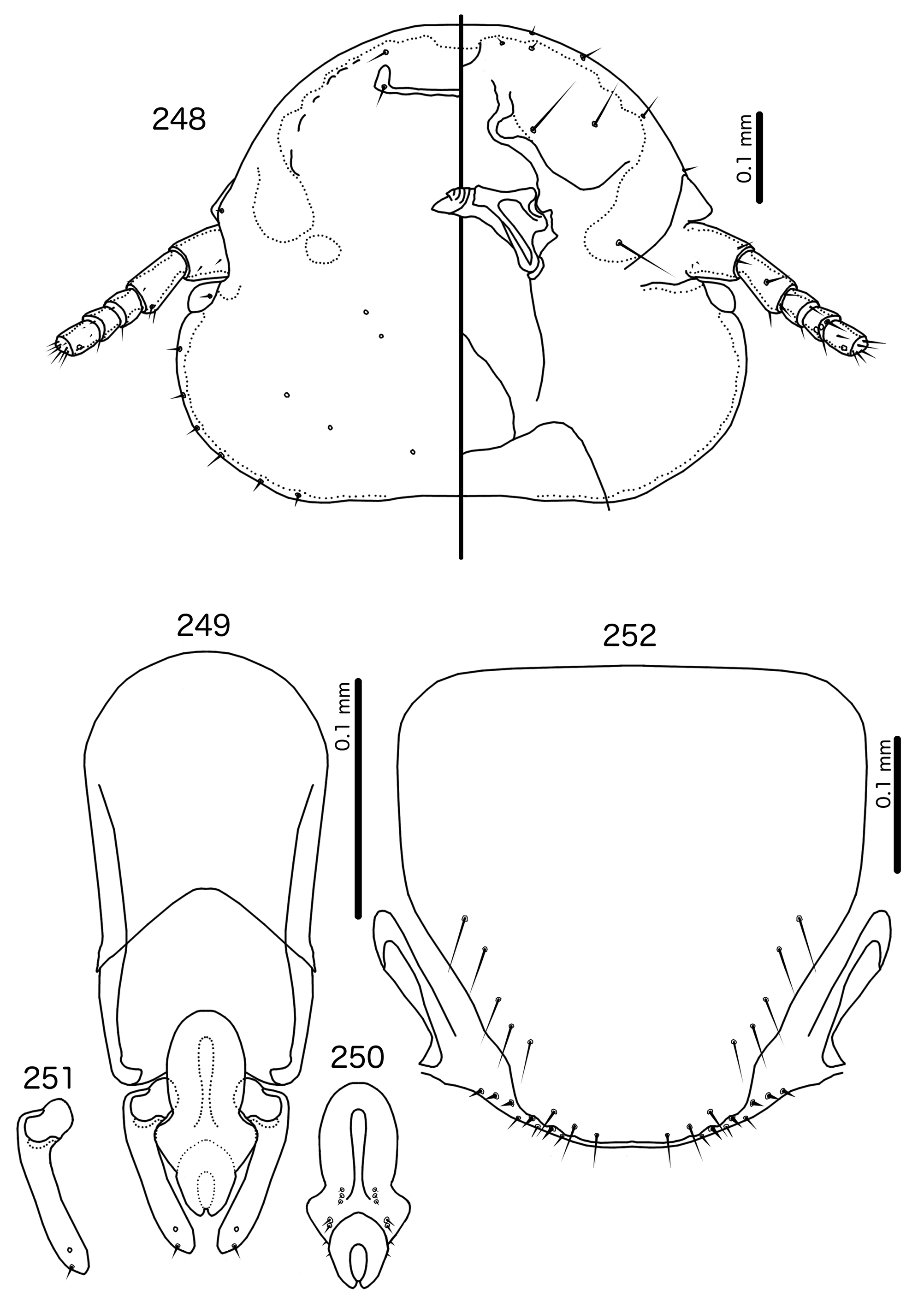
Schizosairhynchus is separated from Manucodicola and Bizarrifrons by the following characters: preantennal area asymmetrical in Bizarrifrons ( Fig. 477 View FIGURES 477 – 478 ) and Manucodicola ( Figs 454 View FIGURES 453 – 456 , 460 View FIGURES 459 – 462 ), but symmetrical in Schizosairhynchus ( Figs 465 View FIGURES 465 – 469 , 472 View FIGURES 472 – 476 ); dorsal anterior plate connected to main head plate in Bizarrifrons ( Fig. 377 View FIGURES 377 – 378 ) and Manucodicola ( Figs 453 View FIGURES 453 – 456 , 459 View FIGURES 459 – 462 ), but completely separated from main head plate and extended posteriorly into a pointed horn that overlaps with main head plate in Schizosairhynchus ( Figs 465 View FIGURES 465 – 469 , 472 View FIGURES 472 – 476 ); ppss of Schizosairhynchus located on the medio-posterior margin of prothorax ( Figs 463–464 View FIGURES 463 – 464 , 470–471 View FIGURES 470 – 471 ), but on the postero-lateral corners in Bizarrifrons ( Fig. 477 View FIGURES 477 – 478 ) and Manucodicola ( Figs 451–452 View FIGURES 451 – 452 , 457–458); posterior margin of pterothorax with median indentation in Schizosairhynchus ( Figs 463–464 View FIGURES 463 – 464 , 470–471 View FIGURES 470 – 471 ), but without such indentation in Manucodicola ( Figs 451–452 View FIGURES 451 – 452 , 457–458) and Bizarrifrons ( Fig. 477 View FIGURES 477 – 478 ); sternal plate II extended laterally in Schizosairhynchus ( Figs 463– 464 View FIGURES 463 – 464 , 470–471 View FIGURES 470 – 471 ), but not in Manucodicola ( Figs 451–452 View FIGURES 451 – 452 , 457–458) or Bizarrifrons ( Fig. 477 View FIGURES 477 – 478 ). The proximal mesosome substantially overlaps with basal apodeme in Schizosairhynchus ( Figs 466 View FIGURES 465 – 469 , 473 View FIGURES 472 – 476 ) and Bizarrifrons ( Fig. 477 View FIGURES 477 – 478 ), but this is not the case in Manucodicola ( Figs 455 View FIGURES 453 – 456 , 461 View FIGURES 459 – 462 ). However, like in Manucodicola ( Figs 455 View FIGURES 453 – 456 , 461 View FIGURES 459 – 462 ), the gonopore is located ventrally and the mesosomal lobes are fused distally in Schizosairhynchus ( Figs 467 View FIGURES 465 – 469 , 474 View FIGURES 472 – 476 ); in contrast, the gonopore is terminal and the mesosomal lobes are not fused distally in Bizarrifrons ( Fig. 378 View FIGURES 377 – 378 ). Rugose nodi and a ventral sclerite are present in Schizosairhynchus ( Figs 467 View FIGURES 465 – 469 , 474 View FIGURES 472 – 476 ), but absent in the two other genera ( Figs 455 View FIGURES 453 – 456 , 461 View FIGURES 459 – 462 , 478 View FIGURES 477 – 478 ). Female Schizosairhynchus ( Figs 469 View FIGURES 465 – 469 , 476 View FIGURES 472 – 476 ) have vos located on subgenital plate, whereas the vos of almost all other genera treated here, including Manucodicola ( Figs 456 View FIGURES 453 – 456 , 462 View FIGURES 459 – 462 ) and Bizarrifrons (not illustrated) are located lateral to the subgenital plate.
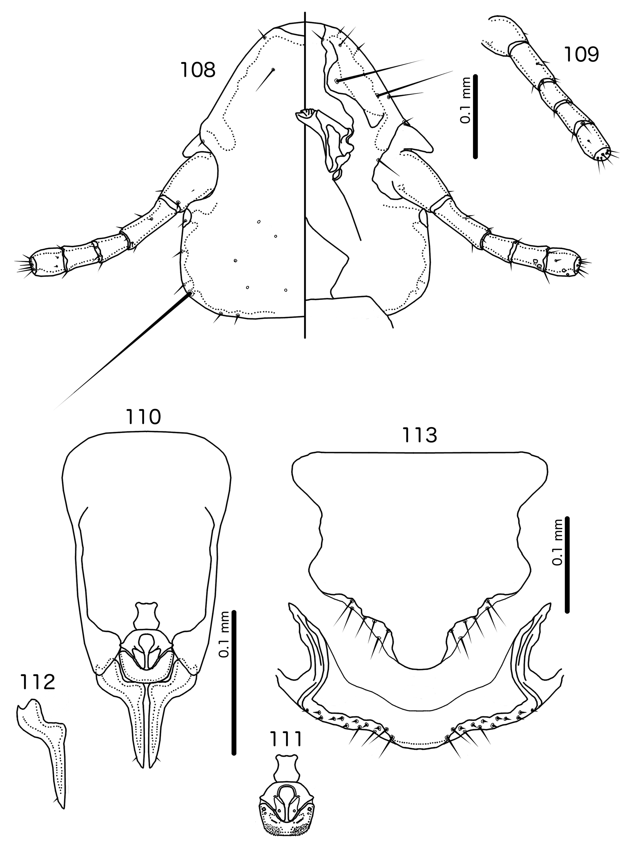
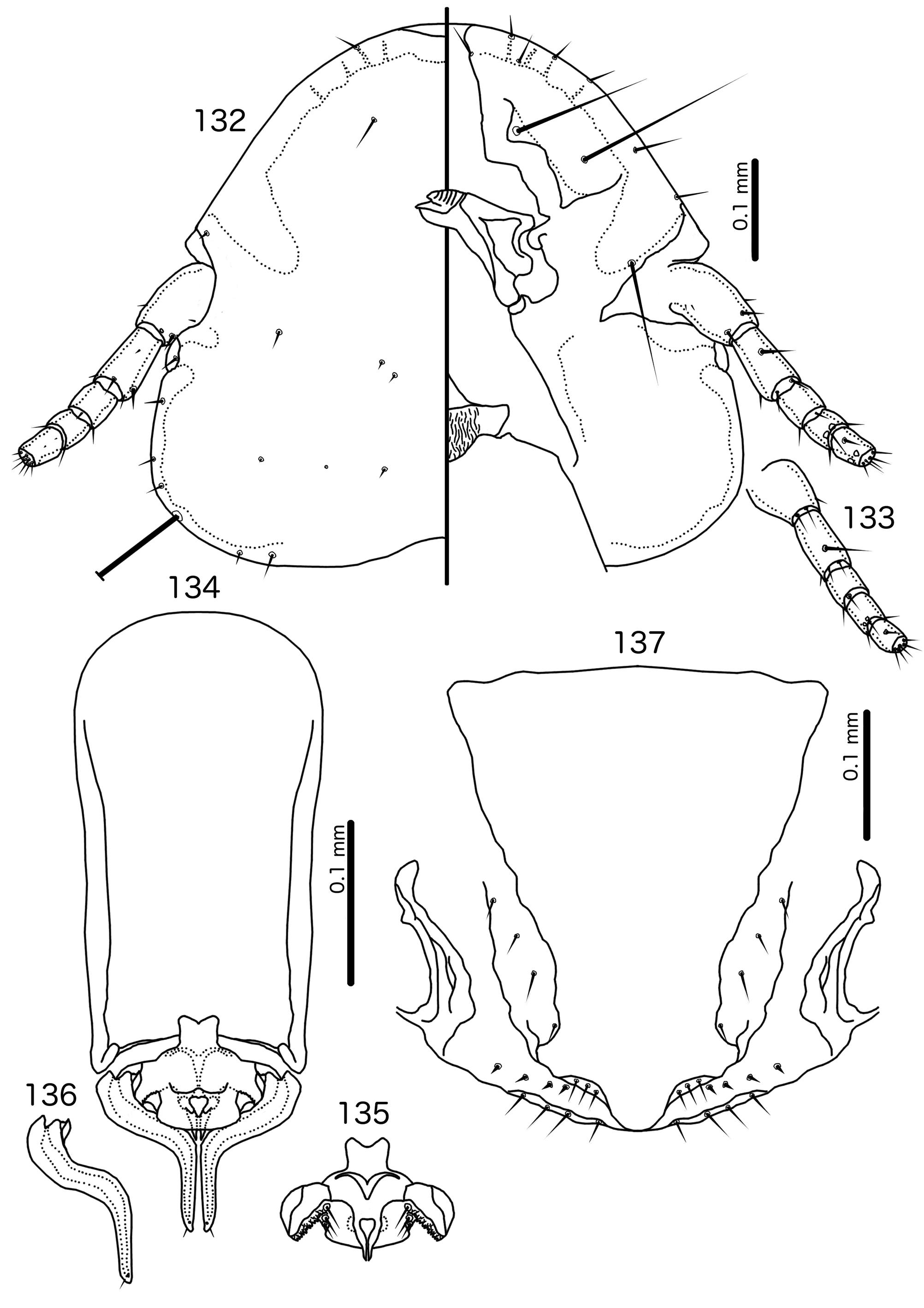
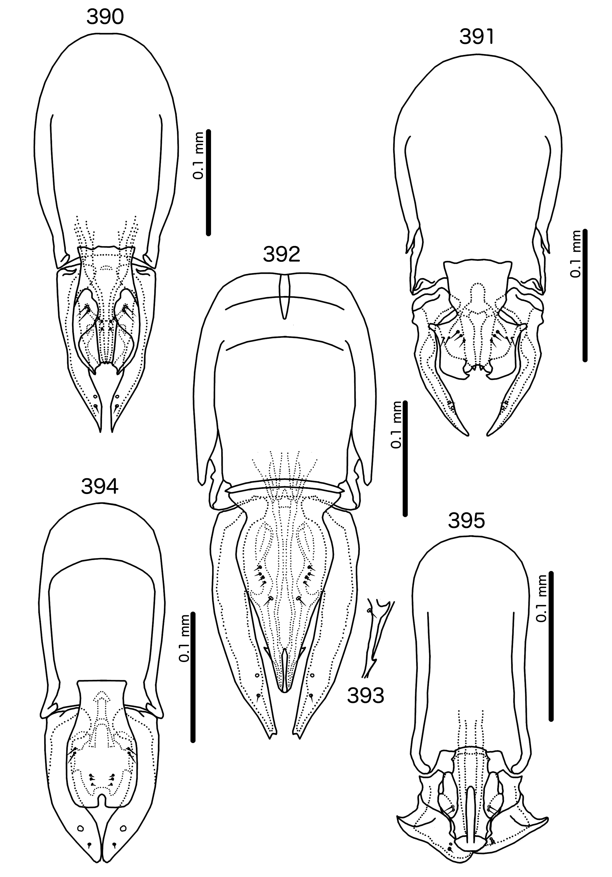
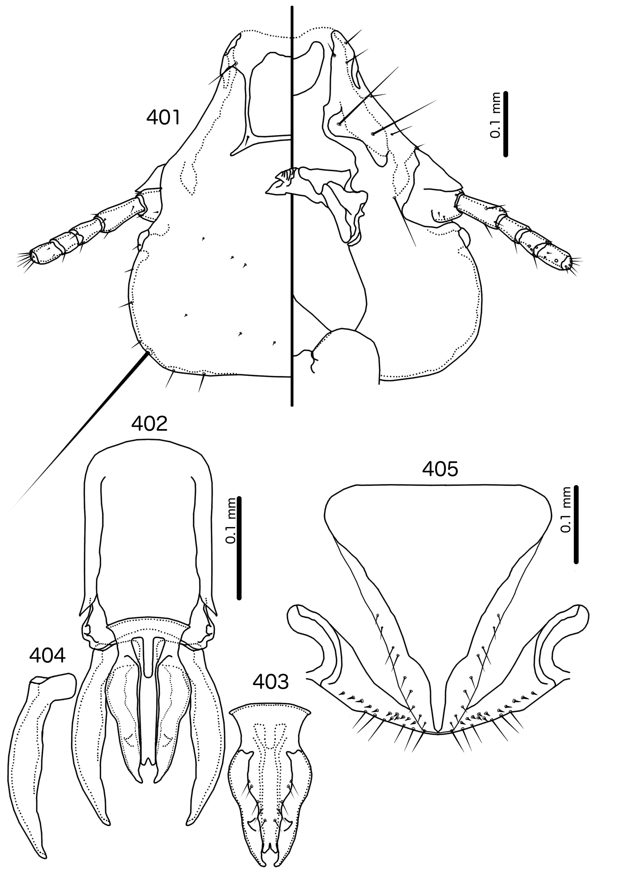
Description. Both sexes. Head bulb-shaped ( Figs 465 View FIGURES 465 – 469 , 472 View FIGURES 472 – 476 ), preantennal area narrow. Frons deeply concave, hyaline. Hyaline margin present laterally, continuous with anterior fleshy lobes. Marginal carina widely interrupted medianly. Premarginal carina absent. Ventral carinae visible to anterior end of head, extending farther anterior than marginal carina. Dorsal preantennal suture continuous with hyaline margin, extending posteriorly to position of ads, and medianly continuous, separating dorsal anterior plate from main head plate. Dorsal anterior plate elongated, anterior margin concave, posterior margin with distinct hardened extension that covers suture and continues posterior to suture, overlapping with main head plate. Ventral anterior plate absent. Head setae as in Fig. 465 View FIGURES 465 – 469 , 472 View FIGURES 472 – 476 ; as3 absent; ads and dsms often thorn-like; pts and pns sensilla, often hard to see. Coni long, slender, reaching beyond distal margin of scapes. Antennae monomorphic. Temporal carinae visible; mts 3 only macrosetae. Gular plate small, shape varying between species.
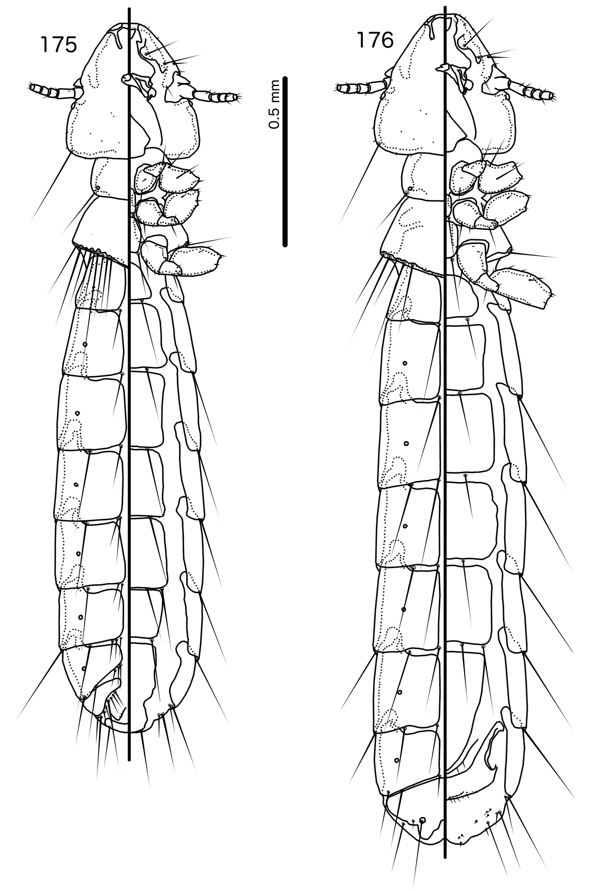
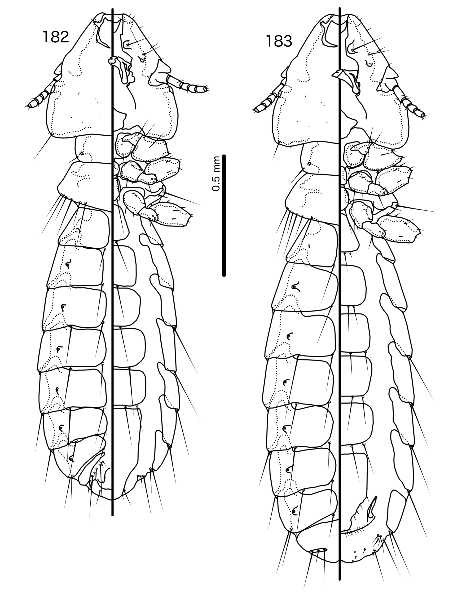
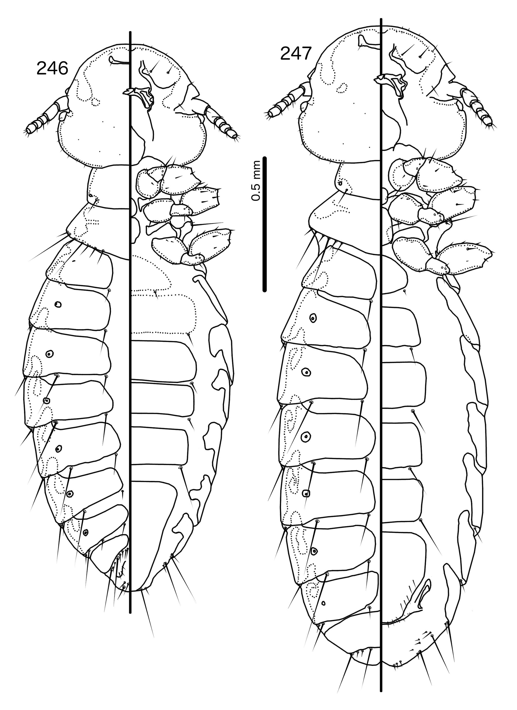
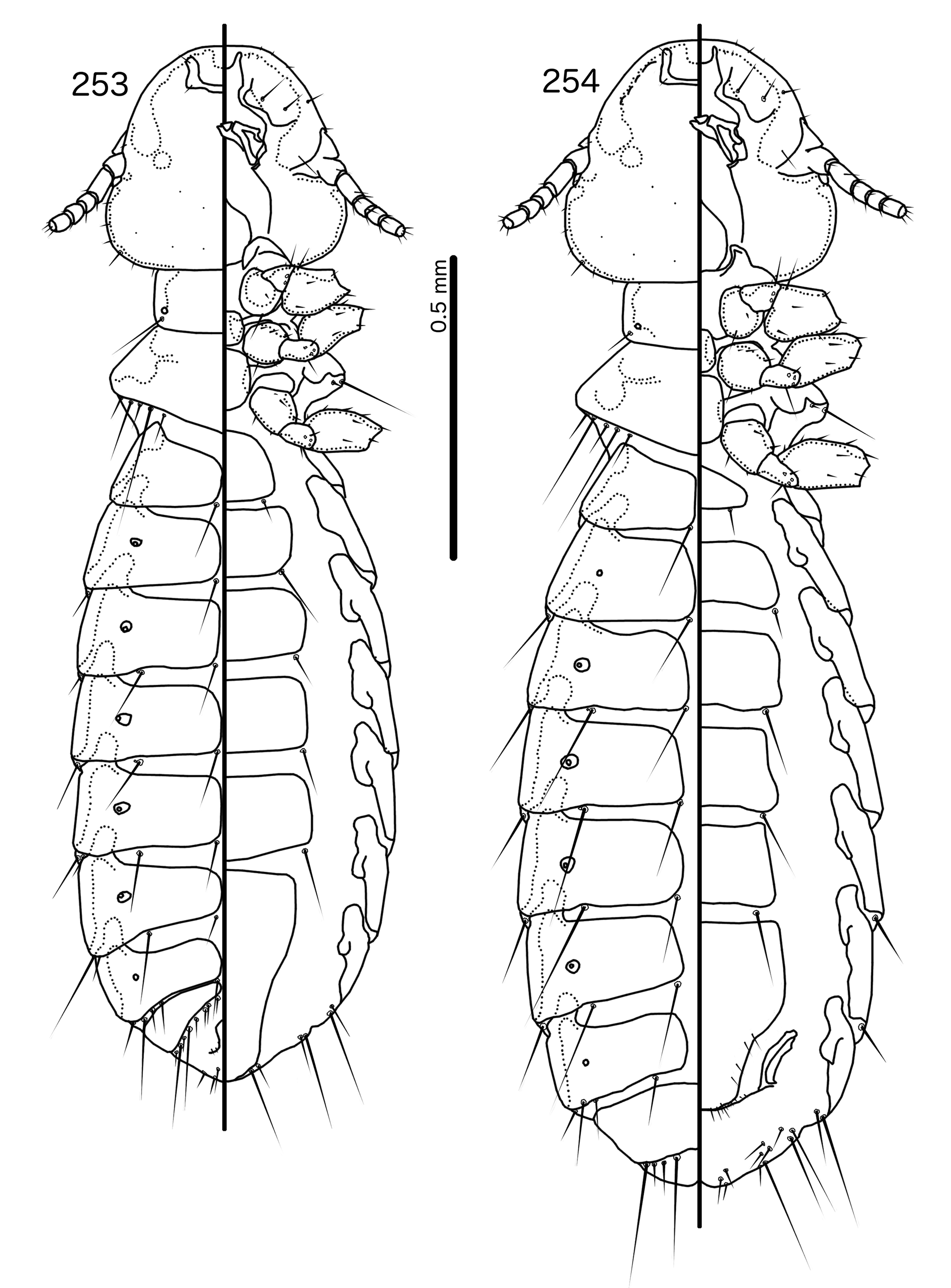
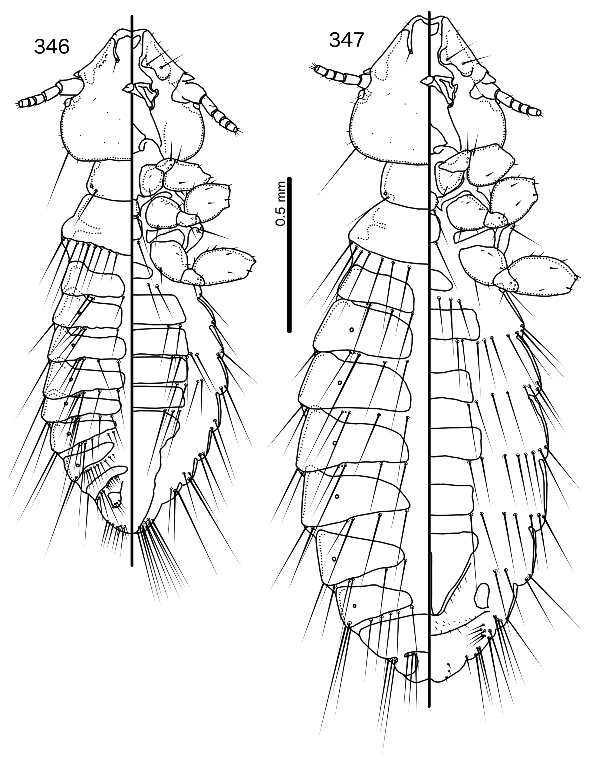
Prothorax rounded pentagonal, with posterior margin convergent to blunt median point ( Figs 463–464 View FIGURES 463 – 464 , 470– 471 View FIGURES 470 – 471 ); ppss on medio-posterior margin. Proepimera with large, blunt median ends. Pterothorax rounded crescentshaped; lateral margins convex, divergent; posterior margin rounded. Posterior margin of pteronotum narrowly indented at midline; indentation more extensive in male than in female and may continue for more than half the length length; mms narrowly interrupted medianly. Meso- and metasterna not fused, very small; 1 seta on posterolateral corner on each side of each plate. Metepisterna slender; median ends blunt. Leg chaetotaxy as in Fig. 25 View FIGURES 25 , except fI-p2 absent; fII-a3 and fIII-a2 dorsal. Many leg setae long and spike-like.
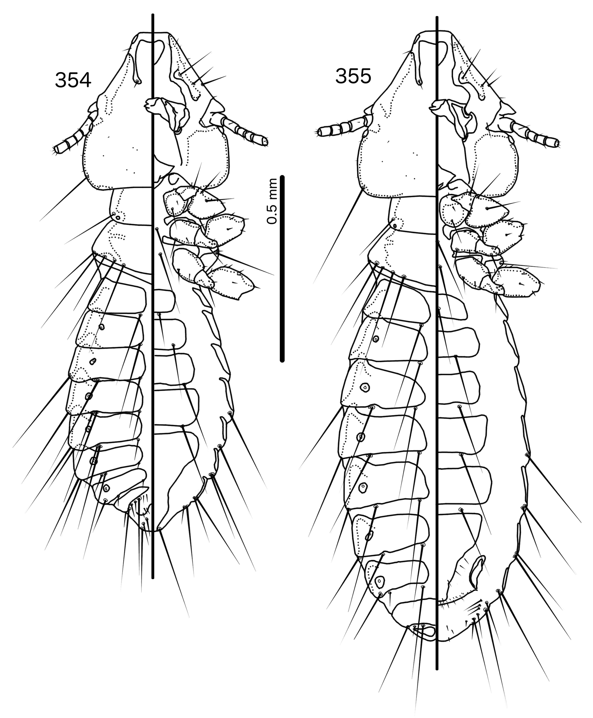
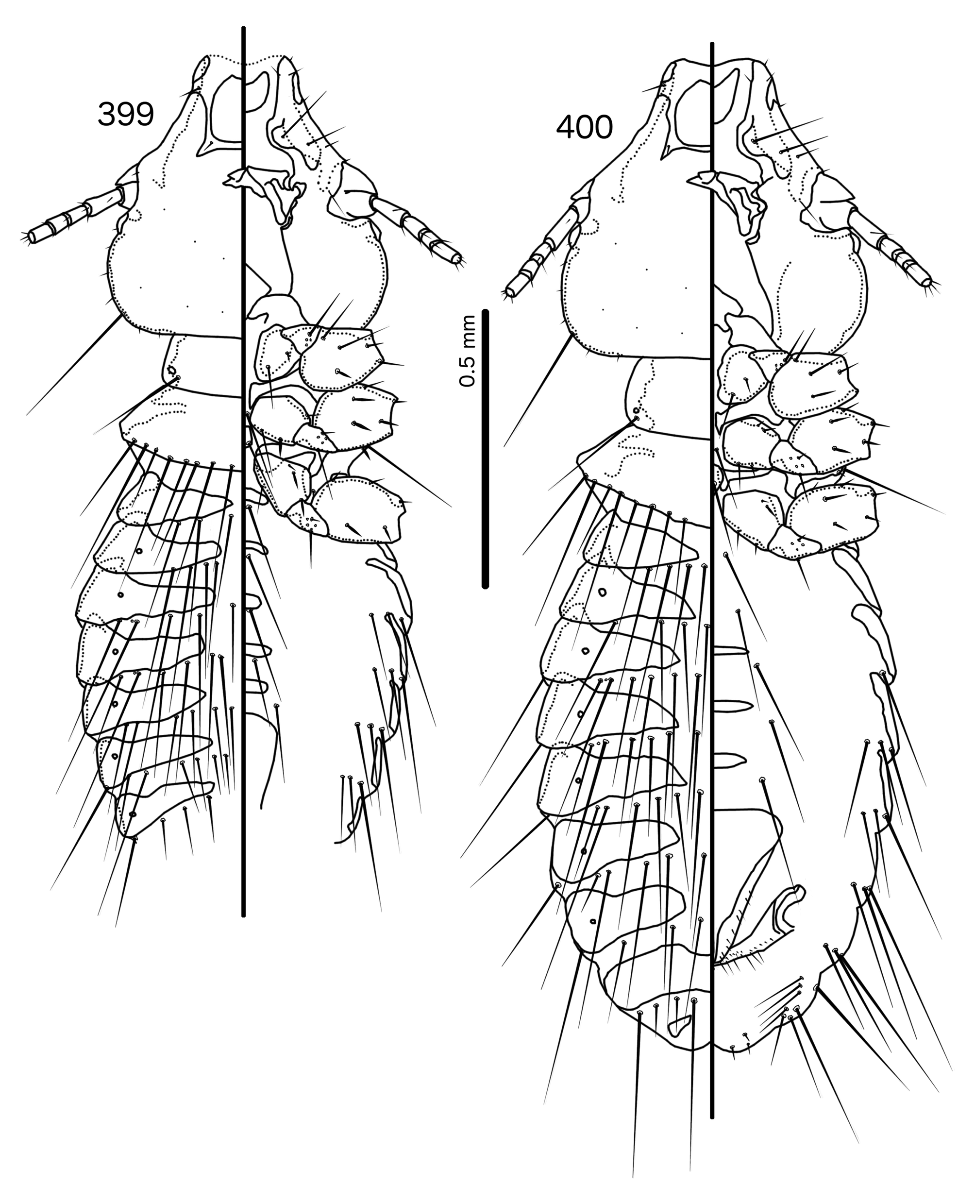
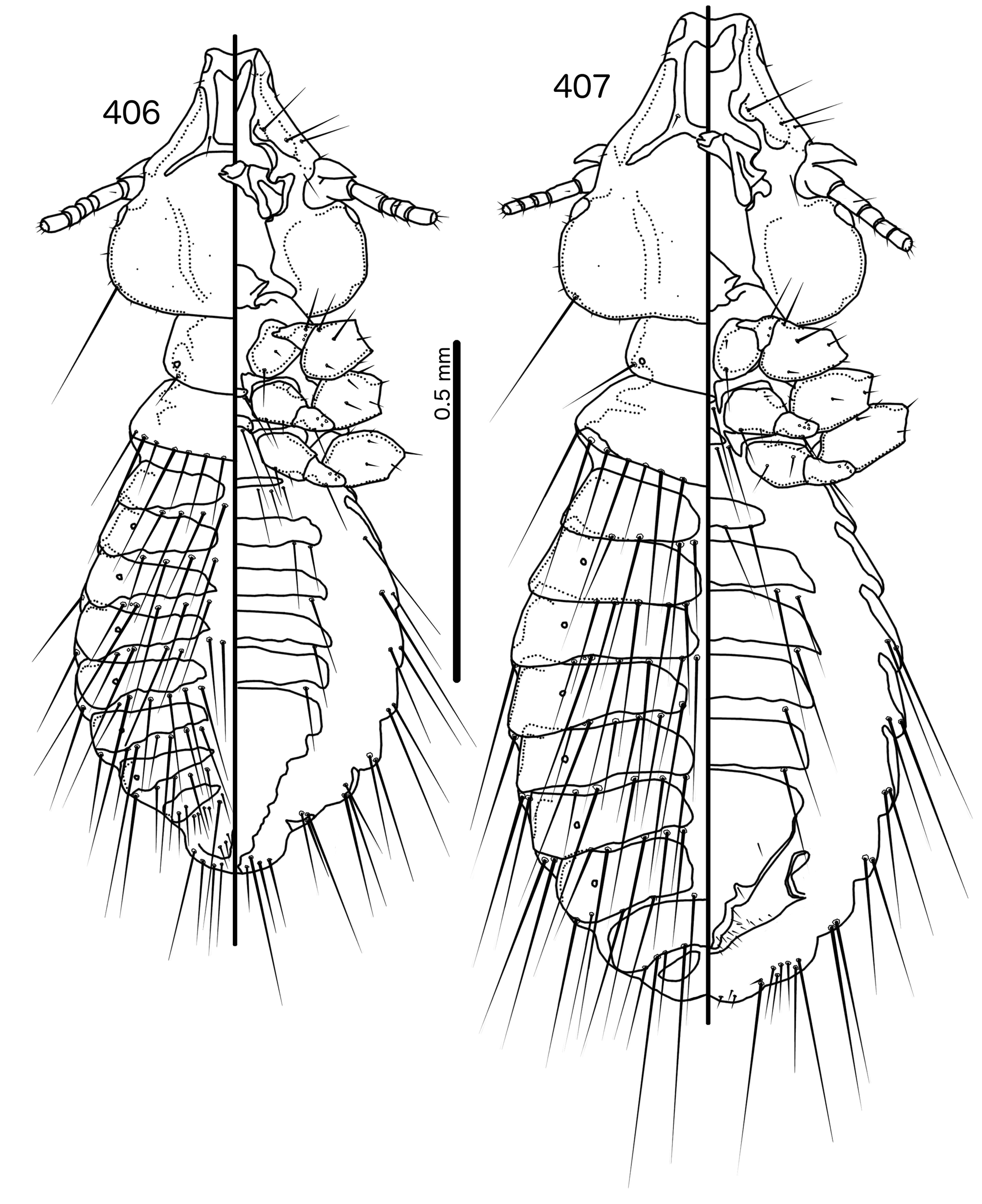
Abdomen ( Figs 463–464 View FIGURES 463 – 464 , 470–471 View FIGURES 470 – 471 ) almost circular in male, oblong in female. Tergopleurites triangular, more blunt in females than in males; tergopleurites II–IX+X in male and tergopleurites II–VIII in females moderately divided medianly. Sternal plate II in both sexes large, transversally continuous; sternal plates III–VI in both sexes small, crescent-shaped, medianly continuous; at least some sternal plates with small lateral accessory plates. Pleural incrassations moderate. Tergopleurites moderately extended onto ventral surface. Re-entrant heads large. Male subgenital plate trapezoidal, reaching posterior margin of abdomen and wrapping around to dorsal side; accessory sternal plates present lateral to subgenital plate on segments VII–VIII. Female subgenital plate roughly triangular, reaching vulval margin, either not flaring ( Fig. 476 View FIGURES 472 – 476 ) or flaring into partial cross-piece ( Fig. 469 View FIGURES 465 – 469 ); accessory sternal plates present lateral to subgenital plate on segment VII. Abdominal chaetotaxy as in Table 2, and Figs 463–464 View FIGURES 463 – 464 , 470–471 View FIGURES 470 – 471 . Vulval margin (Fig, 469, 476) with few slender vms, numerous thorn-like vss; vos located on and following lateral margins of the subgenital plate; distal vos situated median to vss.
Male genitalia ( Figs 466–468 View FIGURES 465 – 469 , 473–475 View FIGURES 472 – 476 ) prominent. Basal apodeme trapezoidal ( Fig. 473 View FIGURES 472 – 476 ) or rectangular ( Fig. 466 View FIGURES 465 – 469 ). Proximal mesosome rectangular, overlapping basal apodeme. Anterior margin may be thickened partially ( Fig. 474 View FIGURES 472 – 476 ) or entirely ( Fig. 467 View FIGURES 465 – 469 ) Gonopore small, ventral, open distally ( Figs 467 View FIGURES 465 – 469 , 474 View FIGURES 472 – 476 ). Mesosomal lobes large, bulging laterally, fused distal to gonopore. Rugose nodi may be present in postero-lateral corners; 2 ames microsetae on each side antero-lateral to gonopore; 2 pmes microsetae laterally on each side on lateral bulges of mesosomal lobes. Parameral heads ( Figs 468 View FIGURES 465 – 469 , 475 View FIGURES 472 – 476 ) folded, oblique. Parameral blades strongly curved medianly; pst1 sensilla, central; pst2 microsetae, central, near pst1.
Host distribution. Based on the three known species and undescribed material, Schizosairhynchus is limited to starlings of the genera Aplonis Gould, 1836 , Basilornis Bonaparte, 1850 , Mino Lesson, 1827 , and Sarcops Walden, 1875 . These four host genera are all members of the “South Asian/Pacific Starlings ” clade ( Lovette & Rubenstein 2007), and it is likely that Schizosairhynchus also parasitise other starlings belonging to this clade, such as species of Scissirostrum Lafresnaye, 1845 , Ampeliceps Blyth, 1842 , Streptocitta Bonaparte, 1850 , Enodes Temminck, 1839 , and Gracula Linnaeus, 1758 . Additional collections are required to determine the full host range of Schizosairhynchus .
Geographical range. South-East Asia and Australasia.
Etymology. The genus name is derived from Greek “ skhizein ” for “to split”, the Okinawan martial arts weapon the sai, and Greek “ rhunkhos ” for “bill”. A sai consists of a long baton (“ monouchi ”) flanked on both sides with shorter prongs (“ yoku ”), which are often curved and distally pointed. A sai split medianly is reminiscent of the shape of the preantennal area ( Figs 463–464 View FIGURES 463 – 464 , 470–471 View FIGURES 470 – 471 ) of members of this genus. Gender: masculine.
Remarks. No representative of Schizosairhynchus was included in the phylogeny of Bush et al. (2016), but structure of the male genitalia suggests Schizosairhynchus may be most closely related to Manucodicola and Bizarrifrons . The collection of fresh, sequenceable material in the future will help clarify the relationships of Schizosairhynchus within the Brueelia -complex.
Included species
* Schizosairhynchus erysichthoni new species
* Schizosairhynchus minovenator new species
* Schizosairhynchus philippensis ( Tandan & Kumar, 1969: 205) n. comb. [in Sturnidoecus ]


No known copyright restrictions apply. See Agosti, D., Egloff, W., 2009. Taxonomic information exchange and copyright: the Plazi approach. BMC Research Notes 2009, 2:53 for further explanation.
|
Kingdom |
|
|
Phylum |
|
|
Class |
|
|
Order |
|
|
Family |
Schizosairhynchus Gustafsson & Bush
| Bush, Sarah E. 2017 |
Schizosairhynchus philippensis ( Tandan & Kumar, 1969: 205 )
| Tandan 1969: 205 |
Sturnidoecus
| Eichler 1944: 81 |