
Turdinirmoides Gustafsson & Bush, 2017
|
publication ID |
https://doi.org/10.11646/zootaxa.4313.1.1 |
|
publication LSID |
lsid:zoobank.org:pub:A5Fdfba5-F992-44A8-84C2-1756C943C19B |
|
DOI |
https://doi.org/10.5281/zenodo.5296923 |
|
persistent identifier |
https://treatment.plazi.org/id/832187E9-FFE5-FFB2-FF74-67F0FB58FCD4 |
|
treatment provided by |
Plazi |
|
scientific name |
Turdinirmoides Gustafsson & Bush |
| status |
gen. nov. |
Turdinirmoides Gustafsson & Bush , new genus
Degeeriella Neumann, 1906: 60 ( in partim). Brueelia Kéler, 1936a: 257 ( in partim).
Type species. Degeeriella grandalae Clay, 1936: 912
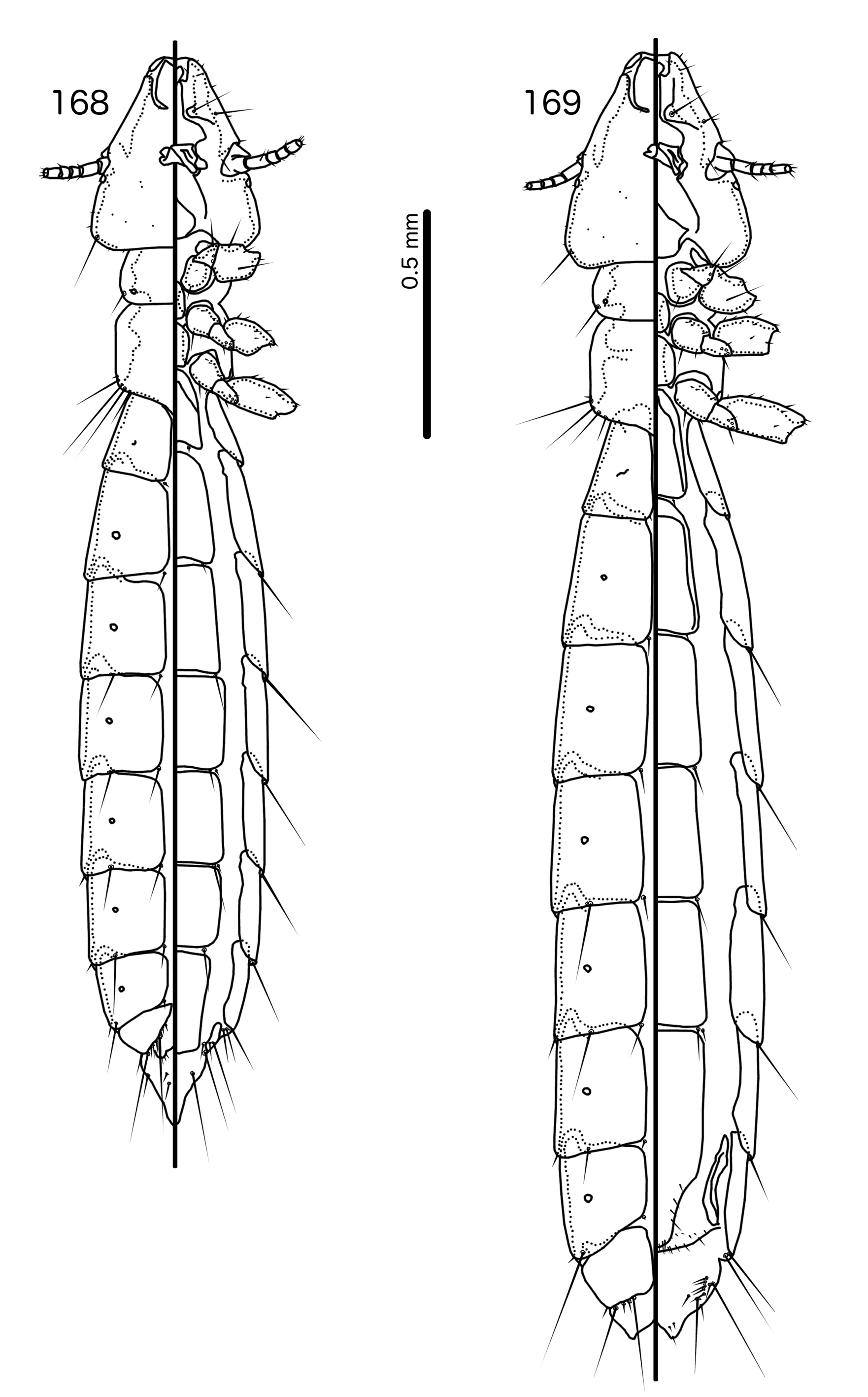
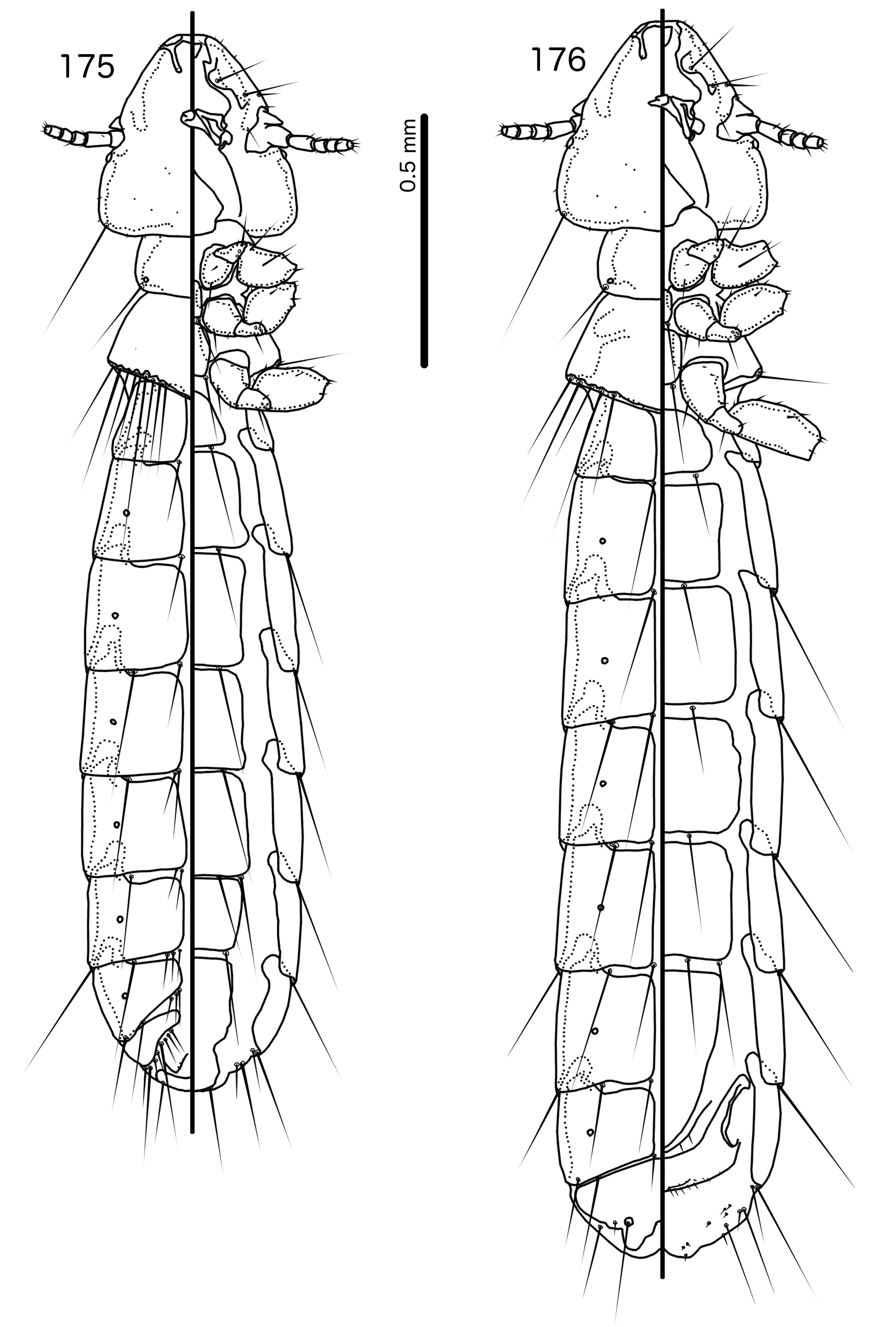
Diagnosis. Turdinirmoides n. gen. is most similar to Aratricerca n. gen. The most conspicuous similarity between the two genera is the division of the male subgenital plate into sternal plate VII and a posterior subgenital plate, with sts on sterite VII. Within the Brueelia -complex, this character is found only in these two genera, suggesting a close relationship. However, in Aratricerca ( Figs 168–169 View FIGURES 168 – 169 ) the pterothorax has parallel lateral margins, female tergopleurite IX+X is not fused with tergopleurite XI, and the vulval margin does not have a detached cross-piece, whereas in Turdinirmoides ( Figs 175–176 View FIGURES 175 – 176 ) the pterothorax is pentagonal, female tergopleurite IX+X is fused to tergopleurite XI, and there is a detahced cross-piece. For more details regarding similarities and differences between these two genera, see the diagnosis of Aratricerca above.
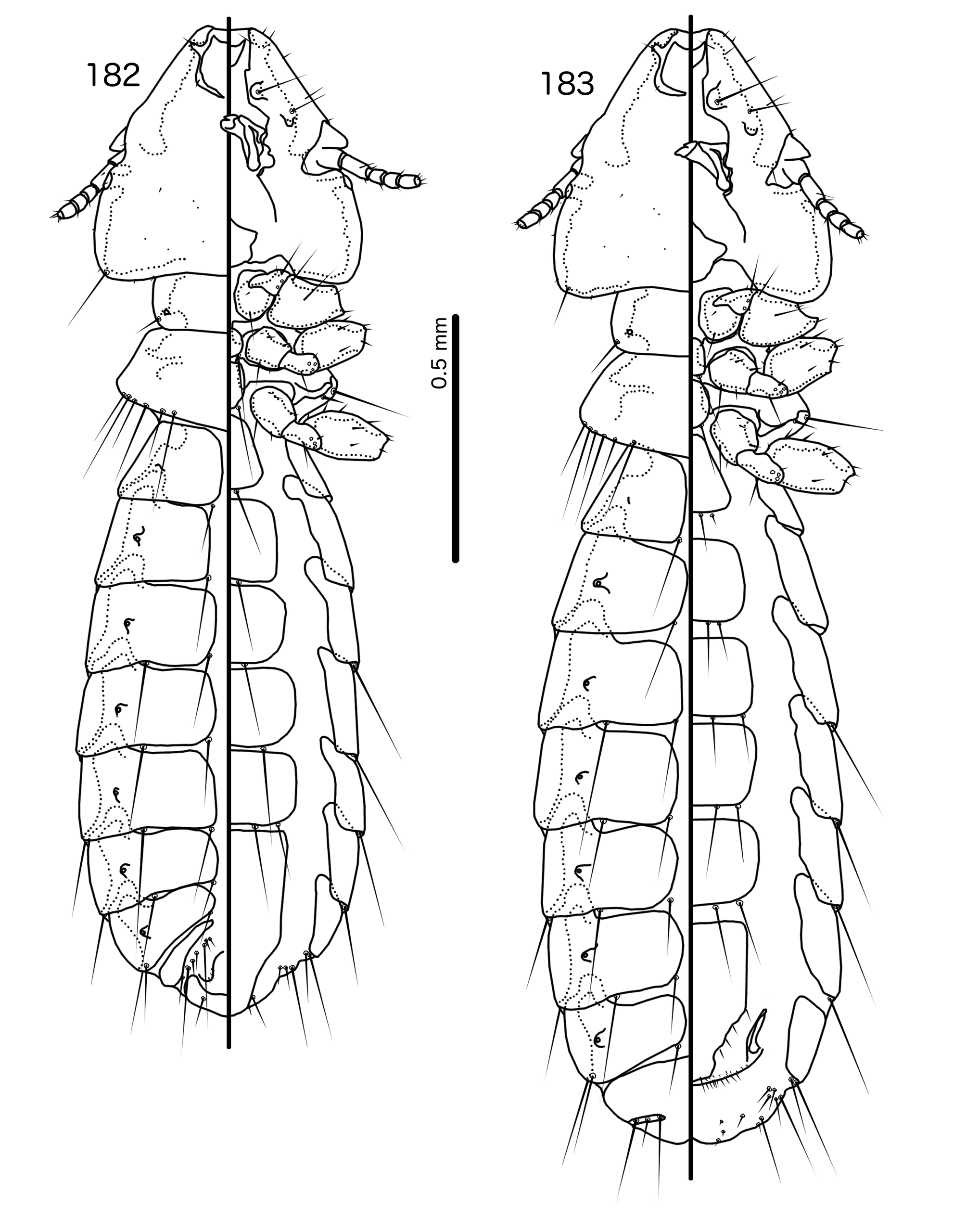
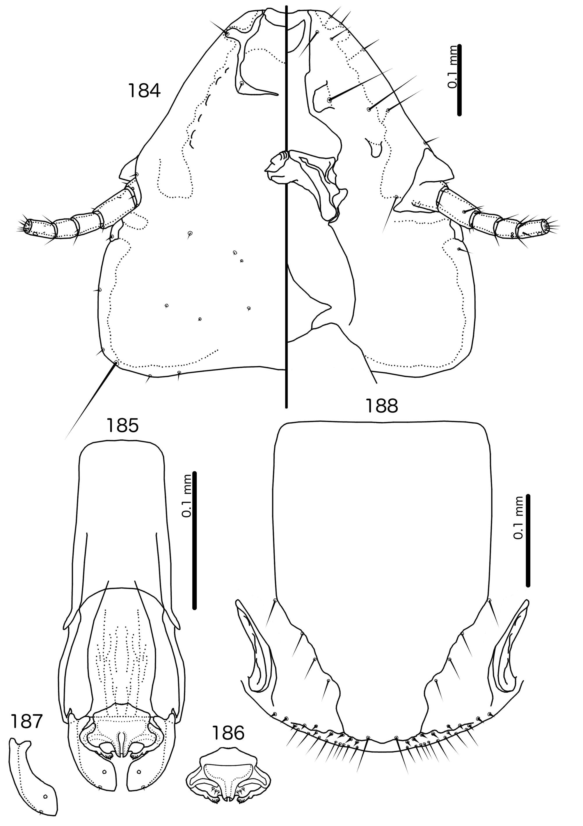
Preantennal structure and general habitus of Turdinirmoides are also similar to those of Turdinirmus . As in Turdinirmus ( Figs 182–183 View FIGURES 182 – 183 ), but unlike in Aratricerca ( Figs 168–169 View FIGURES 168 – 169 ), both the mesosternum and the metasternum of Turdinirmoides ( Figs 175–176 View FIGURES 175 – 176 ) have one seta on each side. However, both pos and pns are present in Turdinirmus ( Fig. 184 View FIGURES 184 – 188 ), but absent in Turdinirmoides ( Fig. 177 View FIGURES 177 – 181 ). In both Turdinirmus ( Figs 182–183 View FIGURES 182 – 183 ) and Turdinirmoides ( Figs 175–176 View FIGURES 175 – 176 ) sternal plate VI has more than one sts in both sexes, but in male Turdinirmus ( Fig. 182 View FIGURES 182 – 183 ) there is no separation between sternal plate VII and the more distal subgenital plate, and there are no sts on segment VII; in Turdinirmoides the male subgenital plate is divided at segment VII, and there are sts on segment VII ( Fig. 175 View FIGURES 175 – 176 ). The female subgenital plate of Turdinirmus ( Figs 188 View FIGURES 184 – 188 , 195 View FIGURES 191 – 195 ) reaches or approaches the vulval margin, and there is no cross-piece, whereas the vulval margin in Turdinirmoides ( Fig. 181 View FIGURES 177 – 181 ) has a detached crosspiece. The male genitalia of Turdinirmus ( Figs 185–187 View FIGURES 184 – 188 , 192–194 View FIGURES 191 – 195 ) and Turdinirmoides ( Figs 178–180 View FIGURES 177 – 181 ) are structurally similar. However, while the parameral heads of both genera are broadly bifid, those of Turdinirmoides form U-shaped folds, whereas those of Turdinirmus do not. pst2 is a microseta situated marginally in Turdinirmus , but a sensillus situated submedianly in Turdinirmoides The distal ridges of the mesosomal lobes are marginal in Turdinirmus , but more central in Turdinirmoides , and the ventral rugose areas are more extensive on the ventral surface and along the distal margin of the mesosome in Turdinirmoides than in Turdinirmus .
Description. Both sexes. Head broad, flat-dome shaped ( Fig. 177 View FIGURES 177 – 181 ). Marginal carina interrupted submedianly. Dorsal preantennal suture arising from interruptions, reaching ads and dsms, not separating dorsal anterior plate from main head plate posteriorly; laterally suture reaches margin of head, but does not interrupt marginal carina entirely. No displaced section of marginal carina present at clypeo-labral suture. Ventral anterior plate present. Ventral carinae with finger-like median protrusion; carinae clearly delimited anterior to pulvinus but not continuous with marginal carina. Head setae as in Fig. 177 View FIGURES 177 – 181 ; avs2–3 of similar length; pos and pns absent. Coni small. Antennae monomorphic. Temporal carinae not visible; mts 3 only macrosetae. Gular plate roughly triangular.
Prothorax rectangular, lateral margins convex ( Figs 175–176 View FIGURES 175 – 176 ); ppss on postero-lateral corner. Proepimera hammer-shaped medianly. Pterothorax pentagonal; lateral margins divergent and posterior margin convergent to median point ( Figs 175–176 View FIGURES 175 – 176 ). Meso- and metasterna not fused, one seta on postero-lateral corner on each side of each plate. Metepisterna hammer-shaped medianly. mms moderately divided medianly. Leg chaetotaxy as in Fig. 25 View FIGURES 25 , except fI-p2, fI-v4, fII-v2, fIII-v2 absent.
Abdomen ( Figs 175–176 View FIGURES 175 – 176 ) slender and elongatedly oval. Terminal end of abdomen rounded in male, shallowly divided in female. Abdominal chaetotaxy as in Table 2 and 6. Tergopleurites square-shaped; tergopleurites II– IX+X in male and tergopleurites II–VIII in females narrowly divided medianly. Female tergopleurite IX+X fused with tergopleurite XI. Sternal plates quadratic, not approaching pleurites. Pleural incrassations broad, overlapping and forming “rails” along lateral margins of abdomen. Re-entrant heads moderate to large. Male subgenital plate divided into sternal plate VII and subgenital plate on segments VIII–XI, with setae on posterior margin of sternal plate VII. Female subgenital plate roughly triangular, not approaching vulval margin ( Fig. 181 View FIGURES 177 – 181 ). Detached crosspiece present. Vulval margin with broad sclerotized cross-piece not connected to subgenital plate.
Male genitalia as in Figs 178–180 View FIGURES 177 – 181 . Proximal mesosome trapezoidal. Gonopore ( Fig. 179 View FIGURES 177 – 181 ) as convergent thickenings, open distally and proximally. Mesosomal lobes angular or rounded, with ridge across mid-length and distal part rugose; 2 pmes sensilla anterior to ridge just lateral to gonopore. Parameral heads ( Fig. 180 View FIGURES 177 – 181 ) folded into U-shapes, without accessory sclerite. Parameral blades broad, rounded; pst1–2 both sensilla.
Species Sex ps aps psps tps ss sts
To. grandalae M III–VIII – IV–VIII V–VIII II–VIII II–VII F III–VIII – IV–VIII – II–VIII II–VI To. hrabali M III–VIII – IV–VII VII–VIII II–VIII II–VI F III–VIII – IV–VIII – – II–VI Host distribution. Turdinirmoides is widely distributed, and we have seen material from Muscicapidae , Acanthizidae , Rhipiduridae , Prunellidae , and Paramythiidae .
Geographical range. Known only from South Asia.
Etymology. Turdinirmoides is a reference to the genus Turdinirmus (below), which is superficially similar to this genus, especially in the preantennal area. The ending “ -oides ” from Greek “ eidos I’ for ”likeness”. Gender: feminine.
Remarks. No member of this genus was included in the phylogeny of Bush et al. (2016). Although morphological characters suggest a close relationship with Aratricerca , the exact relationship between Turdinirmoides and the preceding three genera lacking pos is still unclear.
Included species
* Turdinirmoides grandalae ( Clay, 1936: 912) n. comb. [in Degeeriella ] Turdinirmoides hrabali (Najer & Sychra [in Najer et al.], 2012c: 65) n. comb. [in Brueelia ] [1]
[1] The description of this species ( Najer et al. 2012c: 65) states that male abdominal sterna II–VII have setae laterally, as is the case in Turdinirmoides and Aratricerca , however the setae of sternum VII are not present in the illustrations of Najer et al. (2012). There are considerable differences between this species and To. grandalae in the abdominal chaetotaxy of both sexes, and the male subgenital plate does not appear to be divided in To. hrabali . Given the small number of species involved, and the interspecific variation in abdominal chaetotaxy seen in the closely related Resartor ( Table 5) we place Brueelia hrabali in Turdinirmoides based on non-setal characters and the stated presence of setae on sternal plate VII in males, but recognise that when a larger number of species in this complex are known and have been adequately described and sequenced, the systematics of Turdinirmoides and related genera may need further revision.


No known copyright restrictions apply. See Agosti, D., Egloff, W., 2009. Taxonomic information exchange and copyright: the Plazi approach. BMC Research Notes 2009, 2:53 for further explanation.
|
Kingdom |
|
|
Phylum |
|
|
Class |
|
|
Order |
|
|
Family |
Turdinirmoides Gustafsson & Bush
| Bush, Sarah E. 2017 |
Turdinirmoides grandalae ( Clay, 1936: 912 )
| Najer 2012: 65 |
| Clay 1936: 912 |
Degeeriella
| Keler 1936: 257 |
| Neumann 1906: 60 |