
Neochlamisus bebbianae ( Brown, 1943 ) II
|
publication ID |
https://doi.org/10.1111/j.1096-3642.2007.00343.x |
|
persistent identifier |
https://treatment.plazi.org/id/862A87D5-FFBF-FFFA-00F7-FBA7FBB52930 |
|
treatment provided by |
Felipe |
|
scientific name |
Neochlamisus bebbianae ( Brown, 1943 ) II |
| status |
|
Neochlamisus bebbianae ( Brown, 1943) II :
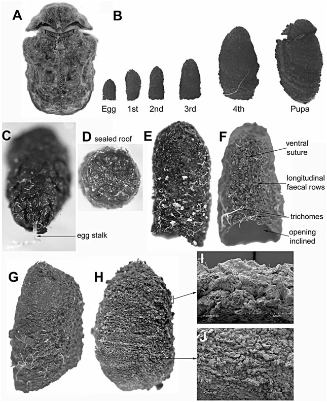
Alnus host form ( Fig. 6 View Figure 6 )
Egg case ( Fig. 6B–D View Figure 6 ): Size ( N = 9): L, 1.37–1.68 mm; W at roof, 0.89–0.94 mm. Colour: brownish black. Shape: bell-like; symmetrical in lateral aspect; ventral, lateral, and dorsal surfaces not apparent. Apex: dome-like; attachment stalk, present, creamy brown, translucent, not coiled. Roof: thin-walled, shallowly convex; internal and external surface textures similarly rough; flange, thin and ragged. External wall: coarse, with faeces arranged as overlapping plates; plates with apical margins exposed and arcuate; shallow uneven longitudinal ridges present; ridges evenly spaced, edges ragged. Trichomes: apparent within matrix, simple, arranged randomly and horizontally. Internal surface: texture lacking apparent plates and ridges; narrow space between case wall and egg.
Larva-I case ( Fig. 6E View Figure 6 ): Size ( N = 5): L, 1.45–1.70 mm; W at base opening, 1.09–1.7 mm. Colour: unevenly dark brown, with larval addition in basal section and in ventral wall darker, almost blackish. Egg case: generally intact; egg stalk present or absent. External surface of instar-I section: with faeces deposited in rows. Ventral wall: distinct in late instar I with insertion of ventral suture and faeces; suture extending subapically through egg case to basal margin. Lateral and dorsal walls: as in egg case, basally, with narrow addition of faecal rows; rows arranged transversely. Base opening: transverse, margin simple. Internal surface: relatively smooth; plates and rows not apparent. Wall thickness: evenly thin; apex approximately two times thicker than wall.
Larva-II case ( Fig. 6F View Figure 6 ): Size ( N = 9): L, 2.75–2.77 mm; W at base opening, 1.23–1.60 mm. Colour: unevenly dark brown. Shape: tubular; apex, dome-like; egg stalk, absent. Egg case: apparent, accounting for half of the total case length; shape, position, and external texture of plates and weak longitudinal ridges, generally intact; flange of original egg case somewhat abraded. Larval-built case section: distinct from maternal section in colour (blackish), surface texture (coarser, in rows), and trichomes (more protuberant). External surface of larval sections: coarse with fine and even faecal rows. Trichomes: present, sparse, deeply embedded into faecal matrix, some more protuberant in larval section. Ventral wall: with suture extending from apex through egg case to base opening; suture and adjacent faecal insertion protuberant. Walls: evenly thick, slightly thicker at base. Base opening: slightly oblique, margin simple. Internal surface: smooth; plates, ridges, and rows not apparent.
Larva-III case ( Fig. 6B View Figure 6 ): Size ( N = 8): L, 3.03– 3.06 mm; W at base opening, 1.62–1.87 mm. Case III resembles case II, with larval section II similar to larval section I.
Larva-IV case ( Fig. 6G View Figure 6 ): Size ( N = 8): L, 3.27– 3.73 mm; W at base opening, 2.06–3.40 mm. Colour: light brown in egg section, dark brown/black in larval sections. Shape: tubular, asymmetrical in lateral aspect, ventral wall longer than dorsal wall; apex forming elongate dome. Egg and larval sections: distinguishable; external plates of egg case not apparent, longitudinal ridges vague; larval section with fine rows. Ventral wall with suture extended from apex to basal margin; ventral field quadriform, not triangular as in earlier instars. Base opening: transverse, margin simple. Internal surface texture: relatively smooth, instar sections not distinct. Wall thickness of egg case and sections I–III evenly narrow; wall of instar IV approximately twice as thick. Trichomes: present, sparse, deeply embedded in faecal matrix.
Pupal case ( Fig. 6H–J View Figure 6 ): Size ( N = 7): L, 5.42– 5.56 mm; W at base opening, 4.05–4.15 mm. Colour: dark-brown/black, base lighter brown. Shape: barrellike, asymmetrical in lateral view with ventral wall shorter than dorsal wall. Egg case: occupying c. 1/8th of the total case length, shape completely distorted, split hemispherically and inclined ventrad; surface plates not apparent. External surface: with fine faecal rows arranged longitudinally on ventral wall, obliquely on lateral wall, and transversely on dorsal wall. Ventral wall: with suture extended from apex to base opening; larval insertions coarse, occupying most of ventral wall. Base opening: oblique, margin simple, completely sealed; seal flattened and discoid, texture rough externally and internally, comprised of faeces and trichomes; exuviae IV positioned internally on top of seal. Trichomes: present, sparse, incorporated deeply into matrix.
No known copyright restrictions apply. See Agosti, D., Egloff, W., 2009. Taxonomic information exchange and copyright: the Plazi approach. BMC Research Notes 2009, 2:53 for further explanation.