
Anastrepha tenella Zucchi
|
publication ID |
https://doi.org/10.11646/zootaxa.3911.3.7 |
|
publication LSID |
lsid:zoobank.org:pub:75CC87E7-799B-4EC4-BEB1-30F22912F721 |
|
DOI |
https://doi.org/10.5281/zenodo.6104294 |
|
persistent identifier |
https://treatment.plazi.org/id/884F8781-7E44-FFF4-FF19-E4CDD703A5B7 |
|
treatment provided by |
Plazi |
|
scientific name |
Anastrepha tenella Zucchi |
| status |
|
Anastrepha tenella Zucchi View in CoL
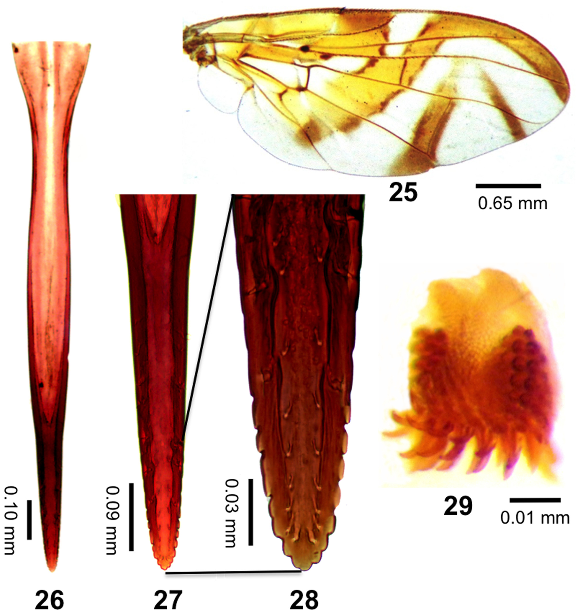
Figs. 25–29 View FIGURES 25 – 29
Anastrepha tenella Zucchi, 1979:40 View in CoL (description, wing, aculeus tip, Brazil, Bahia, Cruz das Almas); Zucchi, 1978 (in PhD thesis); Norrbom et al., 1999a:337 (in catalog); Norrbom et al., 1999b:83 (classification); Zucchi 2000: 22 (in key); Norrbom et al. 2012 (interactive key).
Diagnosis. This species can be recognized from other species of Anastrepha by the following combination of characters: aculeus 1.49–1.50 mm long; in ventral view, lateral margins broadened at midlength and gradually tapering distally; aculeus tip 0.40–0.45 mm long, 0.27–0.28 times aculeus length, 0.09 mm wide; aculeus tip with 12–13 serrations, including 7 small sized serrations distally, and 5–6 poorly defined serrations basally.
Description. Mostly yellow to orange. Setae dark brown.
Head. Yellow to orange except blackish ocellar tubercle. Facial carina, in profile, concave. 3–4 frontal setae; 2 orbital setae, posterior seta slightly more slender than anterior one; ocellar setae weak, small. Antenna not extended to ventral margin of face. Palpus in lateral view dorsally curved, evenly setose.
Thorax. Integument mostly yellow to orange with following areas white: postpronotal lobe; medial vitta with posterior end ovoid (including acrostichal setae); paired sublateral scutal vitta from transverse suture to posterior margin, including base of intra-alar seta; and entire scutellum; dorsal margins of anepisternum and katepisternum. Postpronotal seta on posterior half of postpronotal lobe. Scuto-scutellar suture without darker band or spot. Subscutellum and mediotergite entirely yellow to orange. Mesonotum 2.5–2.96 mm long; 2.1 mm width. Postpronotal lobe, scutum and scutellum entirely microtrichose; scutal setulae yellowish. Setae dark brown. Acrostichal, dorsocentral and intra-alar setae well developed. Katepisternal seta yellowish, well developed but much smaller and weaker than anepimeral seta. Legs entirely yellow to orange. Fore femur with posterodorsal and ventral rows of well developed setae.
Wing ( Fig. 25 View FIGURES 25 – 29 ). Length 6.50–6.51 mm, width 2.82 mm, ratio 2.31. Veins dark brown. Apex of vein R1 at 0.54 wing length, proximal to level of anterior end of crossvein r-m. Cell c 1.34 times as long as pterostigma, pterostigma 2.81–3.10 times as long as wide. Ratio of costa length between apices of Sc and R1/length between apices of R1 and R2+3 0.45. Crossvein r-m at 0.64 distance from bm-cu to dm-cu on vein M. Vein M moderately curved apically, not reaching S-band; cell r4+5 0.82–0.91 times as wide at apex as at level of dm-cu. Vein R2+3 not sinuous. Cell bcu with distal lobe moderately long, length of bcu 1.56–1.57 time as long as anterior margin, lobe 0.68–0.71 time as long as vein A1+Cu2. Pattern mostly orange and moderate brown. Cell c mostly or entirely infuscated to subhyaline. C-band mostly orange; most of pterostigma moderately brown. C-band broadly extending to vein M in cell br along cell bm; covering base of cell r2+3; orange area posterior to pterostigma broad, extending distally into cells r1 and r2+3, except anterior margin distally in cell r1 and narrow distal margin in both cells. C-band and S-band connected along vein R4+5. Basal hyaline area between C-band and S-band extended to vein R4+5. Cell bm entirely hyaline. Cell r1 basomarginal hyaline spot triangular to quadrate, its apex aligned proximal to crossvein r-m. S-band mostly orange; base without posterior extensions in middle of cell cu1 or in cell a1; distal margin of band brown posteriorly in cells r4+5, dm cell cu1; distal section brown along costal margin, distal part of cell r2+3 and in cell r4+5. S-band distal section at apex of vein R2+3 0.54–0.57 times width of cell r2+3; without marginal hyaline band or spots in cell r2+3 or near apices of R2+3 or R4+5. Subapical hyaline area in radial cells distal to r-m extending anteriorly to vein R2+3. V-band proximal arm as dark as apical half of S-band; extending 0.52 distance from apex of vein Cu1 to apex of vein A1+Cu2, not connected anteriorly to S-band. V-band distal arm complete; connected to proximal arm. Apex of V-band not ext ended to vein M, hyaline area present between band and vein M. Area surrounding apex of lobe of cell bcu with microtrichia similar in density to area anterodistal to it along vein Cu1. Area between S-band and V-band entirely microtrichose in cells dm and cu1.
Abdomen. Pale brown. Tergites without markings, entirely microtrichose; setulae pale brown.
Female terminalia ( Figs. 26–29 View FIGURES 25 – 29 ). Oviscape entirely yellow to orange brown; straight; entirely microtrichose; 2.0 mm long; length ratio (oviscape length/mesonotum length) 0.8; spiracle at basal 0.38. Eversible membrane ( Fig. 29 View FIGURES 25 – 29 ) with 35–50 denticles (all sclerotized) in triangular to semicircular or suboval pattern. Aculeus ( Fig. 26 View FIGURES 25 – 29 ) 1.49–1.50 mm long; in ventral view extreme base expanded ( 0.21 mm wide); lateral margins broadened at midlength and distal gradually tapering. Aculeus length/oviscape length 0.75. Aculeus tip ( Figs. 27, 28 View FIGURES 25 – 29 ) 0.17–0.18 mm long. Aculeus tip length/aculeus length 0.27–0.28; width at base 0.09 mm, 4.45–4.78 times as long as wide; lateral margins gradually tapering; distal 0.17–0.18 mm with 12–13 serrations (7 small sized serrations distally, and 5–6 poorly defined serrations basally); serrate part not extending onto dorsal side basally, serrated part 0.40–0.45 times length of tip. Spermathecae sclerotized.
Host. Unknown.
Distribution. Known only from Brazil, state of Bahia.
Type Data. Holotype female, Brazil: Bahia: Cruz das Almas, McPhail trap, no collection date, A. S. Nascimento (coll), ( MZSP) (examined).
Other specimen examined. BRAZIL: Bahia: Itaberaba, McPhail trap, 02.VI.2009, M. C. A. Nunes, 1♀ ( ESALQ).
| MZSP |
Sao Paulo, Museu de Zoologia da Universidade de Sao Paulo |
No known copyright restrictions apply. See Agosti, D., Egloff, W., 2009. Taxonomic information exchange and copyright: the Plazi approach. BMC Research Notes 2009, 2:53 for further explanation.
|
Kingdom |
|
|
Phylum |
|
|
Class |
|
|
Order |
|
|
Family |
|
|
Genus |
Anastrepha tenella Zucchi
| Uramoto, Keiko, Zucchi, Roberto A. & Norrbom, Allen L. 2015 |
Anastrepha tenella
| Zucchi 2000: 22 |
| Norrbom 1999: 337 |
| Norrbom 1999: 83 |
| Zucchi 1979: 40 |