
Urnalana flammea, Galil, Bella S. & Ng, Peter K. L., 2015
|
publication ID |
https://doi.org/10.11646/zootaxa.4027.4.1 |
|
publication LSID |
lsid:zoobank.org:pub:3B32D183-45BA-41F9-82A9-C9C108D53899 |
|
DOI |
https://doi.org/10.5281/zenodo.6102280 |
|
persistent identifier |
https://treatment.plazi.org/id/8870B305-E32E-4C35-9699-FE0388AC8B38 |
|
treatment provided by |
Plazi |
|
scientific name |
Urnalana flammea |
| status |
sp. nov. |
Urnalana flammea View in CoL sp. nov.
( Figs. 5 View FIGURE 5 F, 21, 22A–G, 23C, D, I–M) Material examined. Holotype: male ( 9.3 mm) (MNHN-IU-2013-1341), stn PR227, Madang Resort, 05°12.5'S 145°48.5'E, 10.12.2012. Paratypes: 1 female ( 7.6 mm) ( ZRC 2015.275), stn PR14, Biliau I., 05°12'S 145°48.1'E, 2–3 m, 9.11.2012; 1 male (9.0 mm) 1 ovigerous female ( 10.7 mm) (MNHN-IU-2013-13123) stn PR28, Madang Resort, 05°12.5'S 145°48.5'E, 13.11.2012; 1 male ( 9.2 mm) ( ZRC 2015.276), stn PR245, Tabad I., 05°08.2'S 145°48.7'E, 12 m, 12.12.2012.
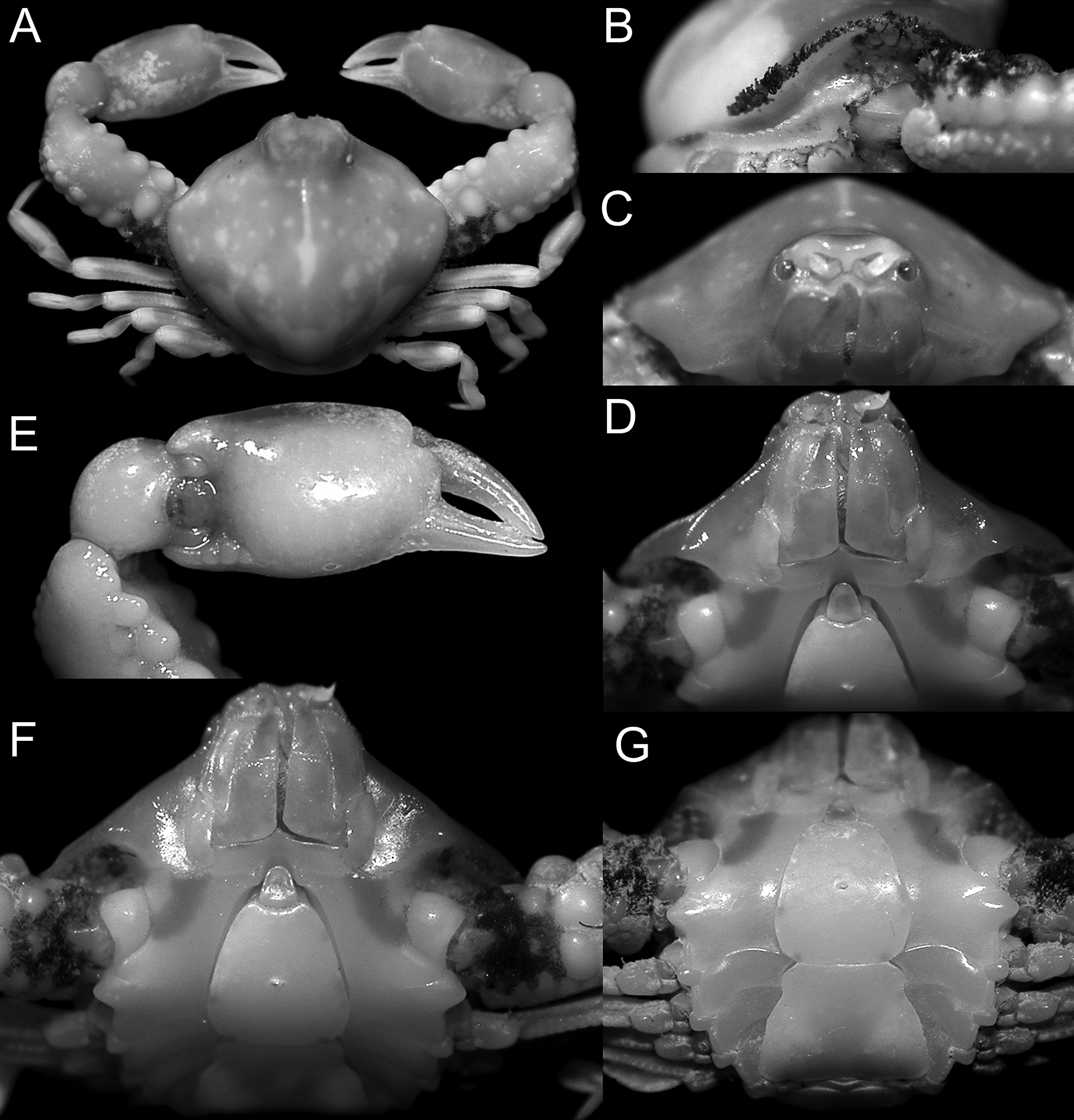
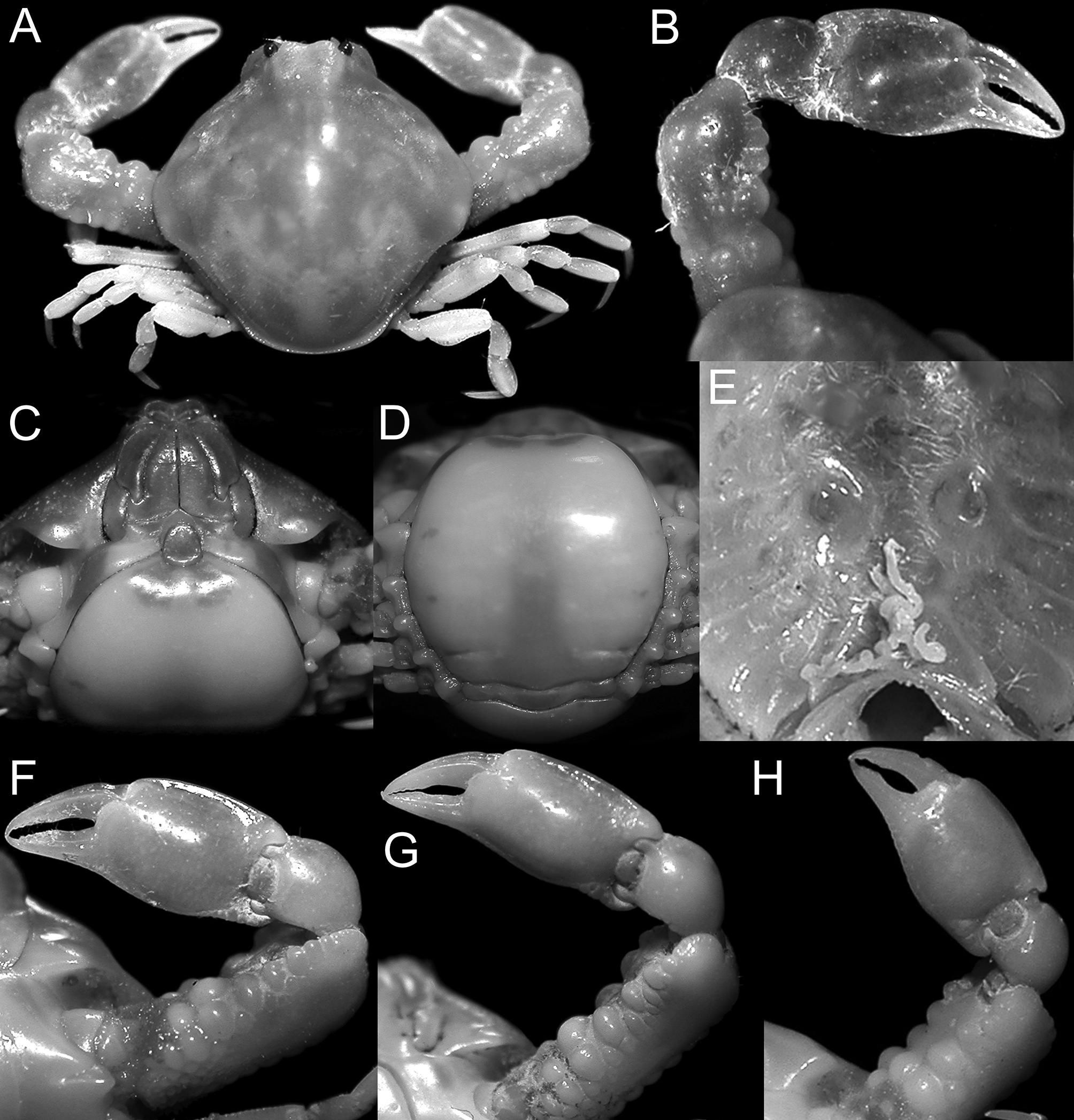
Description. Carapace subpentagonal, urn-shaped, globose; regions of carapace ill defined ( Fig. 21 View FIGURE 21 A). Dorsal surface of carapace sparsely setose, punctate. Front somewhat produced, upturned, medially concave, lateral lobes rounded ( Fig. 21 View FIGURE 21 D). Postfrontal region laterally concave. Anterolateral margin convex, smooth, medially inflated in dorsal view; swollen distal margin of pterygostomian region invisible in dorsal view. Lateral angle of carapace rounded, overhanging thoracic sinus, lacking setose fringe ( Fig. 21 View FIGURE 21 B). Thoracic sinus deep, anteriorly defined by vaulted margin of pterygostomian region ( Fig. 21 View FIGURE 21 D, G); cluster of minute granules overgrown by club-like setae anteriorly; row of pearliform granules on ventral surface of epibranchial angle; another row above base of first pair of ambulatory legs. Epimeral ledge visible over its entire length in dorsal view, continuous with posterior margin, margin beaded, granules larger anteriorly. Posterolateral margin sparsely setose. Posterior margin minutely beaded ( Fig. 21 View FIGURE 21 A).
Antennular fossa sealed by basal antennular article. Antenna short, nestled underneath antennular fossa ( Fig. 21 View FIGURE 21 C). Orbits small, rounded, outer orbital margin with 1 suture. Anterior margin of efferent branchial channel forms part of lower orbital margin, short suture below eye. Eyes retractable. Third maxillipeds concealing trapezoid buccal cavity; smooth, planar, lacking denticle anterolaterally on ischium ( Fig. 21 View FIGURE 21 D), setose fringe lengthwise on endopod of female. Chelipeds subequal, robust, longer in adult male than in female specimens ( Fig. 21 View FIGURE 21 A). Cheliped merus trigonal, merus 2/3 as long as carapace. Dorsal surface proximally with cluster of granules partly obscured by patch of club-shaped setae; anterior margin with 3 large, pearliform granules proximally, row of small granules distally ( Fig. 21 View FIGURE 21 E, 22F, G); posterior margin with row of lozenge-shaped granules, largest medially; ventral surface proximally pitted, granular. Carpus subglobular, granules proximally on inner margin. Palm elongated, 0.7 times as wide as long; dorsal margin of palm distinctly carinate, smooth; inner, outer basal lobe granular, ventral margin with milled carina, continuing into pollex ( Fig. 21 View FIGURE 21 E). Cutting edges of fingers meeting only distally. Ambulatory legs slender, short; meri ventrally concave, meri 1–3 with 2 granular rows on ventral, dorsal surface, merus of fourth pair with single row dorsally, 2 rows ventrally. Carpi of ambulatory legs dorsally carinate, propodi dorsally, ventrally carinate. Dactyli as long as propodi, lanceolate.
Male abdominal cavity deep, elongated, nearly reaching buccal cavity; lateral margin with distinct ridge fitting into suture between abdominal somites. Male abdomen with somite 2 minute, spindle-shaped; somites 3–5 fused; somite 6 large, rounded, narrowing distally, minute median denticle; telson laciniate ( Fig. 21 View FIGURE 21 F, G). Female abdomen with somite 2 yoke-shaped, somites 3–6 fused, greatly enlarged, shield-like; telson laciniate ( Fig. 22 View FIGURE 22. A, B, F C, D). G1 shaft somewhat sinuous, tip slightly swollen, setose. G2 short, curved, apex scoop-like. Vulvae prominent, large, anteriorly reaching suture 5/4; ovoid; interiorly hooded by calcified semi-dome, opening, directed laterally, slit-like, crescentic, bordered by shallow groove ( Fig. 22 View FIGURE 22. A, B, F E).
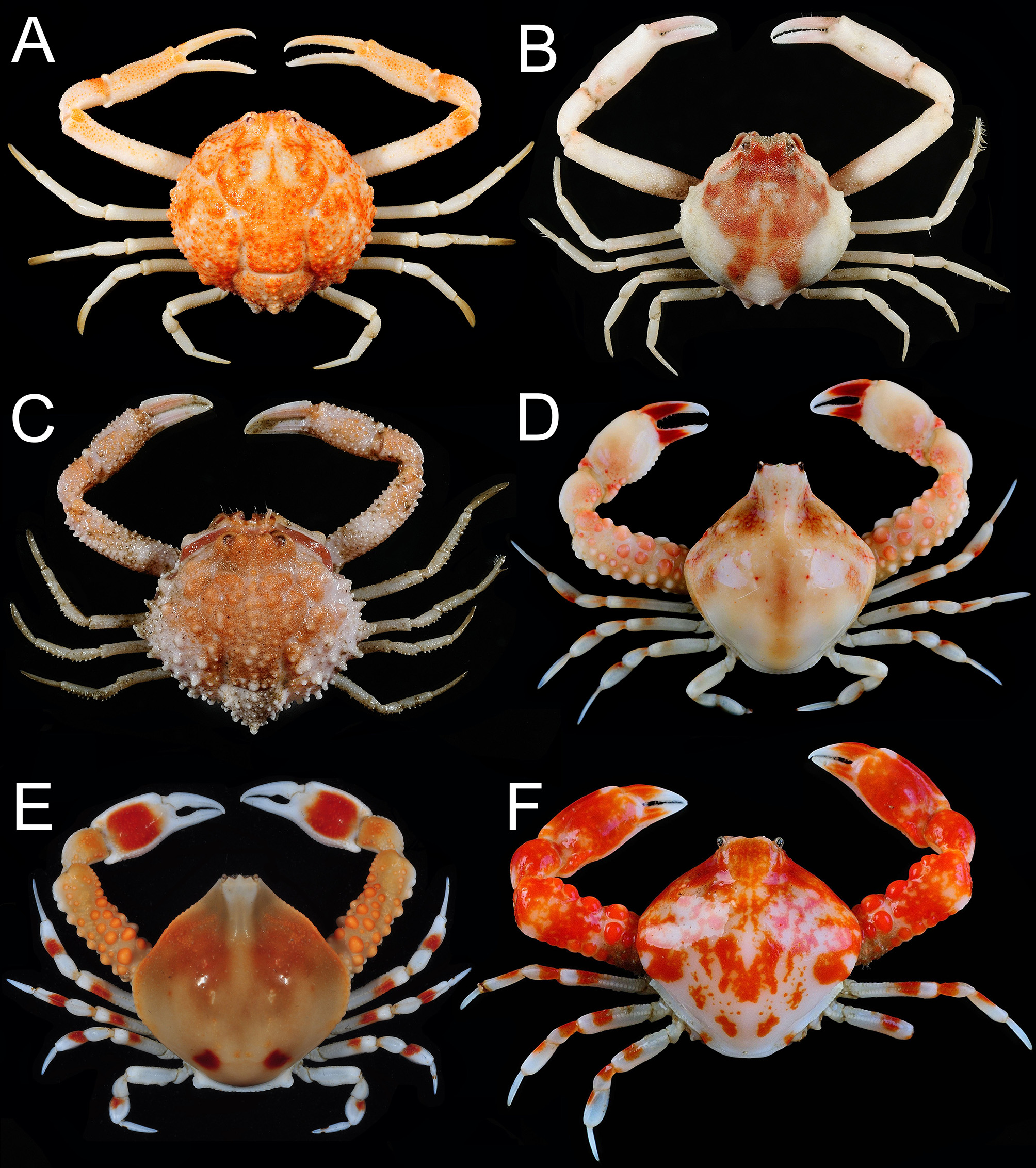
Colour in life. Dorsal surface of carapace and chelipeds bone-coloured, densely set with orange-red markings; finger tips white. Dorsal surface of meri, carpi and propodi of ambulatory legs with orange blotches ( Fig. 5 View FIGURE 5 F). Examination of the ethanol-preserved specimen revealed that the pterygostomian region and third maxillipeds are suffused with pale orange; in male, the sternites and abdomen are pale ivory except for 2 round orange markings on the third sternite and telson; female abdomen with 2 orange patches submedially near distal margin, telson orange.
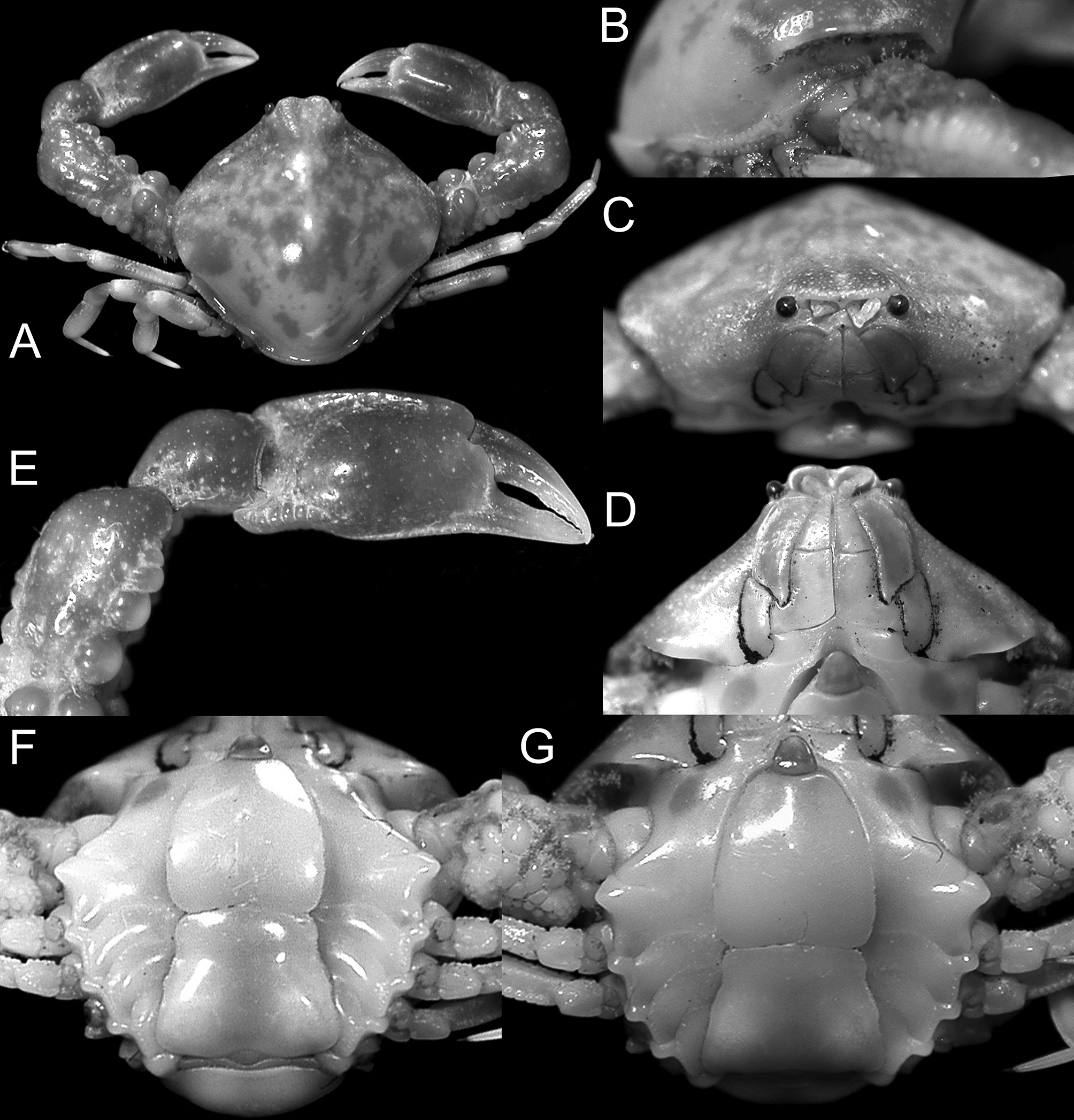
Remarks. The new species is superficially similar to U. purarensis ( Ovaere, 1987) , also described from Madang, Papua New Guinea. The holotype female (and only previously known specimen) was re-examined, and it differs in several key aspects. The merus of the third maxilliped of U. purarensis has a tubercle on the inner distal angle of the ischium ( Fig. 23A View FIGURE 23. A, B ) (merus proportionately longer and the distal margin of the ischium unarmed in U. flammea sp. nov., Fig. 23 View FIGURE 23. A, B C). The ambulatory legs of U. purarensis , notably the meri, are relatively shorter and stouter ( Fig. 23B View FIGURE 23. A, B ) than in to U. flammea ( Fig. 23 View FIGURE 23. A, B D). The merus of the cheliped and chela of U. purarensis ( Figs. 19 View FIGURE 19 E, 20E, 22H) are also proportionately shorter than those of U. flammea sp. nov. ( Figs. 20 View FIGURE 20 A, 21A, E, 22B, F, G), even when females of similar sizes are compared; and the posterior margin of the merus has two diverging rows of granules ( Fig. 19 View FIGURE 19 A) (single row of more lozenge-shaped granules in U. flammea sp. nov., Figs. 21 View FIGURE 21 A, 22A). In addition, the frontal region of U. purarensis is less produced ( Figs. 19 View FIGURE 19 A, 20A) (more projected in U. flammea sp. nov., Fig. 21 View FIGURE 21 A), the posterolateral margin of the pterygostomian region adjacent to the buccal cavity is angled ( Fig. 19 View FIGURE 19 D) (more rounded in U. flammea sp. nov., Fig. 21 View FIGURE 21 D), the posterior carapace margin is proportionately broader in U. purarensis ( Figs. 19 View FIGURE 19 A, 20A) (narrower in U. flammea sp. nov., Figs. 21 View FIGURE 21 A, 22A), the distal part of the fused male abdominal somites 3–5 is noticeably convergent ( Fig. 20 View FIGURE 20 G) (less convergent and relatively broader in U. flammea sp. nov., Fig. 21 View FIGURE 21 F), and the G1 is more sinuous with the distal part distinctly dilated ( Fig. 23 View FIGURE 23. A, B I–L) (less sinuous with the distal part not dilated in U. flammea sp. nov., Fig. 23 View FIGURE 23. A, B E–G).
Etymology. From the Latin, flamma for “flame”, alluding to the flame-coloured markings of the carapace and chelipeds.
Geographical distribution. This species is known only from type location, Papua New Guinea.


| ZRC |
Zoological Reference Collection, National University of Singapore |
No known copyright restrictions apply. See Agosti, D., Egloff, W., 2009. Taxonomic information exchange and copyright: the Plazi approach. BMC Research Notes 2009, 2:53 for further explanation.
|
Kingdom |
|
|
Phylum |
|
|
Class |
|
|
Order |
|
|
InfraOrder |
Brachyura |
|
Family |
|
|
SubFamily |
Leucosiinae |
|
Genus |