
Tanaoa retpela, Galil, Bella S. & Ng, Peter K. L., 2015
|
publication ID |
https://doi.org/ 10.11646/zootaxa.4027.4.1 |
|
publication LSID |
lsid:zoobank.org:pub:3B32D183-45BA-41F9-82A9-C9C108D53899 |
|
DOI |
https://doi.org/10.5281/zenodo.6102266 |
|
persistent identifier |
https://treatment.plazi.org/id/8870B305-E33D-4C0F-9699-F98389768ECD |
|
treatment provided by |
Plazi |
|
scientific name |
Tanaoa retpela |
| status |
sp. nov. |
Tanaoa retpela View in CoL sp. nov.
( Figs. 5 View FIGURE 5 A, 14, 15F–K)
Material examined. Holotype: male (36.5 mm) (MNHN-IU-2013-7961), stn CP3984, Bismarck Sea, northwest Long I., 05°12'S 146°59'E, 500 m, 6.12.2012. Paratypes: 1 male (39.0 mm) ( ZRC 2015.272), stn CP3978, Bismarck Sea, north Bagabag Is., 04°45'S 146°12'E, 456–582m, 5.12.2012. Additional material: BIOPAPUA – 1 female (29.5 mm) (MNHN-IU-2011-3835), stn CP3653, west of New Hanover, 02°13’S 150°23’E, 680–700 m, 28.08.2010; 2 males (38.4 mm, 29.8 mm) (MNHN-IU-2011-914), stn CP3655, west of New Hanover, 02°15’S 150°16’E, 402–440 m, 28.08.2010; 1 male (39.1 mm) (MNHN-IU- 2011-2098), stn CP3669, north of Rabaul, 04°08’S 151°56’E, 382–389 m, 24.09.2010; 1 female (28.7 mm) (MNHN-IU- 2011-2481), stn CP3681, Vitu I., 04°38’S 149°27’E, 564–712 m, 27.09.2010; 1 male (28.6 mm) (MNHN-IU- 2011-2478), stn CP3682, Vitu I., 04°38’S 149°28’E, 515–812 m, 27.09.2010; 3 juveniles (11.7–12.5 mm) (MNHN-IU- 2011-2563), stn DW3748, seamounts near Bougainville, 05°37’S 154°01’W, 398–399 m, 12.10.2010; 1 male (21.4 mm) (MNHN-IU- 2011- 2710), stn CP3695, Feni Is., 02°10’S 147°15’W, 198 m, 14.10.2010; 3 females (19.5–21.0 mm) (MNHN-IU- 2011- 2299), stn CP3760, Is., 03°58’S 153°43’W, 613–660 m, 14.10.2010.
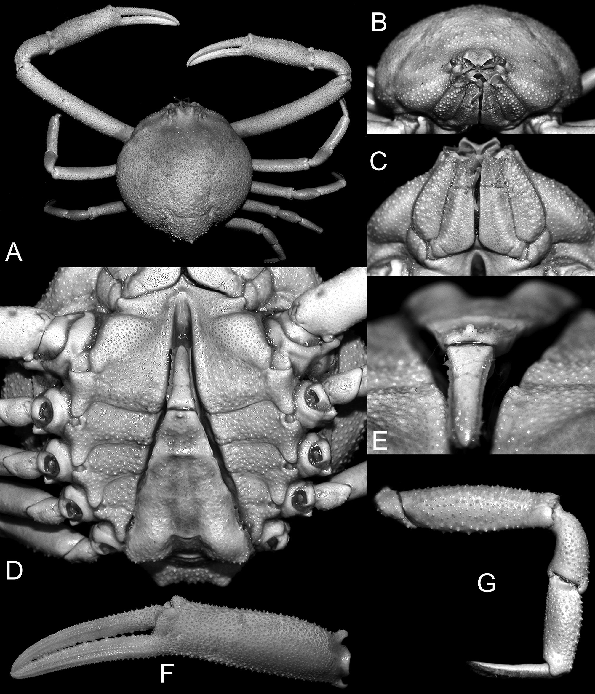
Description. Carapace rounded, slightly wider than long, globose; gastric, cardiac, intestinal regions laterally demarcated by deep grooves ( Fig. 14 View FIGURE 14 A). Dorsal surface covered densely by rounded, pearliform granules of various sizes; 4 pairs of pits along branchiocardiac line, additional pair on mesogastric region ( Fig. 14 View FIGURE 14 A). Intestinal region swollen, anteriorly with rounded median carina, posteriorly with granular tubercle ( Fig. 14 View FIGURE 14 A). Front narrow, slightly produced, upturned, closely set with granules, divided into 2 rounded lobes ( Fig. 14 View FIGURE 14 A–C). Eyes small, retractable within orbit, minutely granulose eyestalk exposed ( Fig. 14 View FIGURE 14 B). Outer orbital margin with 3 sutures. Vshaped gap proximally on ventral margin. Antennular fossae below frontal lobes oblique, antennules obliquely folded, basal antennular operculiform, sealing lower half of antennular aperture when retracted. Antennae small, slender, basal antennal article inserted in orbital hiatus. Postorbital region concave. Anterior margin of efferent branchial channel convex, produced, bilobed, separated by narrow groove from lower orbital margin ( Fig. 14 View FIGURE 14 B, C).
Outer surface of third maxillipeds granular, granules more prominent anteriorly, forming granular ridge mesially on endopod ( Fig. 14 View FIGURE 14 C).
Hepatic margin medially with granular tubercle ( Fig. 14 View FIGURE 14 B, C). Subhepatic region produced, inflated, with horizontal line medially free of granules ( Fig. 14 View FIGURE 14 B, C). Epibranchial margin with 3 equidistant granular tubercles ( Fig. 13 View FIGURE 13 A). Posterolateral margins rounded. Posterior margin of carapace narrow, laterally with 2 prominently granular tubercles ( Fig. 14 View FIGURE 14 A).
Chelipeds slender, subequal, covered with small granules on all articles, including fingers ( Fig. 14 View FIGURE 14 A, F). Cheliped merus, subcylindrical, not nearly as long as carapace; palm subcylindrical, 0.6 as long as merus; fingers longer than dorsal margin of palm, granules arraigned in longitudinal lines, cutting edges denticulate ( Fig. 14 View FIGURE 14 A, F). Ambulatory legs slender; decreasing in size posteriorly; merus, carpus, propodus granular, granules more prominent dorsally; dorsal surface of dactylus setose, dactylar tips corneous ( Fig. 14 View FIGURE 14 A, G).
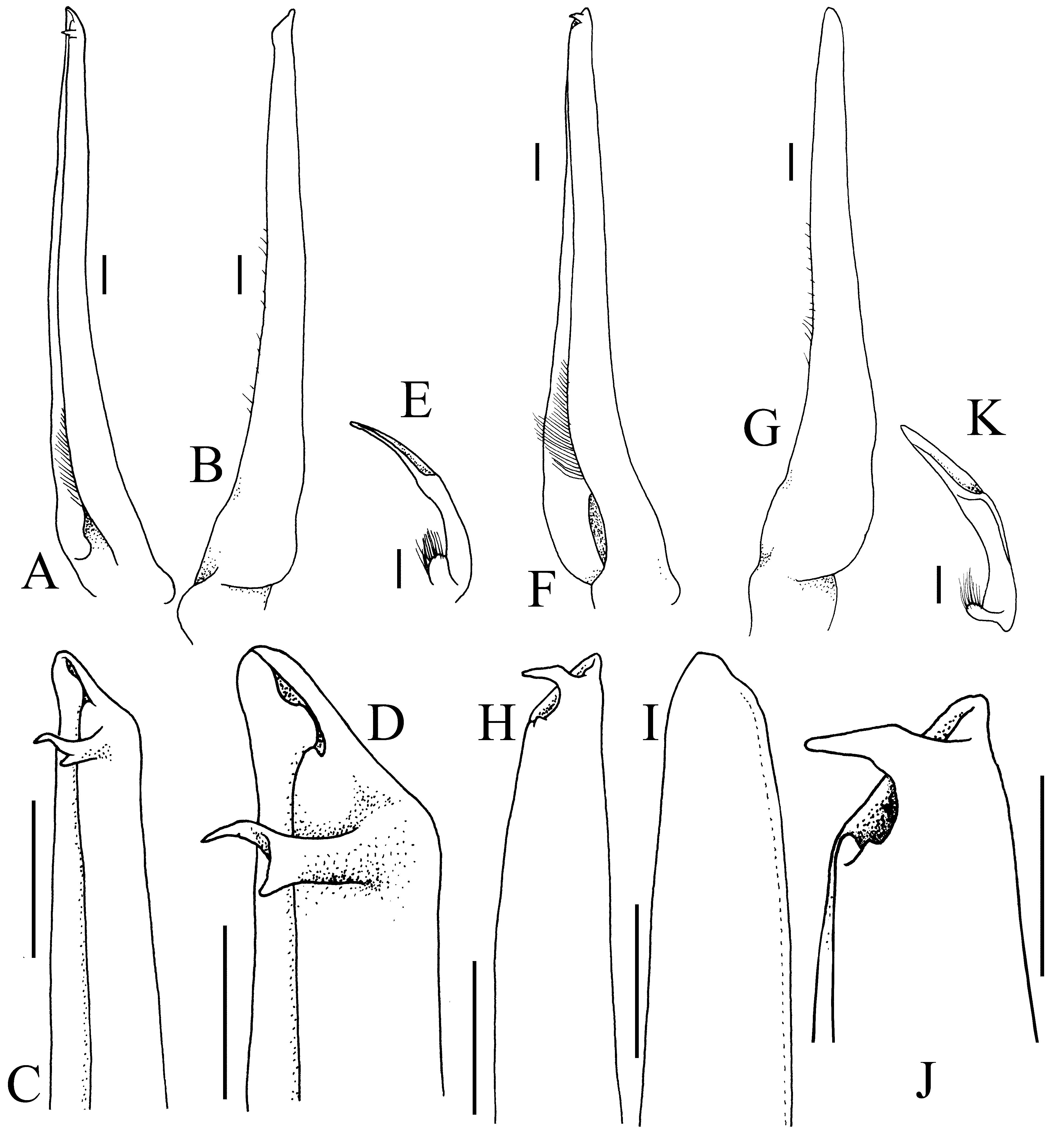
Thoracic sternites set with granules of various sizes; sternite 4 anteriorly inflated ( Fig. 14 View FIGURE 14 D). Male abdominal cavity deep, nearly reaching buccal cavity, anterior margin of cavity ogive, slightly raised ( Fig. 14 View FIGURE 14 D). Male abdomen triangular, elongated; abdominal somites 1, 2 transversely narrow; somite 1 yoke-like, somite 2 medially convex as it fits into cavity of somite 1; somites 3–6 fused, proximo-lateral regions greatly inflated, granular; subterminally with horizontal ridge, anterior margin with quadrate denticle; telson slender, 1/3 as long as fused somites, not reaching tip of abdominal cavity ( Fig. 14 View FIGURE 14 D, E). G1 elongated, slightly sinuous, attenuate, with preapical digitate process perpendicular to tip, opening facing dorsally ( Fig. 15 View FIGURE 15. A – E F–J); G2 short, distally scoop-like ( Fig. 15 View FIGURE 15. A – E K).
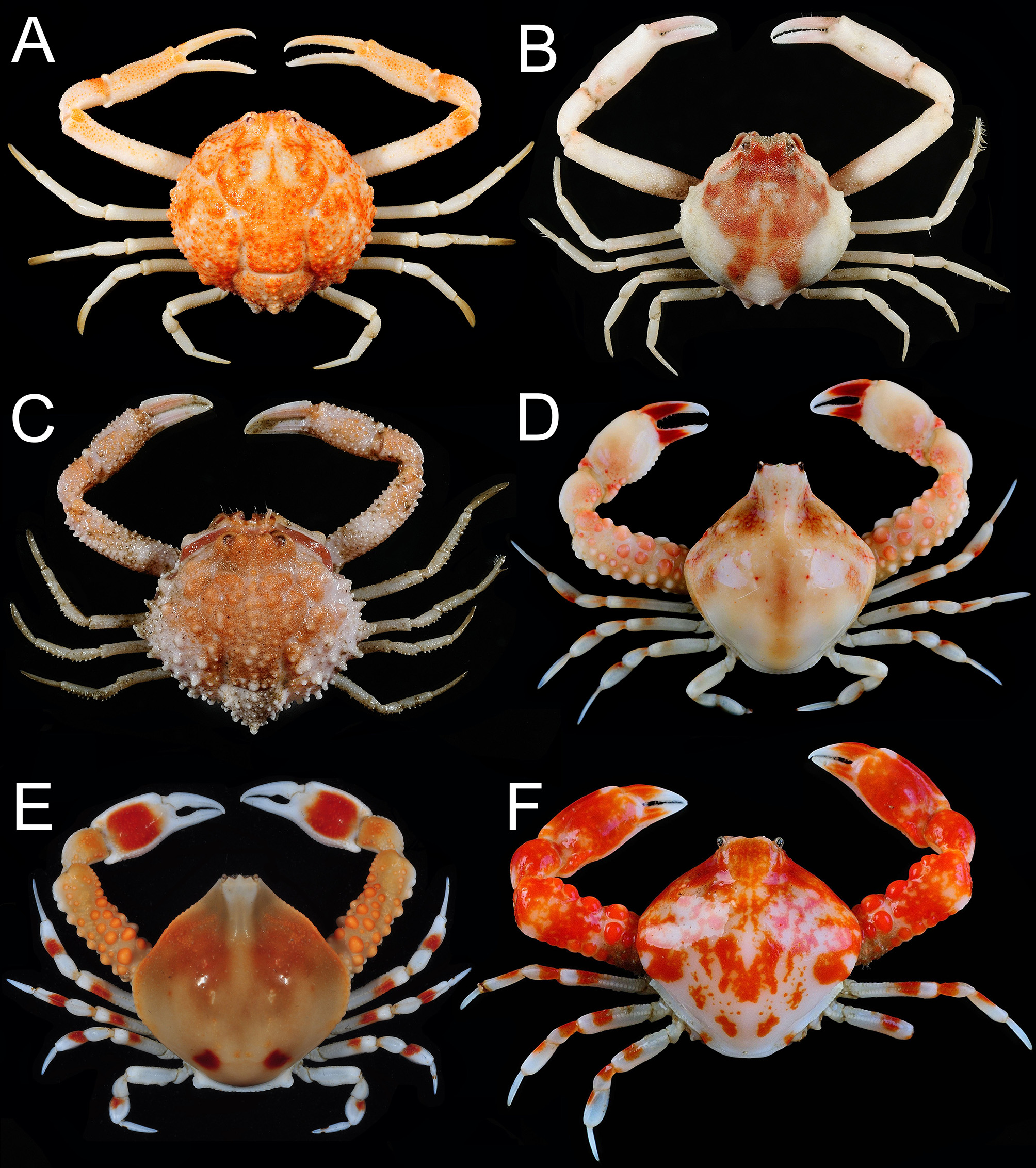
Colour in life. Carapace bright orange-red mottled bone-white. Cheliped merus pale orange, distal part bright orange; palm, fingers mottled pale orange. Ambulatory legs pale ( Fig. 5 View FIGURE 5 A).
Etymology. From retpela for “red” or “orange” in Tok Pisin, the Pidgin language spoken in Papua New Guinea. The name is used as a noun in apposition.
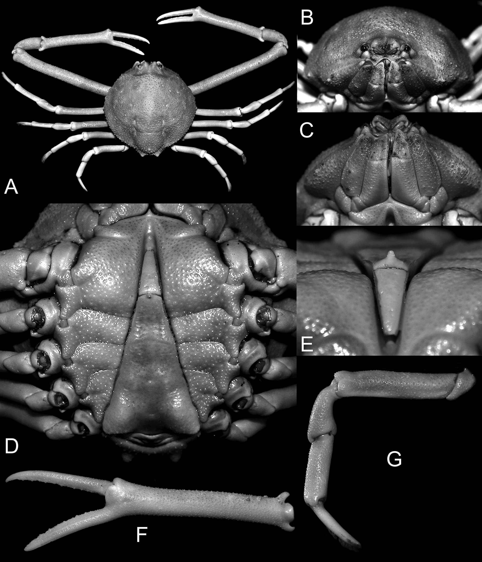
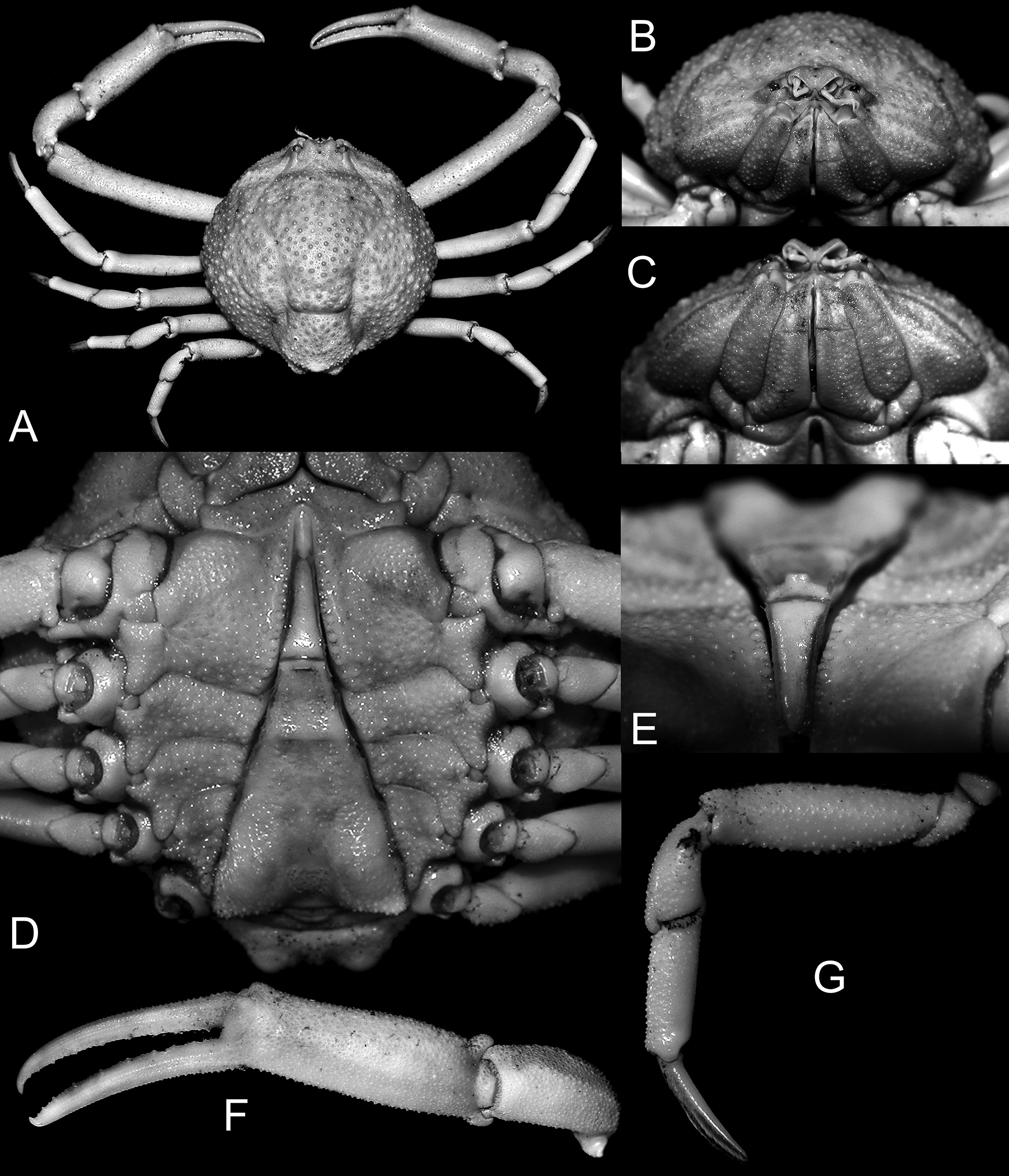
Remarks. Tanaoa distincta , T. serenei , T. kuka sp. nov. and T. retepela sp. nov. are all superficially similar in external appearance. Adults, however, are easy to separate using several reliable characters. Ng & Richer de Forges (2007) appraised the taxonomy of T. distincta and T. serenei , noting that the granulation degree and pattern on the carapace was reliable as a character, and their G1 differ. Among the four species, the granules on the carapace are finest in T. kuka sp. nov. ( Fig. 13 View FIGURE 13 A, B), T. distincta slightly coarser ( Fig. 11 View FIGURE 11 A, B). In T. serenei , the granules are large and densely packed (Fig. 12A, B), while in T. retepela sp. nov. the granules are spaced further apart ( Fig. 14 View FIGURE 14 A, B). Tanaoa kuka sp. nov. is distinguished from the named species by its proportionately elongated chela and palm ( Figs. 13 View FIGURE 13 A, F, 11A, F, 12A, F, 14A, F). With regards to the ambulatory legs, the meri of T. serenei and T. kuka sp. nov. are proportionately the most slender and longest; but whereas in T. serenei , the meri bear sharp granules (Fig. 12A, G) in T. kuka sp. nov. the meri are smoother ( Fig. 13 View FIGURE 13 A, G). The ambulatory meri of T. distincta and T. retepela sp. nov. are proportionately shorter ( Figs. 11 View FIGURE 11 A, G, 14A, G). The G1 of T. distincta is distinct in that the distal part has broad folds or is only subtruncate, lacking subdistal processes (Ng & Richer de Forges 2007: fig. 4A–D). The G1 of T. kuka sp. nov. bears a subdistal bifurcate process ( Fig. 15C, D View FIGURE 15. A – E ). The G1 of T. serenei and T. retepela sp. nov. are superficially similar ( Fig. 15 View FIGURE 15. A – E F–J; Ng & Richer de Forges 2007: fig. 4E, F) in that there is a long subdistal process which tapers to a sharp tip. The overall gonopod structure of T. retepela sp. nov. is nevertheless relatively stouter and the subdistal process relatively shorter ( Fig. 15 View FIGURE 15. A – E F–J) compared to the more slender one of T. serenei which has a relatively longer subdistal process (Ng & Richer de Forges 2007: fig. 4E, F).
Geographical Distribution. This species is known only from the type locality, Papua New Guinea.
| ZRC |
Zoological Reference Collection, National University of Singapore |
No known copyright restrictions apply. See Agosti, D., Egloff, W., 2009. Taxonomic information exchange and copyright: the Plazi approach. BMC Research Notes 2009, 2:53 for further explanation.