
Scalarispongia tubulata, Sandes, Joana, Muricy, Guilherme & Pinheiro, Ulisses, 2016
|
publication ID |
https://doi.org/10.11646/zootaxa.4184.1.10 |
|
publication LSID |
lsid:zoobank.org:pub:1F89E7F4-F460-4F15-88EA-73A92AC88400 |
|
DOI |
https://doi.org/10.5281/zenodo.6085685 |
|
persistent identifier |
https://treatment.plazi.org/id/8B1087F6-FFE4-FFE3-F69D-87480460557C |
|
treatment provided by |
Plazi |
|
scientific name |
Scalarispongia tubulata |
| status |
sp. nov. |
Scalarispongia tubulata View in CoL sp. nov.
( Fig. 4 View FIGURE 4 ; Tab. 1)
Type Specimens : Holotype—MNRJ 17620, Off Estância city (11º21’15.14’’S – 37º06’1.4’’W), Sergipe State, Brazil, 30 m depth, RV “ Oceano I ” Team coll., leg. Petrobras, July 2002 GoogleMaps . Paratype: UFSPOR 223, Off Piaçabuçu city (10º24’30.95’’S – 36º03’6.55’’W), Alagoas State, Brazil, 30 m depth, RV “ Oceano I ” Team coll., leg. Petrobras, July 2003 ( Fig. 1 View FIGURE 1 ). GoogleMaps
Diagnosis. Scalarispongia cushion-shaped with tubular projections of thin walls and surface irregularly microconulose.
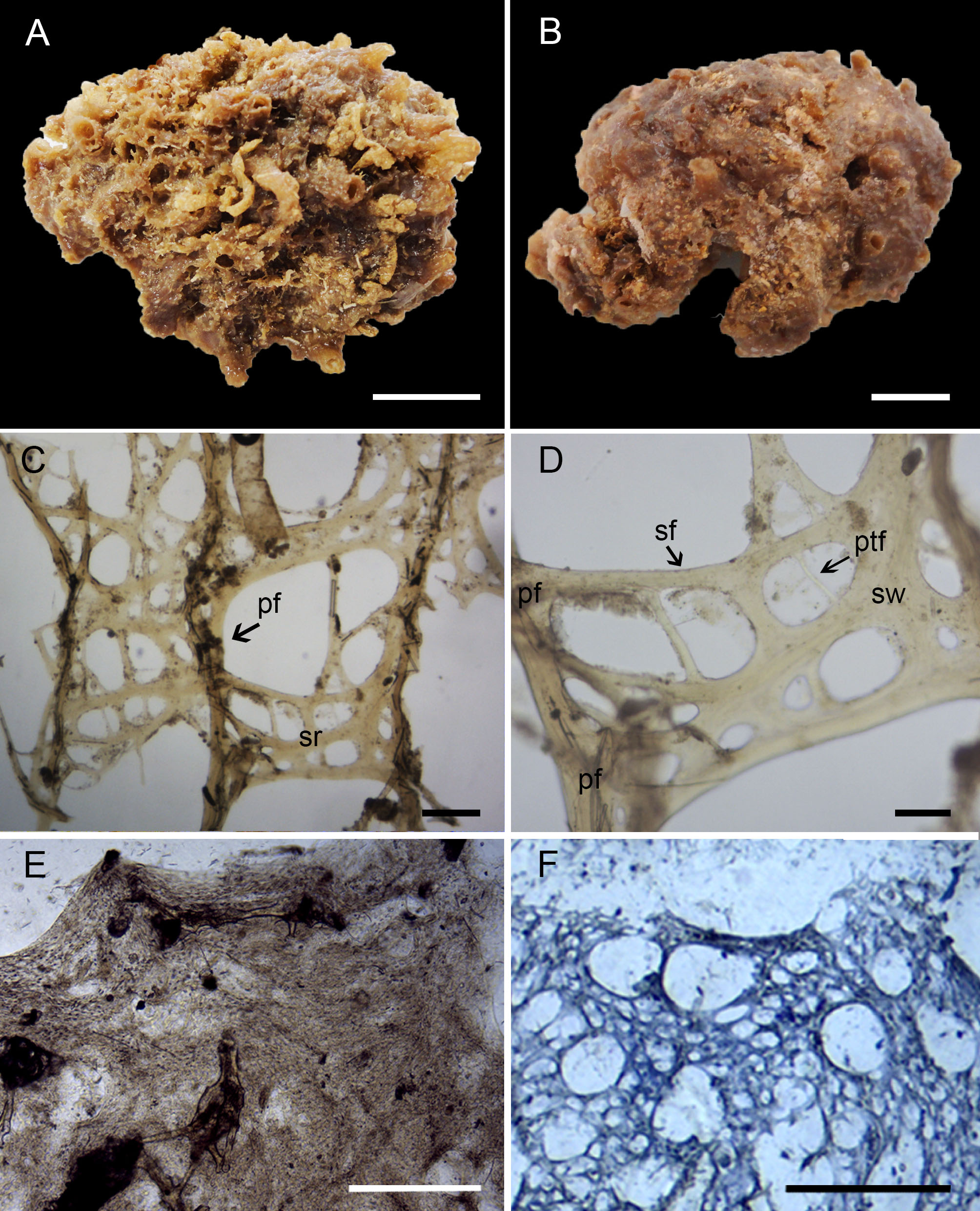
External Morphology ( Fig. 4 View FIGURE 4 A–B). Thick encrusting to cushion-shaped, rounded in the upper part and flat near the base. Holotype measures 5.5 x 2.5 cm (width x height) and paratype measures 6.0 x 1.5 cm (width x height). The color is light brown in ethanol. Color in vivo unknown. Tubular projections are scattered over the surface up to 5 mm high. These projections are fragile and have thin walls. The oscula are located on top of tubes with 1–3 mm in diameter. The surface is irregularly microconulose. The consistency is firm and compressible.
Skeleton ( Fig. 4 View FIGURE 4 C–D) A network of concentrically laminated primary and secondary fibers forms the skeleton ( Fig. 4 View FIGURE 4 C, D). Primary fibers are scattered, axially or totally cored mostly by foreign spicules and debris. Granular pith is not visible. The secondary fiber reticulum is well developed, branching and regular ( Fig. 4 View FIGURE 4 C). All secondary fibers are uncored and occasionally form secondary webs, which are more frequent in the skeleton of the tubes than of the sponge body. In some regions, the secondary fibers connect the primary fibers almost in right angles. Few pseudo-tertiary fibers could be observed, always restricted to the meshes of the secondary reticulum ( Fig. 4 View FIGURE 4 D). Sometimes the fibers are perforated by spicules arranged transversally inside them. Primary fibers are 60–92–140 µm in diameter (n = 12), secondary fibers are 30–64–150 µm in diameter (n = 11) and pseudo-tertiary fibers are 10–25–50 µm in diameter (n = 12). The meshes are circular to oval, 37–82.5–150 µm in diameter, and the secondary webs measure 120–165–230 µm in width (n = 6).
Histology ( Fig. 4 View FIGURE 4 E–F). The ectosome is fibrous, with a layer of elongated microgranular cells, debris and sand grains dispersed outside the fibers ( Fig. 4 View FIGURE 4 E). The choanocyte chambers are diplodal, spherical to oval, with 12.5– 24.4–40 µm in diameter ( Fig. 4 View FIGURE 4 F; n = 15). However, these measurements are probably biased because the fixation in ethanol was not adequate for histological analysis.
Bathymetry and Ecology. The specimens were found at 30 m depth, associated to hydroids and algae.
Geographical distribution. Only known from northeastern Brazil: Sergipe and Alagoas States.
Etymology. The name tubulata refers to the tubular projections typical of the species.
Remarks. Scalarispongia tubulata sp. nov. was allocated in this genus due to the unarmoured surface and regular reticulation with secondary webs, as defined by Cook & Bergquist (2000). However, the new species has a well-developed secondary reticulum similar to Cacospongia species ( Cook, 2007). Compared to the type species Cacospongia mollior Schmidt 1862 , the secondary fiber skeleton of Scalarispongia tubulata sp. nov is regular, with many oval meshes and secondary webs between the primary fibers, whereas the secondary reticulum of C. mollior is irregular and secondary webs are absent ( Cook & Bergquist, 2000; Cook, 2007). The regular reticulation and secondary webs of the new species are more similar to the type species of Scalarispongia , S. scalaris ( Schmidt, 1862) .
Only two species of Scalarispongia and two of Cacospongia were recorded from the Tropical Western Atlantic Ocean. Scalarispongia linteiformis (Lamarck, 1814) was recorded from Greater Antilles and is known only from the original description. It has a ramose shape, thus clearly differing from Scalarispongia tubulata sp. nov. Scalarispongia cincta ( Boury-Esnault, 1973) is transferred here to Thorecta (see above). Cacospongia amorpha Poléjaeff, 1884 and Cacospongia levis Poléjaeff, 1884 were described for the Brazilian coast. Cacospongia amorpha has a massive to rounded shape, oscules flush with the surface and larger, more spaced conules; C. levis has a massive shape, smooth surface and black external color ( Poléjaeff, 1884; Muricy et al., 2011 figs. 8C–D). Scalarispongia tubulata sp. nov. differs from these species by its cushion shape, tubular oscula and microconulose surface (Tab. 1).

No known copyright restrictions apply. See Agosti, D., Egloff, W., 2009. Taxonomic information exchange and copyright: the Plazi approach. BMC Research Notes 2009, 2:53 for further explanation.
|
Kingdom |
|
|
Phylum |
|
|
Class |
|
|
Order |
|
|
Family |
|
|
SubFamily |
Thorectinae |
|
Genus |