
Tokyosoma phialiferum, Mikhaljova, Elena V. & Lim, Kil-Young, 2008
|
publication ID |
https://doi.org/10.5281/zenodo.184776 |
|
DOI |
https://doi.org/10.5281/zenodo.6229645 |
|
persistent identifier |
https://treatment.plazi.org/id/8C058934-C42C-FF95-FF41-FB56FEA99367 |
|
treatment provided by |
Plazi |
|
scientific name |
Tokyosoma phialiferum |
| status |
sp. nov. |
Tokyosoma phialiferum View in CoL sp. n.
Figs 10–11 View FIGURES 10 – 11
Material examined: Holotype: 1 male (ChNU), Andong, Gyeongsangbuk–do, South Korea, 28 October 1990, leg. K.Y. Lim; Paratypes: 2 females (ChNU), same locality as for holotype, 28 October 1990, leg. K.Y. Lim.
Diagnosis: Differs from congeners mainly by the presence large cup–shaped lateral sheath processes of the posterior gonopod colpocoxites and the arched lateral branches of the colpocoxites.
Description: Male. Length 8.0 mm, width about 0.9 mm. Coloration in alcohol pale. Head and collum with feebly marked design of brownish spots. Legs with marbled brown distal part. Eyes black. Antennae brown.
Body with 29 segments. Head densely setose, vertigial suture hardly visible. Eye patches subtriangular, ocelli 20. Collum semicircular. Body width gradually growing until somite 7, body parallel–sided on somites 8–21(22), onward gradually tapering.
Beginning from somite 2(3) somites with normally developed paraterga which gradually grow less distinct toward hind part of body. Metazonital macrochaetae in transverse row on somites 27–28, like elongate triangle on preceding somites. Macrochaetae long, pointed apically, but not very sharply so.
Legs long and slender. Each claw of legs 1–2 at base with two small additional claws dorsally and long setiform outgrowth ventrally. Leg pairs 3–7 not larger than other walking legs. Leg pairs 3–7 with small group of funnel–shaped tarsal papillae apically near claw. Each claw of leg pairs 3–7 with long setiform outgrowth ventrally only i.e. without dorsal additional small claws. Postgonopodal legs (including leg pairs 10 and 11) without tarsal papillae; each claw of them at base with long setiform outgrowth ventrally and two small additional claws dorsally; additional claws gradually missing toward very hind legs. Each claw of very hind legs with long setiform outgrowth ventrally only. Legs 10 and 11 with coxal glands. Coxae 10 and 11 without protruding modifications (excluding the openings of coxal glands).
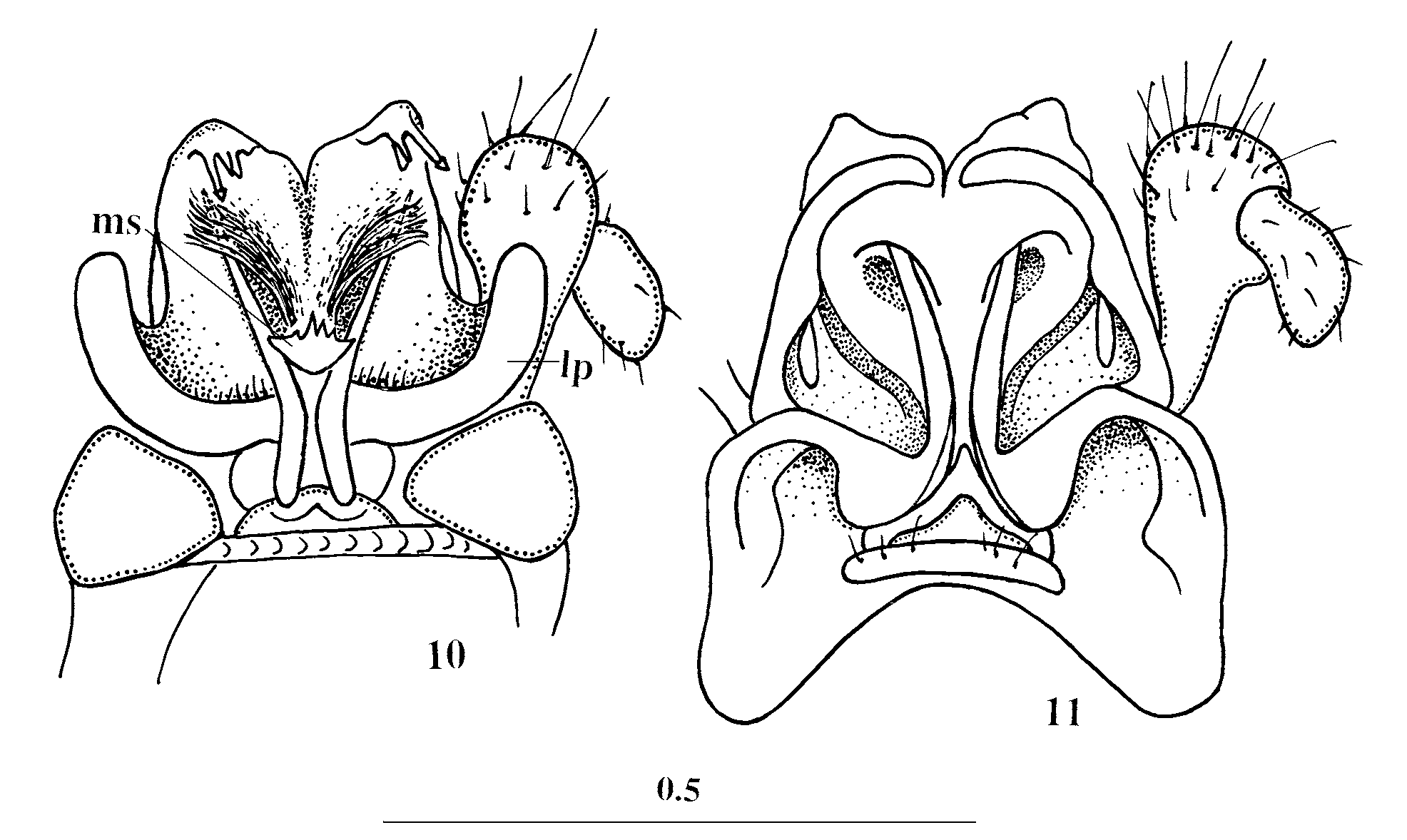
Coxosternum of anterior gonopods with several setae. Anterior gonopod telopodite 1–segmented, subflagelliform, its distal part positioned inside sheaths like bunch of rays ( Fig. 10 View FIGURES 10 – 11 ). Posterior gonopod colpocoxites broad, fused 2/3 extent. Distal part of colpocoxite curved posterad. Mesal sheath processes of posterior gonopod colpocoxites fused medially and forming small single structure (ms) clasping anterior gonopods. Lateral sheath processes of colpocoxites (lp) very large cup–shaped inside set with thin setiform cuticular spinules. Posterior gonopod angiocoxite with globule in posterior view. Posterior angiocoxal processes absent. Angiocoxite depressed centrally in anterior view supply with one long process; distal portion of this process sheathed by colpocoxite on anterior side ( Fig. 11 View FIGURES 10 – 11 ). Each colpocoxite with lateral branch arched distally. Basal part of colpocoxite lateral branch fastened to angiocoxite frontally. Posterior gonopod telopodite with short femur.
Female. Length 8.5–9.0 mm, width about 1.0 mm. Body with 29 segments. Ocelli 22–23. Vulvae not dissected.
Etymology: The specific epithet refers to the cup–shaped lateral sheath processes of colpocoxites of posterior gonopods.
No known copyright restrictions apply. See Agosti, D., Egloff, W., 2009. Taxonomic information exchange and copyright: the Plazi approach. BMC Research Notes 2009, 2:53 for further explanation.
|
Kingdom |
|
|
Phylum |
|
|
Class |
|
|
Order |
|
|
Family |
|
|
Genus |