
Adontorhina keegani Barry & McCormack
|
publication ID |
https://doi.org/ 10.5281/zenodo.177538 |
|
DOI |
https://doi.org/10.5281/zenodo.5665911 |
|
persistent identifier |
https://treatment.plazi.org/id/8C428A6B-2061-B65A-FF3E-FCCA9B3AFA80 |
|
treatment provided by |
Plazi |
|
scientific name |
Adontorhina keegani Barry & McCormack |
| status |
sp. nov. |
Adontorhina keegani Barry & McCormack , new species
( Figures 1–3 View FIGURE 1 View FIGURE 2 View FIGURE 3 )
Type locality. Porcupine Bank, 53° 29.9’N, 13° 59.9’W, 300 m Eastern Atlantic.
Holotype. A complete shell, collected by P.J. Barry (10/11/03), NMINH.2006.57 Measurements (Length x height x breadth) 0.94 mm x 0.7 mm x 0.38 mm.
Paratypes. Three complete shells, as holotype, NMINH.2006.64.1–4. Measurements 0.6 mm x 0.42 mm x 0.3 mm; 0.73 mm x 0.55 mm x 0.35; 0.68 mm x 0.49 mm x 0.33 mm.
Two paratypes prepared for electron microscopy, NMINH.2006.65. Measurements 0.92 mm x 0.7 mm x 0.37 mm. NMW.Z.2007.008. Measurements 0.98 mm x 0.76mm x 0.5 mm.
Etymology. Named after Professor Brendan F. Keegan in recognition of his contribution to marine science studies in Ireland over many years.
Material examined. CEO3 Station 8 52° 59.9’N, 13° 59.9’W, 191.6 m, 4 specimens; CEO3 Station 0 9, 53° 29.9’N, 13° 59.9’W, 300 m, 3 specimens; CEO4 Station 0 5, 52° 59.9’N, 12° 44.9’W, 789 m, 8 specimens.
Distribution. Found in muddy sand on the Porcupine Bank, West of Ireland, on either side of the highest point of the bank. Depth range 300 – 789 m.
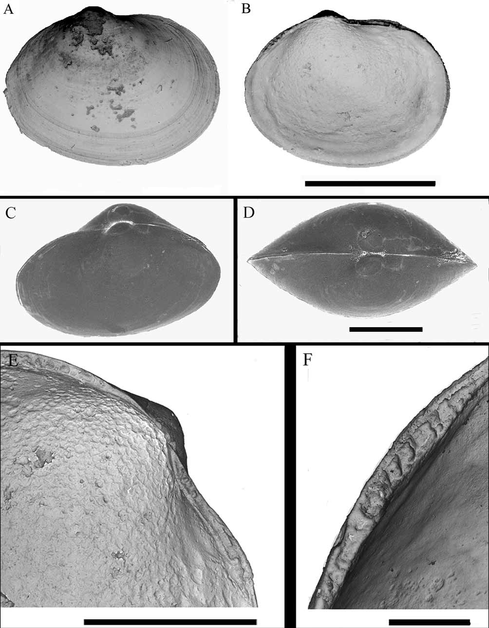
Shell description. Shell minute, maximum length to 0.98mm, fragile, compressed; elongate oval, length / height ratio of 1.2-1.36; inequilateral, anterior end longer; anterodorsal margin straight initially, rising above the horizontal plane before descending into broadly rounded anterior; ventral margin weakly curved until intersected by the weak posterior sulcus; umbones small, sunken, orthogyrate; prodissoconch I approximately 130 µm in diameter; lunule obscure, with raised commissure; escutcheon obscure; periostracum thin, lightly straw coloured; surface smooth near the umbones, thickened commarginal striae towards the margins, radial striae few, confined to the posterior ( Figure 1 View FIGURE 1 A); colour white, transparent in juveniles; ligament mostly internal, on a sunken plate, one third the length of the dorsal margin; hinge plate composed of two sections ( Figure 1 View FIGURE 1 E), anterior section thinner than posterior section. Irregular granules visible in both valves, anterior and posterior to the beak; directly below the beak, hinge plate is not visible.
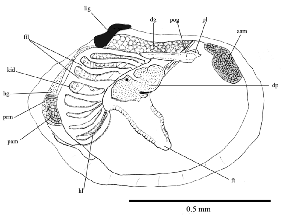
Internal anatomy. Both adductor muscles are relatively large, the posterior muscle is rounded but with a tapered ventral end; both muscles are divided into quick and catch areas ( Figure 2 View FIGURE 2 ); anterior muscle much larger than the posterior. There is a single point of mantle fusion to form the posterior exhalent aperture. The mantle is thin, and contains a small glandular area below the anterior adductor muscle; inner mantle fold not expanded, with a small cluster of gland cells overlain by a thin layer of radial muscle; rejection tract wide and shallow; middle and outer mantle folds very short, forming a shallow periostracal groove. Each gill has a single demibranch, comprised of seven to eight filaments; gill filaments type 2 ( Dufour, 2005); filaments short but laterally expanded with well developed filamentar muscles; latero-frontal cilia well developed; interfilamentar junctions occur. Labial palps small, positioned near the end of the proximal oral groove; groove very long, wide. Oesophagus short, descending into a small stomach. Hindgut loops very high before descending along the posterior margin, through the pericardium, becoming markedly widened as it descends down to the posterior adductor muscle. The lateral pouch is very small (in contrast to most other thyasirids); just visible underneath the anterior end of the gill filaments with one marked indentation in its surface; pouch unlobed, not divided. Digestive gland and kidney large (consistent with the other species in the Thyasiridae ). Foot short and well ciliated, the cilia extending back over the heel; tip of the foot very narrow and pointed; heel very well developed as are the pedal retractor muscles; heel large, extending very far down into the mantle cavity; heel sagittally grooved; pedal retractor muscles well developed.
Differential diagnosis. The distinctive biangulate posterior shell margin separates Adontorhina keegani from other Adontorhina species. Also, A. keegani is markedly smaller than other species of Adontorhina which are usually 1.5 to 3 mm in diameter ( Scott, 1986). The internal anatomy appears reduced compared to other Adontorhina species, with few gill filaments to each demibranch and small lateral pouches. The hindgut of A. keegani is greatly expanded in comparison with most other thaysirid species. Further features which separate A. keegani from other Adontorhina species can be found in Table 1 View TABLE 1 .
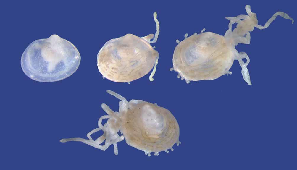
Remarks. Hydroids were found growing on the valves of living specimens of A. keegani ( Figure 3 View FIGURE 3 ). Only one specimen out of fifteen was recorded as being free of epifauna.
Most of the specimens had a disproportionate grouping of hydroids on the posterodorsal margin. The hydroids on the posterior were always the largest and in some cases, grew to double the length of the shell they were attached to. Smaller hydroids were observed on the ventral and anterior margins. The occurrence of this epifauna was limited to the vertical axis of the shell, present only where the margins meet.
Adontorhina Adontorhina Adontorhina Adontorhina Adontorhina cyclia sphaericosa lynnae keegani similis
No known copyright restrictions apply. See Agosti, D., Egloff, W., 2009. Taxonomic information exchange and copyright: the Plazi approach. BMC Research Notes 2009, 2:53 for further explanation.
|
Kingdom |
|
|
Phylum |
|
|
Class |
|
|
Order |
|
|
Family |
|
|
Genus |