
Lepidopora diffusa (Boschma, 1963)
|
publication ID |
https://doi.org/10.11646/zootaxa.3691.1.1 |
|
publication LSID |
lsid:zoobank.org:pub:E98CE6DF-AF3B-4AAA-95CB-8ACD615C9FCC |
|
DOI |
https://doi.org/10.5281/zenodo.5619725 |
|
persistent identifier |
https://treatment.plazi.org/id/955B87C9-A172-DD2F-FF22-F9D7F0662A9F |
|
treatment provided by |
Plazi |
|
scientific name |
Lepidopora diffusa (Boschma, 1963) |
| status |
|
Lepidopora diffusa (Boschma, 1963) View in CoL
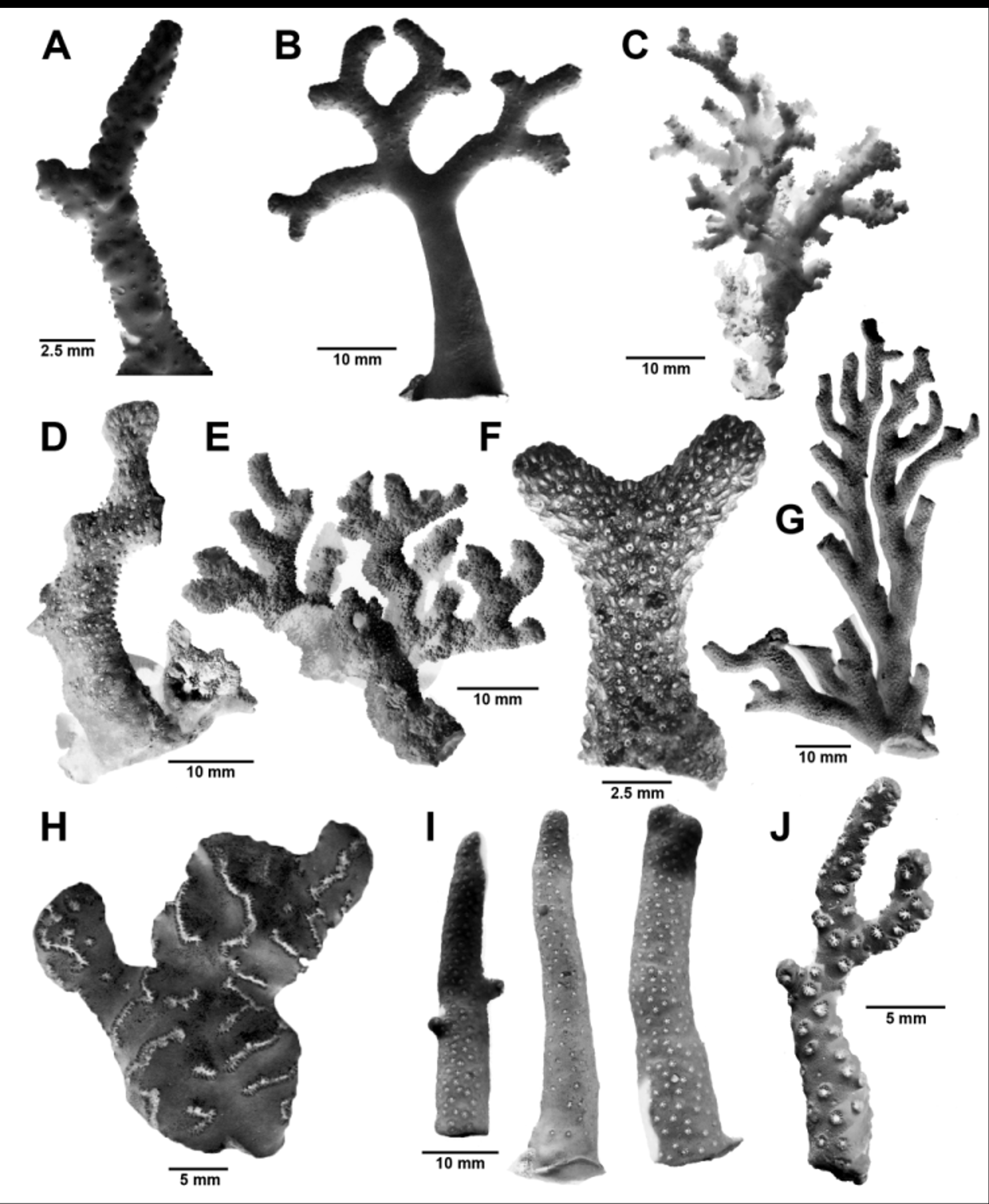
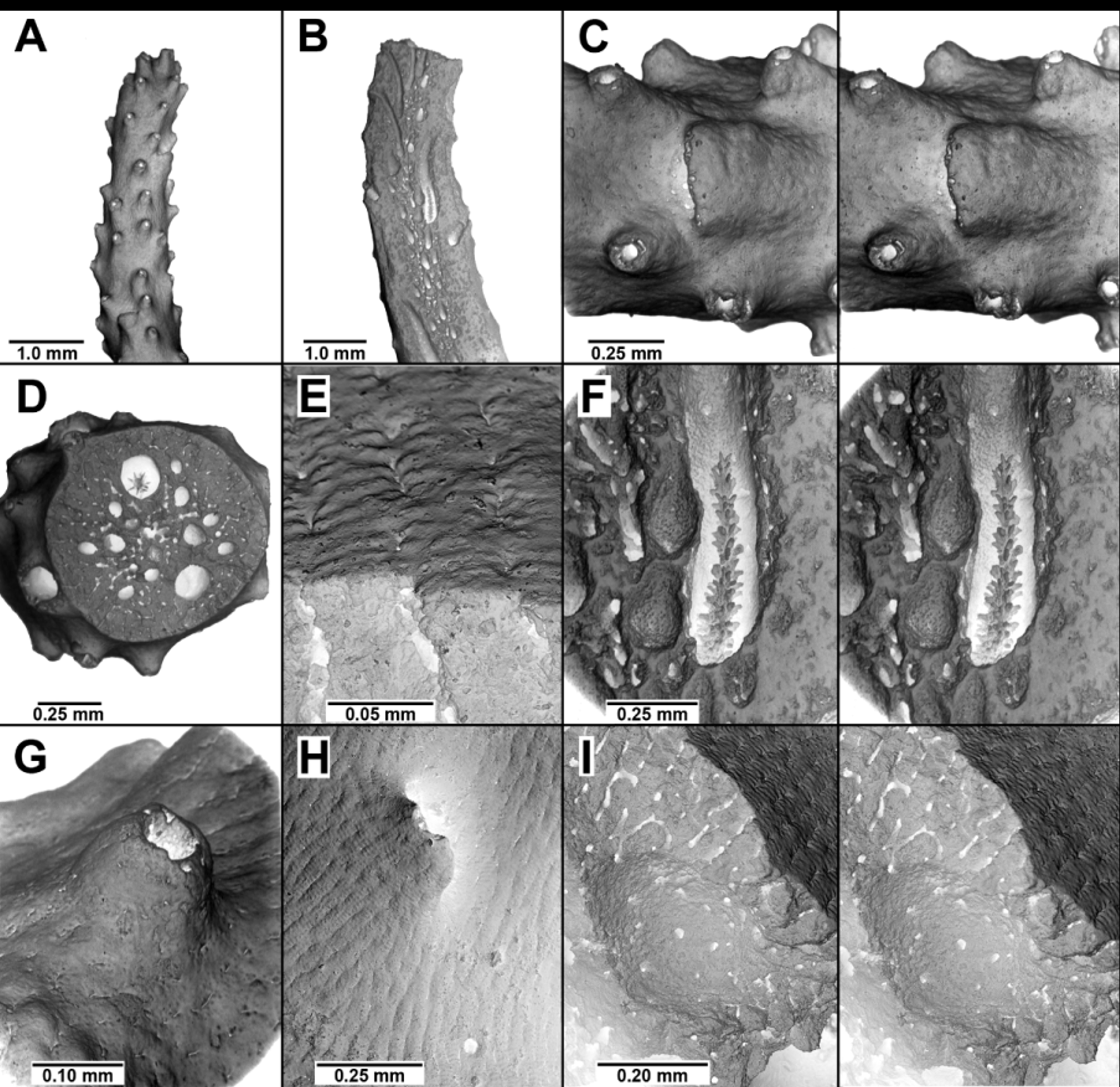
Figs. 1 View FIGURE 1 A–B, 4A–I, 22
Errina (Lepidopora) diffusa Boschma, 1963b: 391 –396, 1 pl., 3 figs.; 1964: 61; 1966b: 112 (mention); 1967: 333; 1968, 207 (diagnosis)—Vervoort & Zibrowius, 1981:16, 27–28 ( lectotype designation).
Lepidopora diffusa: Cairns, 1983b: 428 (new comb.).—Cairns et al., 1999: 44 (listed).
Types and Type Locality. The lectotype was designated by Vervoort & Zibrowius (1981: 28) as the figured specimen (Boschma, 1963b: pl. 1, fig. 2) from UCTES SCD296 (RMNH 13751). Fourteen additional paralectotypes from stations UCTES SCD254 and 296 are deposited at the Naturalis Biodiversity Centre (13801, 13802). Part of one paralectotype from UCTES SCD296 is also deposited at the NMNH as SEM stub 121 (USNM 96200). Type Locality: 33°07.3’S, 28°01’E (off East London, South Africa), 88 m.
Material Examined. Types; MN SM163, 25 fragments, SAM H3149; MN SM185, 2, SAM H3152; PF 808, 4, SAM H1231; PF 7023, 3, SAM H3088; PF 12104, 1, SAM H1236; PF 13061, 4 branches, SAM H3086; PF 13654, 28 branches, SAM H1216, 1 branch, BM 1980.9.19.14, and 1 colony and SEM stub 1665 (USNM 76395); PF 12312, 14 colonies, SAM H3089, and 1 branch, BM 1980.9.19.13.
Description. Colonies are bushy to planar ( Fig. 1 View FIGURE 1 B), the largest corallum (the lectotype) 50 mm in height and 26 mm in width, with a basal branch diameter of 3–4 mm. Branching is sparse, dichotomous, and equal, the branching axils usually 90°. Branches are circular to slightly elliptical in cross section, terminating in slender tips about 1 mm in diameter, the tips blunt rather than pointed; there is no branch anastomosis. The coenosteum is linear-granular in texture, the coenosteal strips 40–65 µm in width, the granules arranged in transverse rows (about 95 rows/mm), each row consisting of 3–5 low rounded granules ( Fig. 4 View FIGURE 4 E). The transverse rows of granules appear similar to a linear-imbricate structure. Colonies are dimorphic in colour, some being a light pink with white branch tips and branch cores, and others homogeneously white.
Gastropores uniformly distributed on branches, circular in shape ( 0.12–0.20 mm in diameter), and usually bordered abaxially by a broad, low lip ( Figs. 4 View FIGURE 4 C, H), which causes the polyp to be directed anteriorly. Gastropore tubes are cylindrical and long, 3 to 4 times the length of the gastrostyle, often seen in branch cross section ( Fig. 4 View FIGURE 4 D) to have the same diameter as the gastropore itself; a ring palisade is absent. The gastrostyle is cylindrical and elongate (L:W = 4.5–5.5), up to 0.7 mm long and about 0.12 mm in maximal diameter. Uniformly spaced blunt spines (up to 45 µm long and 14 µm in diameter) cover the style and are slanted in an upward direction. Dactylopore spines are also uniformly distributed over the branch surface and occasionally arranged in short linear series of 4–10 usually along the branch edge. They are low (up to 0.2 mm tall), apically perforate mounds ( Fig. 4 View FIGURE 4 G), the circular apical pore about 85 µm in diameter and the dactylopore mound being about 0.20 mm in basal diameter and 0.12–0.14 mm in distal diameter. The tubular mounds are usually slanted anteriorly. Dactylopore tubes extend for long distances down the branch centre (axial dactylopores, Fig. 4 View FIGURE 4 B).
Internal cavities just below the branch surface 0.35–0.45 mm in diameter are common, some of which appear to communicate to the surface via very small efferent pores, these cavities presumed to be the male ampullae ( Fig. 4 View FIGURE 4 I). Female ampullae were not observed.
Comparisons. Sixteen species and another two forms (see Zibrowius & Cairns 1992) are known in the genus Lepidopora , however none are known from the Indian Ocean or southeastern Atlantic. Given the insularity of stylasterid species, this is a good indication that L. diffusa is a discrete species. Nonetheless, only five species, including L. diffusa , have abcauline gastropore lips, but the other four species have linearly arranged gastro- and dactylopores, whereas those of L. diffusa are uniformly distributed. Conversely, of the ten species having uniformly distributed gastro- and dactylopores, including L. diffusa , none have gastropore lips.
Remarks. Boschma’s (1963b) original description was fairly complete, the only contributions made herein relate to observations of the male ampullae, details of the coenosteal texture based on better preserved specimens, more detailed corallum illustrations made possible with the SEM, and several additional distributional records.
Distribution. Known from off southeastern South Africa between Richards Bay ( Natal) and Sebastian Bluff, Agulhas Bank (Fig. 22), 47– 101 m.
No known copyright restrictions apply. See Agosti, D., Egloff, W., 2009. Taxonomic information exchange and copyright: the Plazi approach. BMC Research Notes 2009, 2:53 for further explanation.
|
Kingdom |
|
|
Phylum |
|
|
Class |
|
|
Order |
|
|
Family |
|
|
Genus |