
Amphibolips salicifoliae Medianero & Nieves-Aldrey
|
publication ID |
https://doi.org/10.5281/zenodo.275711 |
|
DOI |
https://doi.org/10.5281/zenodo.6201103 |
|
persistent identifier |
https://treatment.plazi.org/id/9702547D-FFB6-FFFF-FF3D-2DAC40F5F86F |
|
treatment provided by |
Plazi |
|
scientific name |
Amphibolips salicifoliae Medianero & Nieves-Aldrey |
| status |
sp. nov. |
Amphibolips salicifoliae Medianero & Nieves-Aldrey sp. nov.
( Figs.5 View FIGURE 5 , 6 View FIGURE 6 , 7 View FIGURE 7 F, 7G & 8G–H)
Type material. Holotype Ƥ ( Fig. 7 View FIGURE 7 F) (in Museo Nacional de Ciencias Naturales, Madrid, Spain, cardmounted. Cat nº 2024). PANAMA, Chiriquí, Volcan Baru 8º 47' 50 8” N, 82º 29' 35 9” W, 1800–2070m; ex gall on leaf of Quercus salicifolia Née (Fagaceae) , gall collected 27.i.2009, insect emerged ii.09, E. Medianero leg. Paratypes: 2Ƥ, 23: same data as holotype. Two paratypes in MNCN, two paratypes in Maestría en Entomología, Universidad de Panamá ( MEUP).
Additionally, 1Ƥ of the type series was dissected for SEM observation (in MNCN).
Etymology. Named after the hosp plant species Quercus salicifolia .
Diagnosis and comments. The coarsely rugose sculpture of head and mesosoma, trapezoid shape of clypeus projecting ventrally, short gula, antenna with 11 flagellomeres, with first flagellomere long, the robust and short mesosoma, with coarse rugose sculpture, the lateral carinae of propodeum slightly divergent, and the shape and setation of the hypopygial spine include the new species within the genus Amphibolips . Additionally, the general structure of the male, especially the 15-segmented antenna with the first flagellomere flattened and slightly expanded dorsally, and the structure of the gall, fit also well with the characters of the Amphibolips species. However, A. salicifoliae differs from all known species of Amphibolips by its simple metatarsal claws. From the other Panamanian Amphibolips species described here, A. salicifoliae differs, besides in claw structure, in its predominantly brown-rufous coloration, almost hyaline wings (F6-F10 at most as long as wide), its broad and smooth posterior notauli, a posterior scutellum margin that is not emarginate, metasomal tergites that are not micropunctate and by the relatively shorter projection of the hypopygial spine.
The Nearctic species A. quercusracemaria (Ashmead, 1881) and A. nubilipennis (Harris, 1841) induce similar spherical leaf galls as the galls of the new species from Panama, although the inner structure of the former is different (Melika pers. comm.). However, A. salicifoliae differs from A. quercusracemaria in at least wing coloration, the number of antennal flagellomeres and the absence of micropunctures on the metasoma. The new species is different from A. nubilipennis in coloration; shape of notauli, smooth scutellar foveae and absent metasomal micropunctures.
Description. Body length 3.77 mm (range 3.58–3.91; N = 3) for females; 3.15 mm (range 2.91–3.4; N = 2) for males. Head, mesosoma, metasoma and coxae of female shining brownish-rufous. Antenna and fore and middle legs, excepting coxae light yellowish hind legs yellowish brown. Forewing hyaline, with some very light infumation, veins dark brown. Male: head and mesosoma black. Metasoma black to dark brown. Mandibles, antenna and legs dark brown, excepting anterior and middle light brown tibiae and tarsi.
Female. Head, wrinkled to coarsely rugose, pubescent, in dorsal view about 2.9 times wider than long. POL 1.3 times longer than OOL, posterior ocellus separated from inner orbit of eye by 2.5 times its longest diameter. Head in anterior view more or less oval ( Fig. 5 View FIGURE 5 A), 1.25 times wider than high. Genae not expanded behind eyes. Vertex, frons, and occiput coarsely rugose; sculpture less strong medially on face; vertex and frons barely pubescent with sparse and shorter setae, occiput with relatively long setae. Clypeus trapezoid, 1.2 times wider than high, shining smooth, moderately pubescent, ventral margin slightly sinuate and projecting over mandibles. Anterior tentorial pits conspicuous; epistomal sulcus and clypeo-pleurostomal lines indistinct. Malar space 0.38 times as height of compound eye. Distance between antennal rim of torulus and compound eye 0.8 times its width including rim. Ocellar plate slightly raised. Head, posterior view ( Fig. 5 View FIGURE 5 B). Gula relatively short; distance between occipital and oral foramina shorter than height of the occipital foramen. Occiput without occipital carina, with some strong transverse wrinkles.. A carina dorso-lateral to occipital foramen present, curved, ventrally continuing pass posterior tentorial pits. Hypostomal sulci separated at hypostoma.
Mouthparts ( Fig. 5 View FIGURE 5 B). Mandibles strong, exposed; with dense setae in base, right mandible with three teeth; left with two teeth. Cardo of maxilla not visible, maxillary stipes about 2.1 times longer than wide. Maxillary palp 5-, labial palp 3-segmented.
Antenna 0.4 times as long as body ( Fig. 6 View FIGURE 6 A); with 11 flagellomeres, flagellum not broadening towards apex; with relatively long, erect setae and elongate placodeal sensilla visible only on F5–F11. Relative lengths of antennal segments: 25:15:49:31:25:26:19:17:15:15:14:13:30. Pedicel, globose, small, 0.6 as long as scape; F1 1.5 times as long as F2 ( Fig. 6 View FIGURE 6 B). F7-F10 as long as wide or slightly transverse; F11 2.0 times longer than wide, 2.3 times as long as F10 ( Fig. 6 View FIGURE 6 C).
Mesosoma. Coarsely rugose, 1.2 times as long as high in lateral view. Pronotum, with rugose sculpture and densely pubescent laterally. Ratio of length of pronotum medially/laterally = 0.23.
Mesonotum. Mesoscutum ( Fig. 5 View FIGURE 5 C), moderately and uniformly pubescent, with moderately strong rugose sculpture. Notauli traceable only in posterior one third of mesoscutum length; deep, very broad, smooth and abruptly terminated. Median mesoscutal impression indistinct. Anteroadmedian signa visible. Transscutal fissure narrow. Scutellum ( Fig. 5 View FIGURE 5 F), rounded, about 0.3 as long as mesoscutum, coarsely rugose, posterior margin not emarginate. Scutellar foveae ovoid, deep and smooth, their anterior margins forming an arc contra to transscutal fissure; posterior margins diffuse about 0.3 as long as scutellum, separated by septum. Scutellum, in lateral view, overlapping dorsellum. Mesopleuron ( Fig. 5 View FIGURE 5 D) sculptured and setose only in medial and basal areas, smooth and bare posterodorsally.
Metanotum ( Fig. 5 View FIGURE 5 E). Metapectal-propodeal complex. Metapleural sulcus reaching posterior margin of mesopectus at mid-height of metapectal-propodeal complex ( Fig. 5 View FIGURE 5 D). Lateral propodeal carinae distinct, slightly divergent ( Fig. 5 View FIGURE 5 E). Median propodeal area smooth and glabrous ( Fig. 5 View FIGURE 5 E).
Legs. Metatarsal claws simple, without basal lobe or tooth ( Fig. 6 View FIGURE 6 G).
Forewing ( Fig. 7 View FIGURE 7 H). As long as body, hyaline, without conspicuous darkened spots or infuscate areas, veins strongly pigmented. Radial cell 3.6 times longer than wide; open along anterior margin; areolet small, triangular. Rs slightly bowed, M nearly straight, not reaching wing margin. Rs+M reaching basalis at its midheight. First abscissa of radius (2r) curved, 2r-m straight. Hair fringe on apical margin short.
Metasoma ( Fig. 6 View FIGURE 6 H). Smooth and shiny; large, as long as head and mesosoma combined; in lateral view 1.12 times as long as high. T3 covering about 2/3 of metasoma; without micropunctures; with sparsed long setae anteromedially. Projecting part of hypopygial spine, beyond attachment of lateral flap, relatively short ( Fig. 6 View FIGURE 6 J); about 0.8 times as long as basal height of spine; in ventral view the spine is about 4 times as long as wide; lateral margins of hypopygial spine with long setae not projecting over apical end of the spine.
Male ( Fig. 7 View FIGURE 7 G). Besides coloration, similar to female except as follows: antenna with 13 flagellomeres ( Fig. 6 View FIGURE 6 D); F1 slightly curved and flattened ( Fig. 6 View FIGURE 6 F). Placodeal sensillae present on all flagellomeres ( Fig. 6 View FIGURE 6 D–F), micropores visible distally on flagellomeres 4–13. Relative length of antennomeres: 13:6:32:23:21:18:16:17:16:16:14:15:14:14:15.
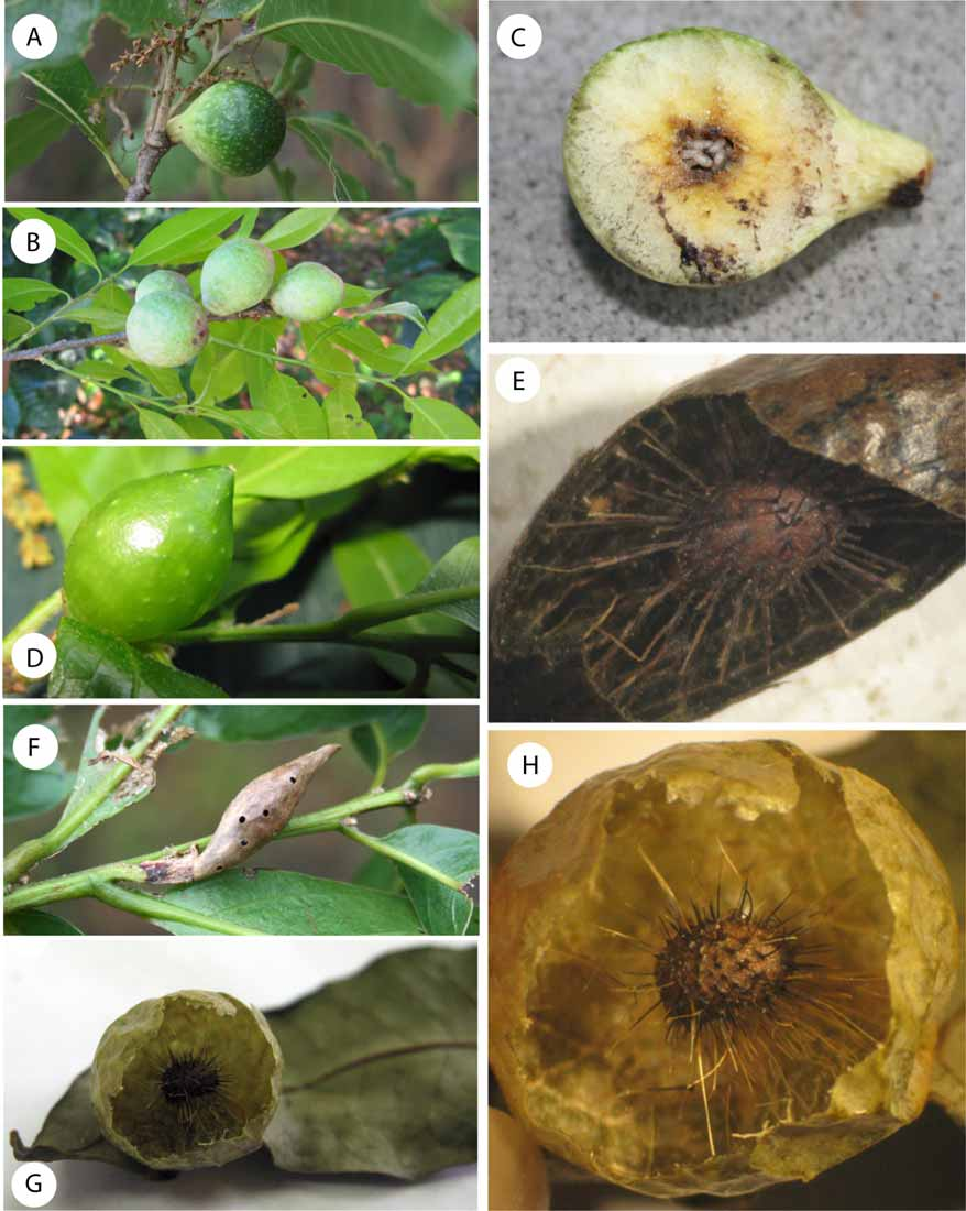
Gall ( Fig. 8 View FIGURE 8 G–H) Regular spherical, smooth, greenish when fresh, dark brown and very glossy when dry. The outer shell is thin; internally the larval cell is central and supported by thin hair-like radiating filaments ( Fig. 8 View FIGURE 8 H). When dry the gall is very brittle, and may be easily crushed with the fingers. Diameter of the gall measures 30 mm on average.
Distribution. A. salicifoliae was found between 1870–2680 m a.s.l. at the single site of Volcan Baru, Panama. In comparison with A. castroviejoi is a rare species.
Biology. Only the sexual generation of A. salicifoliae is known, inducing galls on the underside of Quercus salicifolia leaves. Galls are found between January–April, during the dry season, and the insects emerge in the same season. This species displays a remarkable sexual dimorphism in coloration.


| MNCN |
Museo Nacional de Ciencias Naturales |
No known copyright restrictions apply. See Agosti, D., Egloff, W., 2009. Taxonomic information exchange and copyright: the Plazi approach. BMC Research Notes 2009, 2:53 for further explanation.
|
Kingdom |
|
|
Phylum |
|
|
Class |
|
|
Order |
|
|
Family |
|
|
Genus |