
Amphibolips aliciae Medianero & Nieves-Aldrey
|
publication ID |
https://doi.org/10.5281/zenodo.275711 |
|
DOI |
https://doi.org/10.5281/zenodo.6201101 |
|
persistent identifier |
https://treatment.plazi.org/id/9702547D-FFBB-FFFA-FF3D-2AF947B9FB1A |
|
treatment provided by |
Plazi |
|
scientific name |
Amphibolips aliciae Medianero & Nieves-Aldrey |
| status |
sp. nov. |
Amphibolips aliciae Medianero & Nieves-Aldrey sp. nov.
( Figs.3 View FIGURE 3 , 4 View FIGURE 4 , 7 View FIGURE 7 D–E & 8D–F)
Type material. Holotype Ƥ ( Fig. 7 View FIGURE 7 D) (in Museo Nacional de Ciencias Naturales, Madrid, Spain, card mounted. Cat nº 2023). PANAMA, Chiriquí, Carretera de Volcancito, Boquete 8º 46' 23 7” N, 82º 27' 19 7” W, 1404 m; ex gall on twigs of Quercus salicifolia Née (Fagaceae) , gall collected 25.i.2009, insect emerged 25.i.09, E. Medianero leg. Paratypes: 23, same data as holotype, but one 3 ex gall collected 12.i.2008, insect emerged ii.08, E. Medianero leg. One paratype in MNCN, one paratype in Maestría en Entomología, Universidad de Panamá ( MEUP).
Etymology. Named after our good friend, the botanist Alicia Ibañez, in memory of unforgettable field sampling experiences in Panama.
Diagnosis and comments. Amphibolips aliciae is characterized by complete notauli, broad posteriorly; sub-quadrate deep scutellar foveae; the scutellum only slightly emarginated posteriorly, micropunctures on metasomal T3 very faint, hypopygial spine long; wings are not heavily smoked, only with a smoky brown patch at the base of the radial cell. The new species closely resembles A. castroviejoi in coloration, and most morphological characters but differs in some features as stated in the identification key, especially in the morphology of mesoscutum and scutellum and coloration of forewings. Furthermore, the inner structure of their respective galls is different. From the related sexual forms described by Kinsey from Mexico ( Kinsey, 1937), A. aliciae is readily distinguishable mainly by less heavily infuscate forewing. By this late morphological character, the new species resembles some Nearctic species of Amphibolips , namely A. melanocera Ashmead, 1885 ; A. cookii Gillette, 1888 ; A. globulus Beutenmüller, 1909 ; A. acuminata Ashmead, 1896 , but differs from all of them by distinct combinations of several features, such as type of gall, legs and metasoma coloration, mesoscutum and scutellum sculpture type, shape and sculpture of the scutellar foveae, number of male antennal segments, and other morphological characters ( Beutenmüller 1909).
Description. Female. Body length (measured from anterior margin of head to posterior margin of metasoma) 4.58 mm (N = 1) for females; 4.08 mm (range 4.0–4.16; N = 2) for males. Head and mesosoma of female shiny and black. Metasoma, reddish brown. Antenna black with five last flagellomeres clearer. Legs black, with tibiae dark brown and tarsi light brown. Forewing slightly smoky brown, with veins dark brown and large brown patch at base of radial cell. Male with coloration similar to female.
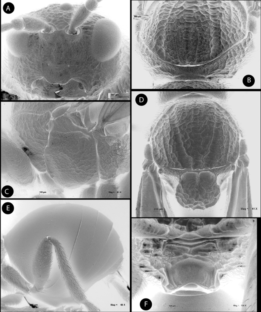
Female. Head, coarsely reticulate-rugose, pubescent, in dorsal view about 3 times wider than long. POL 0.8 times longer than OOL, posterior ocellus separated from inner orbit of eye by 2.2 times its longest diameter. Head in anterior view ( Fig. 3 View FIGURE 3 A) more or less oval, 1.17 times wider than high. Genae slightly expanded, mostly smooth and pubescent. Vertex, frons, face, and occiput reticulate-rugose, moderately pubescent. Clypeus, trapezoidal, smooth and moderately pubescent; ventral margin projecting over mandibles, its margin slightly sinuate. Anterior tentorial pits visible; epistomal sulcus and clypeopleurostomal lines indistinct; some irradiating strigae from clypeus visible, although obscured by coarse sculpture. Malar space 0.6 times height of compound eye. Distance between antennal rim of torulus and compound eye 0.6 times width of antennal socket including rim. Ocellar plate not raised. Mouthparts. Mandibles strong, exposed, with dense setae in base.
Antenna ( Fig. 4 View FIGURE 4 A) as long as 1/2 body length; with 11 flagellomeres; flagellum not broadening towards apex; with erect setae and elongate placodeal sensilla visible on F2–F11, placodeal sensillae increasing in number towards apex, on F6-F11 closely arranged in two rows ( Fig. 4 View FIGURE 4 C); 1–2 pores visible on apex of F4–F10 ( Fig. 4 View FIGURE 4 C). Relative lengths of antennal segments: 22:16:52:32:19:16:12:15:15:15:15:15:35. Pedicel, globose, small, 0.7 as long as scape; F1 1.6 times as long as F2 ( Fig. 4 View FIGURE 4 B). F6-F10 longer than broad; ultimate flagellomere 2.5 times longer than broad, 2.3 times as long as F10.
Mesosoma. In lateral view as high as long, with strong coarse rugose sculpture. Pronotum ( Fig. 3 View FIGURE 3 B), densely pubescent and with strong coarse sculpture in lateral areas. Ratio of length of pronotum medially/ laterally = 0.3. Pronotal plate indistinct.
Mesonotum. Mesoscutum ( Fig. 3 View FIGURE 3 D), moderate and uniformly pubescent, with strong reticulate-rugose sculpture. Notauli, complete, deep and wide, wider and strongly converging posteriorly, crossed by transversal rugae of the mesoscutum sculpture. Median mesoscutal impression faint in the coarse sculpture. Anteroadmedian signa visible. Transscutal fissure straight, narrow. Scutellum ( Fig. 3 View FIGURE 3 D), rounded, narrower basally, about 0.3 as long as mesonotum, strongly reticulate-rugose and slightly emarginate at tip, moderately pubescent. Scutellar foveae more or less square, large, about 0.4 as long as scutellum, smooth, separated by a septum, the anterior margin marked, and posteriorly the margin more diffuse. Axillula moderately pubescent, their anterior and posterior margins marked. Mesopleuron ( Fig. 3 View FIGURE 3 C) coarsely rugose and moderately pubescent; posterodorsal area less coarsely rugose and hairless.
Metanotum ( Fig. 3 View FIGURE 3 F). Metapectal-propodeal complex. Metapleural sulcus reaching posterior margin of mesopectus at about mid-height of metapectal-propodeal complex ( Fig. 3 View FIGURE 3 C). Lateral propodeal carinae distinct, broad, slightly divergent. Median propodeal area with rugose sculpture, and some hairs ( Fig. 3 View FIGURE 3 F). Nucha dorsally smooth.
Legs strong, densely pubescent; metatarsal claw with strong basal acute lobe, the secondary tooth measuring less than 1/3 of length of apical tooth ( Fig. 4 View FIGURE 4 D).
Forewing ( Fig. 7 View FIGURE 7 E). As long as body, veins strong and very pigmented. Radial cell 3.3 times longer than wide; open along anterior margin; areolet small, triangular, closed and distinct. Rs slightly bowed, M nearly straight, not reaching wing margin. R1 depigmented. Rs+M reaching basalis about at its mid-height. First abscissa of radius (2r) and 2r-m straight. Wing fringe on distal margin short.
Metasoma ( Fig. 3 View FIGURE 3 E). In greater part smooth and shiny, large, as long as head and mesosoma combined, in lateral view 1.14 times longer than high. Second metasomal tergitecovering about 2/3 of metasoma, posteriorly with a band of fine micropunctures hardly visible. Following tergites with micropunctures well visible. Anteroventral area of T3 with a patch of long setae. Projecting part of hypopygial spine long; in lateral view 4 times as long as high; lateral margins of hypopygial spine with long setae but do not reach apex of spine or form an apical tuft of setae.
Male. Similar to female except as follows: Antenna with 13 flagellomeres ( Fig. 4 View FIGURE 4 E); F1 slightly curved, flattened and expanded distally ( Fig. 4 View FIGURE 4 G). Placodeal sensillae on F1-F13 ( Figs. 4 View FIGURE 4 F & 4G). Relative length of antennomeres: 12:4:35:24:23:22:20:18:18:18:20:19:17:15:14. Metasoma smaller; T2 0.5 of metasoma length.
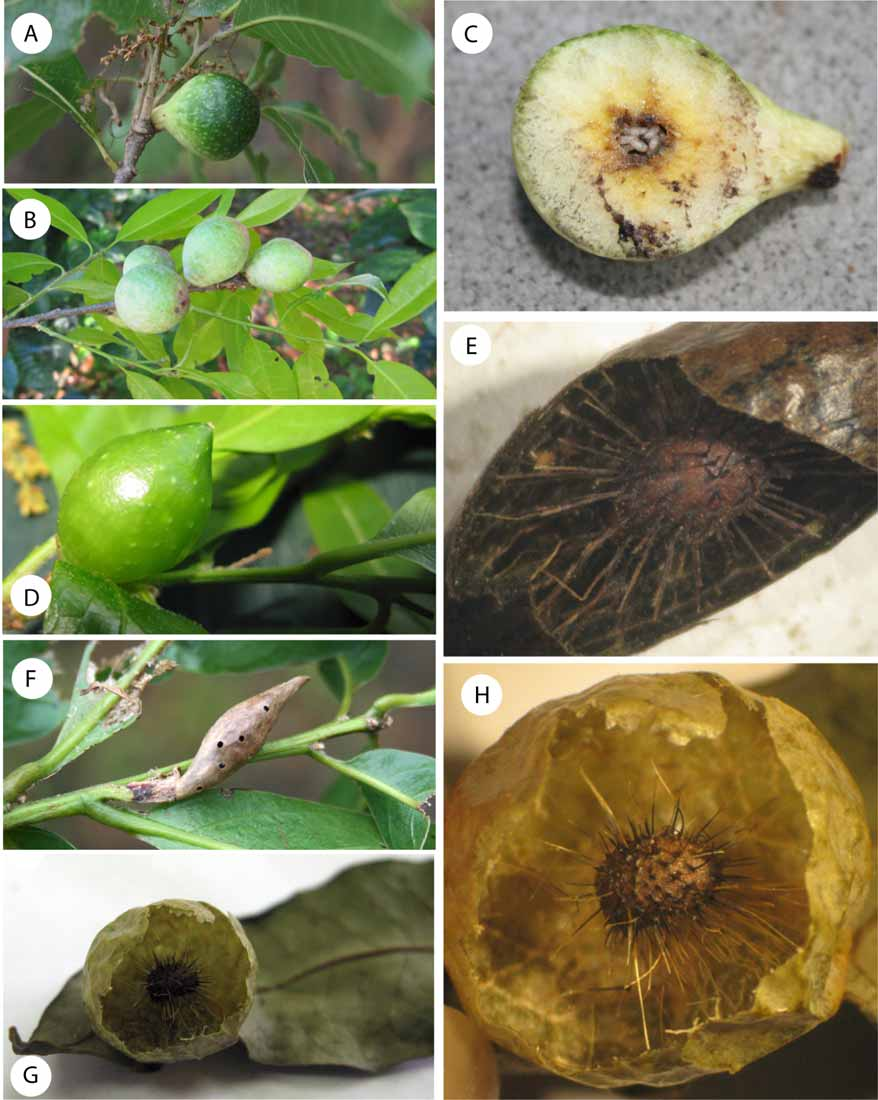
Gall. ( Figs. 8 View FIGURE 8 D–F) Elongate, ovate or spindle-shaped, with a very short nipple at the apex ( Fig. 8 View FIGURE 8 D). Greenish when fresh, coffee brown and very glossy when dry ( Fig. 8 View FIGURE 8 F). The outer shell is very thin, and internally there is a single central larval cell held in place by very thin hair-like filaments ( Fig. 8 View FIGURE 8 E). When dry the gall is very brittle, and may be easily crushed with the fingers ( Fig. 8 View FIGURE 8 E). On average, the gall measures 2.5 mm long. Externally the gall is similar to the gall of A. castroviejoi . However the inner structure is different, the later being soft, juicy while the gall of A. aliciae is composed of radiating filaments supporting the central larval chamber.
Distribution. A. aliciae was found between 1000–2681 m a.s.l.at Chiriqui, Panama. Galls are rare.
Biology. Only the sexual generation of A. aliciae is known, inducing galls on twigs of Quercus salicifolia Née and likely other Quercus species (section Lobatae). The galls are found between December-April during the dry season in Panama. Adults emerged in January and February.


| MNCN |
Museo Nacional de Ciencias Naturales |
No known copyright restrictions apply. See Agosti, D., Egloff, W., 2009. Taxonomic information exchange and copyright: the Plazi approach. BMC Research Notes 2009, 2:53 for further explanation.
|
Kingdom |
|
|
Phylum |
|
|
Class |
|
|
Order |
|
|
Family |
|
|
Genus |