
Amphibolips castroviejoi Medianero & Nieves-Aldrey
|
publication ID |
https://doi.org/10.5281/zenodo.275711 |
|
DOI |
https://doi.org/10.5281/zenodo.6201098 |
|
persistent identifier |
https://treatment.plazi.org/id/9702547D-FFBF-FFF7-FF3D-2FA14772FCEA |
|
treatment provided by |
Plazi |
|
scientific name |
Amphibolips castroviejoi Medianero & Nieves-Aldrey |
| status |
sp. nov. |
Amphibolips castroviejoi Medianero & Nieves-Aldrey sp. nov.
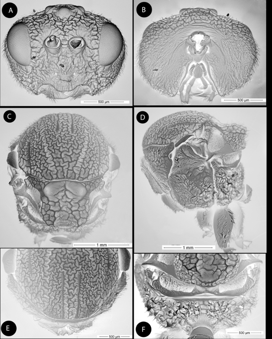
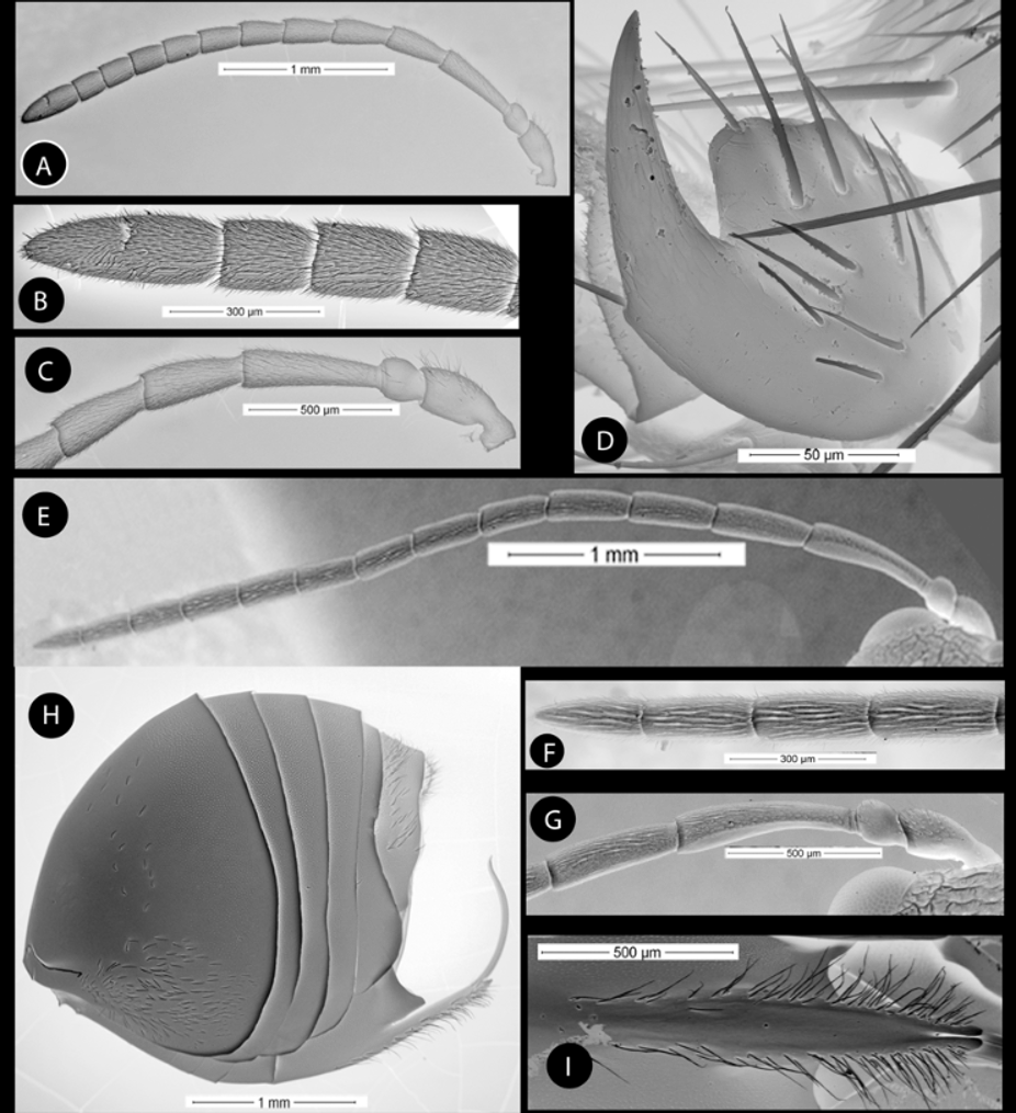
( Figs.1 View FIGURE 1 , 2 View FIGURE 2 , 7 View FIGURE 7 C, 7F & 8 A–C)
Type material. Holotype Ƥ ( Fig. 7 View FIGURE 7 A) (in Museo Nacional de Ciencias Naturales, Madrid, Spain ( MNCN), card-mounted. Cat. nº 2022). PANAMA, Chiriquí, Carretera de Volcancito, Boquete 8º 43' 23 07” N, 82º 27' 19 07” W, 1404 m; ex gall on twigs of Quercus salicifolia Née (Fagaceae) , gall collected 28.i.2008, insect emerged ii.08, E. Medianero leg. Paratypes: 13 same data as holotype; 1Ƥ, 13: same data as holotype, but collected 12.i.2008, insect emerged i.08. One paratype in MNCN, two paratypes in Maestría en Entomología, Universidad de Panamá ( MEUP).
Additionally, 1Ƥ paratype of the type series was dissected for SEM observation (in MNCN).
Etymology. Named after Dr. Santiago Castroviejo, dear colleague and friend, a recently deceased eminent botanist who worked for many years in the Flora of Coiba National Park ( Panama).
Diagnosis and comments. Closely allied to A. dampfi Kinsey , from Mexico, being similar in color and a majority of morphological characters. Males of the two species share a similar forewing coloration pattern, which is almost entirely smoky, with a clear crossing band extending from the radial cell to the discoidal cell. The species differ mainly in the sculpture of the thorax. A. dampfi have a very coarse sculpture, forming a series of small, rectangular spaces ( Kinsey 1937: p. 429), whereas the sculpture of the thorax is very irregular, even shapeless, in A. castroviejoi . The new species has a wide band extending across the forewing from the tip of radial cell to posterior part the apical margin ( Fig.7 View FIGURE 7 C), whereas the band does not extend as far across the ventral margin of the wing in A. dampfi ( Kinsey, 1937) . Additionally, A. castroviejoi have complete notauli, only lost in the coarse surface in anterior one third, and the anteroadmedian signa visible. In A. dampfi , the notauli are indicated but nearly lost in sculpture and the anteroadmedian signa are less visible.
Description. Body length (measured from anterior margin of head to posterior margin of metasoma) 5.0 mm (N = 2) for females; 4.45 mm (range 4.33–4.58; N = 2) for males. Head and mesosoma of female shiny and black. Metasoma, clypeus, mandibles, antenna and legs rufo-piceous; with scape, pedicel, F1, F2, coxae and femora more darkened. Forewing almost entirely and very heavily smoky, especially in medial half of basal and radial cells, with a wide clear band extending across wing from tip of radial cell to the apical area between the medial and cubital veins; small clear clouds present on Cu-a and R1 +Sc. Male with coloration similar to female, but legs uniformly rufo-piceous.
Female. Head, coarsely rugose, pubescent; in dorsal view about 2.6 times wider than long. POL 1.1 times longer than OOL, posterior ocellus separated from inner orbit of eye by 2.5 times its longest diameter. Head in anterior view ( Fig. 1 View FIGURE 1 A) transversely ovate, 1.29 times wider than high, gena slightly broadened behind eye. Vertex, frons, lower face, gena, and occiput with strong reticulate-rugose sculpture, irradiating carinae from clypeus not discernible; head moderately pubescent, with relatively long setae, except vertex and frons with sparse and shorter setae. Clypeus trapezoid, 1.5 times wider than high, shiny and smooth, moderately pubescent, ventral margin strongly projecting over mandibles and slightly sinuate. Anterior tentorial pits well visible; epistomal sulcus and clypeo-pleurostomal lines slightly distinct. Malar space 0.6 times height of compound eye. Toruli situated slightly below mid-height of compound eye; distance between antennal rim and compound eye 0.7 times width of antennal socket including rim. Ocellar plate slightly raised. Occipital carina lacking; strong transverse wrinkles present on occiput. A carina, dorso-laterad to occipital foramen, present, which is long and continues ventrally past posterior tentorial pits. Gula short; distance between occipital and oral foramina less than height of occipital foramen ( Fig. 1 View FIGURE 1 B). Hypostomal sulci well separate at oral fossa.
Mouthparts ( Fig. 1 View FIGURE 1 B): mandibles strong, exposed; with dense setae in base, right mandible with three teeth; left with two teeth. Cardo of maxilla not visible, maxillary stipes about 3.3 times longer than wide. Maxillary palp five-segmented. Labial palp three-segmented.
Antenna ( Fig. 2 View FIGURE 2 A), of moderate length, as long as 1/2 body length; with 13 antennomeres; flagellum not broadening towards apex; with relatively long, erect setae, and elongate placodeal sensilla hardly visible ( Fig. 2 View FIGURE 2 B). Relative lengths of antennal segments: 17:12:38:26:25:22:19:18:16:15:15:13:28. Pedicel ( Fig. 2 View FIGURE 2 C), globose, small, 0.7 as long as scape; F1 1.4 times as long as F2. F6-F10 longer than wide, F11 2.7 times longer than wide, 2.1 times as long as F10 ( Fig. 2 View FIGURE 2 B). Placodeal sensillae on F8-F11 disposed in one row of 2–4 sensillae in half dorsal area of each flagellomere.
Mesosoma. Strongly, coarsely rugose, in lateral view as high as long. Pronotum, moderately pubescent; lateral surface of pronotum with strong rugose sculpture; moderately pubescent, with relatively long setae. Ratio of length of pronotum medially/laterally = 0.24. Pronotal plate indistinct dorsally ( Fig. 1 View FIGURE 1 E).
Mesonotum ( Fig. 1 View FIGURE 1 C). Mesoscutum barely pubescent and with strong rugose-reticulate sculpture. Notauli distinct posteriorly and medially, broad and convergent posteriorly, crossed by transversal rugose sculpture; median mesoscutal impression indistinct, lost in the coarse sculpture. Anteroadmedian signa clearly visible. Transscutal fissure narrow, well-visible, deeply impressed. Scutellar foveae ellipsoidal, deep, about 1/3 as long as scutellum, smooth and separated by a septum; their anterior and posterior margins marked. Scutellum ( Fig. 1 View FIGURE 1 C) subquadrate from above, about 0.4 as long as mesoscutum, strongly reticulate-rugose and deeply emarginate at posterior margin, emargination reaching posterior one third of scutellum length; in lateral view extending posteriorly slightly over the dorsellum. Axillula moderately pubescent, their anterior and posterior margins marked. Mesopleuron coarsely rugose and moderately pubescent, excepting the posterodorsal area ( Fig. 1 View FIGURE 1 D).
Metanotum ( Fig. 1 View FIGURE 1 F). Metapectal-propodeal complex. Metapleural sulcus reaching posterior margin of mesopectus at about mid-height of metapectal-propodeal complex ( Fig. 1 View FIGURE 1 D). Lateral propodeal carinae indistinct, slightly divergent anteriorly, ( Fig. 1 View FIGURE 1 F). Median propodeal area rugose and densely pubescent. Nucha rugose.
Legs. Densely pubescent; femora and tibiae robust; metatarsal claws with strong triangular basal lobe or teeth ( Fig. 2 View FIGURE 2 D).
Forewing ( Fig. 7 View FIGURE 7 C): As long as body, radial cell 3.4 times longer than wide; open along anterior margin; areolet small, ovoid, closed and distinct. R1, Rs and M nearly straight, not reaching wing margin. Rs+M reaching basalis at its mid-height. First abscissa of radius (2r) and 2r-m curved. Apical margin with short hair fringe.
Metasoma ( Fig. 2 View FIGURE 2 H), large as long as head and mesosoma combined, in lateral view as high as wide. Second metasomal tergite covering about two third of metasoma, with band of micropuntures clearly visible in posterior one third; punctures visible on subsequent tergites; ventral area of second metasomal tergite moderately pubescent. Projecting part of hypopygial spine long ( Fig. 2 View FIGURE 2 I); about 3 times as long as wide in ventral view; laterally with long setae, longer than spine width but not forming an apical patch.
Male ( Fig. 7 View FIGURE 7 B). Similar to female except as follows: Antenna with 13 flagellomeres ( Fig. 2 View FIGURE 2 E); F1 slightly curved, posteriorly flattened and expanded apically ( Fig. 2 View FIGURE 2 G). Placodeal sensillae present on flagellomeres 1– 1 3, i n c r e a s i n g i n n u m b e r t o w a r d s a p e x (F i g. 2F). R e l a t i v e l e n g t h o f a n t e n n o m e r e s: 15:9:35:27:23:23:19:19:17:17:17:16:15:14:13. Metasoma smaller than in female; T2 0.7 of metasoma length.
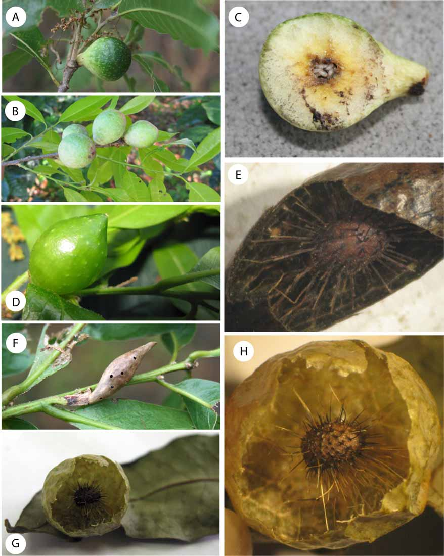
Gall ( Fig. 8 View FIGURE 8 A–C)., Irregularly spherical or globose, a bit elongated at its basis, monothalamic, with smooth and mottled surface. Light green when fresh ( Fig. 8 View FIGURE 8 A) and light cream when mature ( Fig. 8 View FIGURE 8 B); the outer shell is thin but firm. Internally it is of a soft, uniformly spongy consistence, filling the entire gall ( Fig. 8 View FIGURE 8 C). The larval cell is rounded and is embedded in the soft internal substance. Diameter 58 to 45 mm (on average 54 x 43 mm). Formed in twigs of Quercus salicifolia Nee. The gall most closely resembles that of Amphibolips murata Weld, 1957 known from Florida ( USA).
Distribution. A. castroviejoi was found between 1000–2681 m a.s.l. at Chiriqui, Panama.
Biology. Only the sexual generation is known, inducing galls on Quercus salicifolia and likely on other Quercus species (section Lobatae). The galls are found between December and May, during the dry season in Panama. The insects studied emerged in January and February.

| MNCN |
Museo Nacional de Ciencias Naturales |
No known copyright restrictions apply. See Agosti, D., Egloff, W., 2009. Taxonomic information exchange and copyright: the Plazi approach. BMC Research Notes 2009, 2:53 for further explanation.
|
Kingdom |
|
|
Phylum |
|
|
Class |
|
|
Order |
|
|
Family |
|
|
Genus |