
Ljania bipapillata, Thor, 1898
|
publication ID |
https://doi.org/10.5281/zenodo.210800 |
|
DOI |
https://doi.org/10.5281/zenodo.6170935 |
|
persistent identifier |
https://treatment.plazi.org/id/994787FF-FF82-FFCA-FF06-2267FA878870 |
|
treatment provided by |
Plazi |
|
scientific name |
Ljania bipapillata |
| status |
|
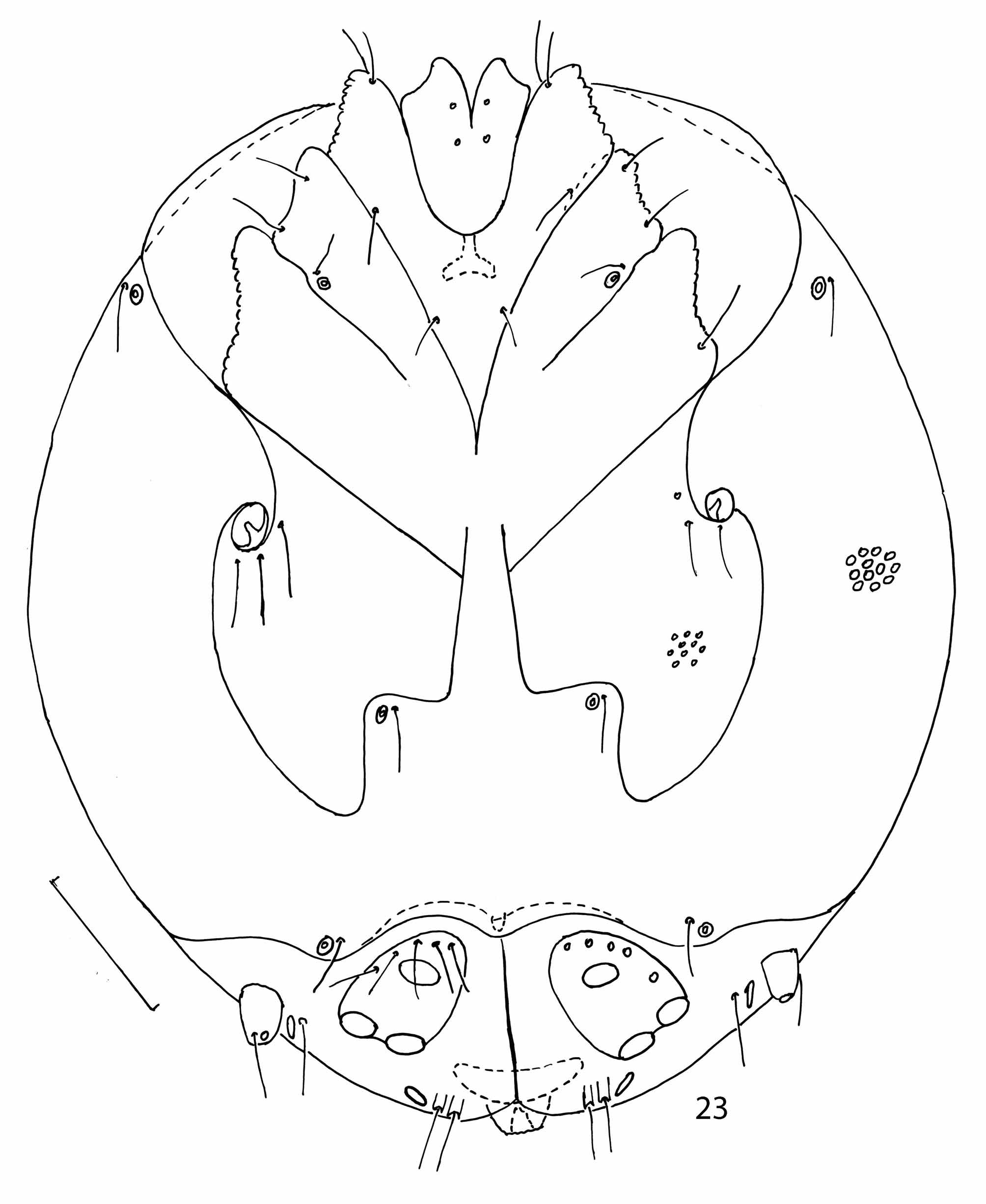
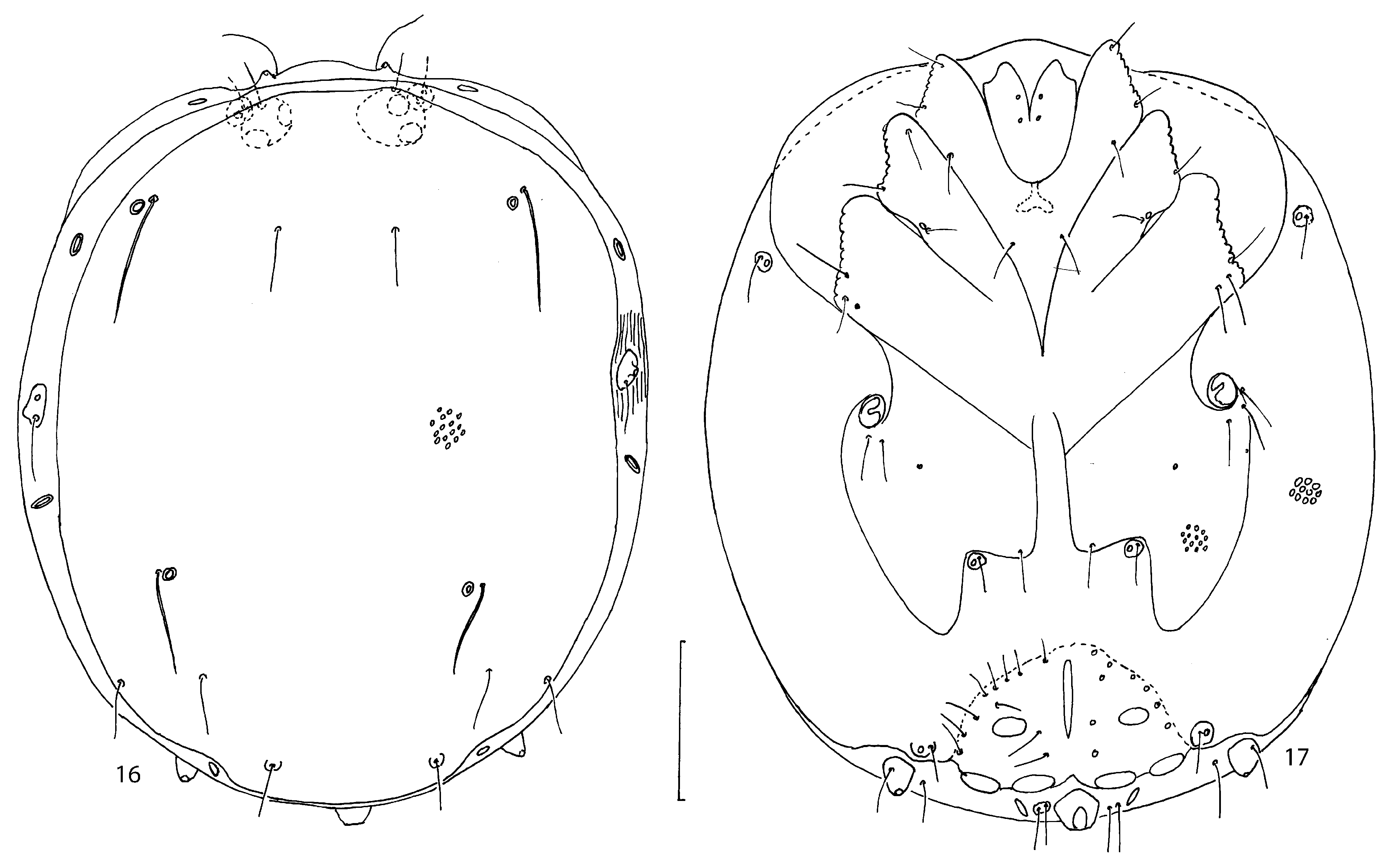
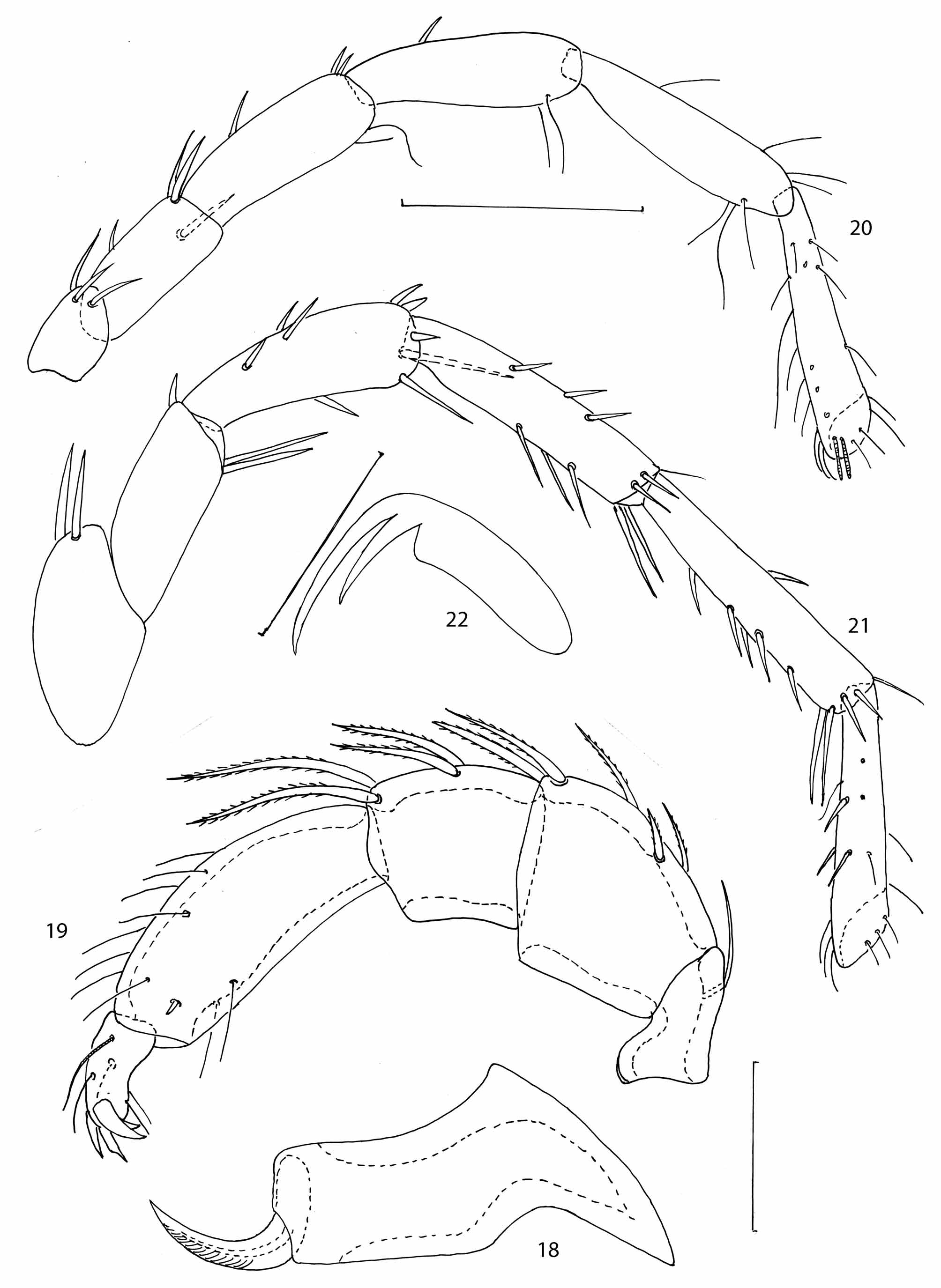
( Figs 16–23 View FIGURES 16 – 17 View FIGURES 18 – 22 View FIGURE 23 )
Material examined. 2 males and 8 females: 2 females, Yaroslavl Province, Nekouz District, Kamenka stream (inflow of Sit’ River) near village Sit’–Pokrovskoe, 29.09.1973 and 10.10.1974, leg. P.V. Tuzovskij; 1 male, Russia, North Caucasus, Krasnodar Territory, Seversk District, Ubin River near settlement Ubinskaya, 28 0 5 1976, leg. P.V. Tuzovskij; 1 male, 6 females, Russia, Republic Komi, the Pechora River basin near Northern polar circle, 11. 0 7. 1969, leg. O.S. Tsember.
Diagnosis. Adults: idiosoma nearly circular (L/W ratio 1.03–1.15), with convex anterior margin between setae Vi, medial margins of Cx-IV well separated, genital field of male with 9–11 pairs of fine setae, anteromedial margin of Cx-IV and its posteromedial portion approximately equal in length; dorsoproximal setae on P-3 considerably shorter than dorsodistal setae, equal in length as, or longer than dorsal margin of segment; ventral setae on P-4 located distally, leg claws with long external clawlet and short internal clawlet.
Morphology. Both sexes. Body flat, wide and nearly circular (L/W ratio 1.03–1.15), with slightly convex anterior margin between setae Vi. Setae Si without glandularia Si at distal end of dorsal shield.
Anterior ends of Cx–I slightly extending beyond anterior margin of idiosoma ( Fig. 17 View FIGURES 16 – 17 ). Suture line between Cx–I/II and Cx-III/IV complete; suture line between Cx–II/III incomplete, obliterated medially. Medial margins of Cx–IV and posteromedial portions of Cx–III separated, anteromedial portions of Cx–III fused completely. Cx–IV anteromedial and posteromedial margins approximately equal in length. Chelicera ( Fig. 18 View FIGURES 18 – 22 ) with large basal segment and short stylet. Basal segment of chelicera with acute dorsal hump, cheliceral stylet crescent-shaped.
P–3 with dorsoproximal setae shorter than dorsodistal ones, both dorsodistal setae somewhat longer than dorsal L of segment; P–4 ventral setae near distal end of segment..
Legs: See Figs 20-22 View FIGURES 18 – 22 . Claws of all legs with two unequal clawlets, external clawlet longer than internal one.
Male. Genital field with 9–11 fine setae on each side, all acetabula oval, small and approximately subequal in size; distance between anterior acetabulum and both posterior acetabula longer than maximum acetabulum diameter.
Measurements (n=2). L of idiosoma 485–495, W 420–440; L of dorsal shield 460–470, W 375–390; L of anteromedial portion of Cx– IV 55 –60; L of posteromedial portion 54–60; L of genital field 78–82, W 130–145; L of genital acetabula (ac. 1–3): 18–24, 24–30, 20–30; L of capitulum 78–80; L of basal cheliceral segment 75–80, L of cheliceral stylet 28–30; L of pedipalpal segments (P–1–5): 23–24, 42–45, 36 –42, 60–65, 24–28; L of leg segments I–Leg–1–6: 35–42, 48–60, 75–80, 84–90, 95–100, 105–115; II–Leg–1–6: 35–42, 45–50, 50–57, 63–72, 72– 80, 83–87; III–Leg–1–6: 40–45, 55–60, 58–60, 84–87, 95–98, 85–95; IV–Leg–1–6: 65–72, 84–90, 85–90, 105– 110, 110–118, 105–110.
Female. Dorsal shield wide (L/W ratio 1.12–1.17). Genital plates L/W ratio 1.1–1.2 ( Fig. 23 View FIGURE 23 ).
Measurements (n=8). L of idiosoma 570–630, W 520–610; L of dorsal shield 545–585, W 465–520; L of anteromedial portion of Cx– IV 63 –67; L of posteromedial portion 62–65; L of genital plate 72–78, W 60–72; L of genital acetabula (ac. 1–3): 18–21, 24–27, 24–28; L of capitulum 90; L of basal segment of chelicera 100–110, L of cheliceral stylet 45–48; L of pedipalpal segments (P–1–5): 25–30, 55–63, 45–50, 78–85, 30; L of leg segments I– Leg–1–6: 40–45, 70–75, 95–110, 105–110, 110–115, 115–120; II–Leg–1–6: 40–45, 50–55, 55–60, 75–80, 88–93, 95–100; III–Leg–1–6: 50–55, 60–65, 70–75, 95–100, 105–110, 110–115; IV–Leg–1–6: 78–85, 78–85, 100–110, 125–130, 130–140, 115–125.
Deutonymph. See Viets (1936).
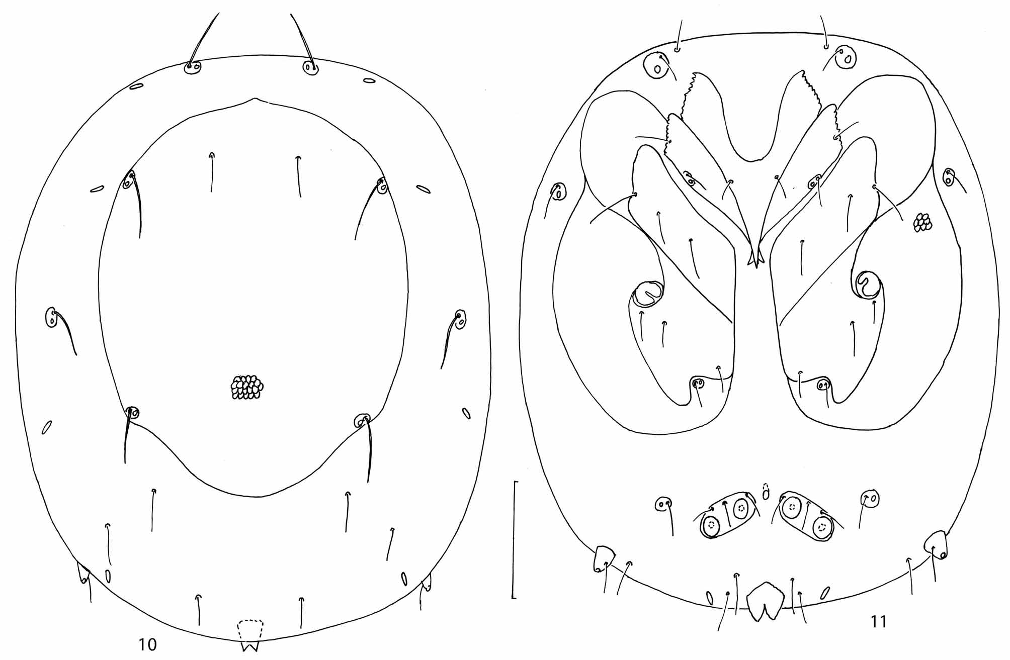
Remarks. The deutonymph of L. bipapillata differs from the deutonymph of L. macilenta in the anterior coxal group with wide posterior end and genital plates with convex anterior and posterior margins ( Viets 1936). In contrast, the anterior coxal group in the deutonymph L. macilenta has an acute posterior end and two short lateral apodemes ( Fig. 10 View FIGURES 10 – 11 ) and the genital plates have straight anterior and posterior margins ( Fig.11 View FIGURES 10 – 11 ).
Larva. Martin (2000) described in detail the larva of L. bipapillata from Himmelreichbach ( Germany), and Smith (1984) described a larva of Ljania sp. nr. bipapillata collected from streams throughout western North America. As there is no marked difference between the two taxa, it is probable that they represent the same species ( Martin 2000).
Habitat. Streams, rivers, brooks.
Distribution. Europe ( Viets 1936, 1956; Lundblad 1968; Viets 1978); Russia: North Caucasus, Yaroslavl Province ( Tuzovskij 1977, 1990), Komi Republic ( Solovkina & Tsember 1971); North America ( Smith 1984).
No known copyright restrictions apply. See Agosti, D., Egloff, W., 2009. Taxonomic information exchange and copyright: the Plazi approach. BMC Research Notes 2009, 2:53 for further explanation.
|
Kingdom |
|
|
Phylum |
|
|
Class |
|
|
Order |
|
|
Family |
|
|
Genus |