
Euepedanus vietnamicus, Tchemeris, 2020
|
publication ID |
https://doi.org/ 10.11646/zootaxa.4858.3.8 |
|
publication LSID |
lsid:zoobank.org:pub:B48283E5-D2D7-4134-B7C5-0FF74C736BFA |
|
DOI |
https://doi.org/10.5281/zenodo.4412514 |
|
persistent identifier |
https://treatment.plazi.org/id/39C412B2-02F2-4645-A6E5-33155CDC7819 |
|
taxon LSID |
lsid:zoobank.org:act:39C412B2-02F2-4645-A6E5-33155CDC7819 |
|
treatment provided by |
Plazi |
|
scientific name |
Euepedanus vietnamicus |
| status |
sp. nov. |
Euepedanus vietnamicus sp. nov.
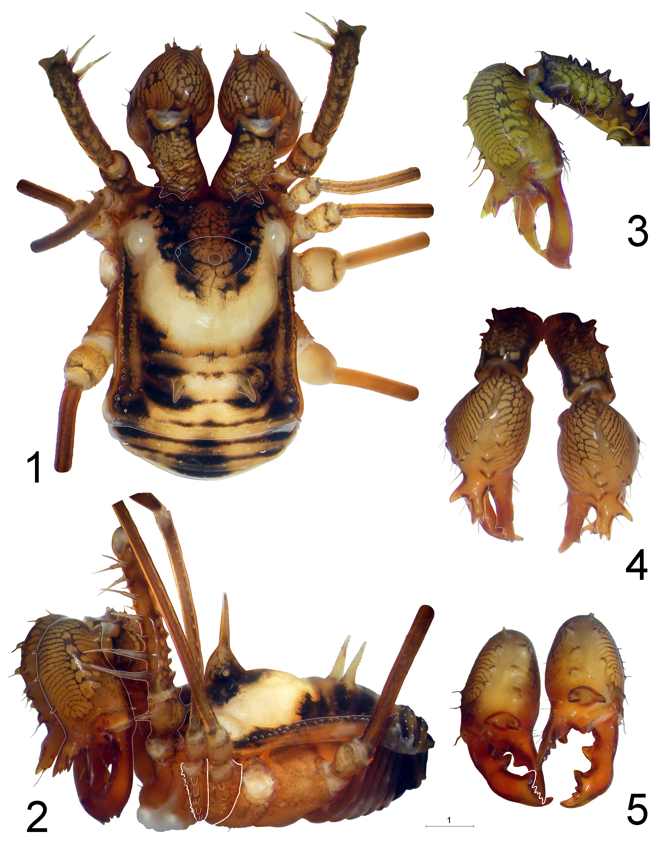
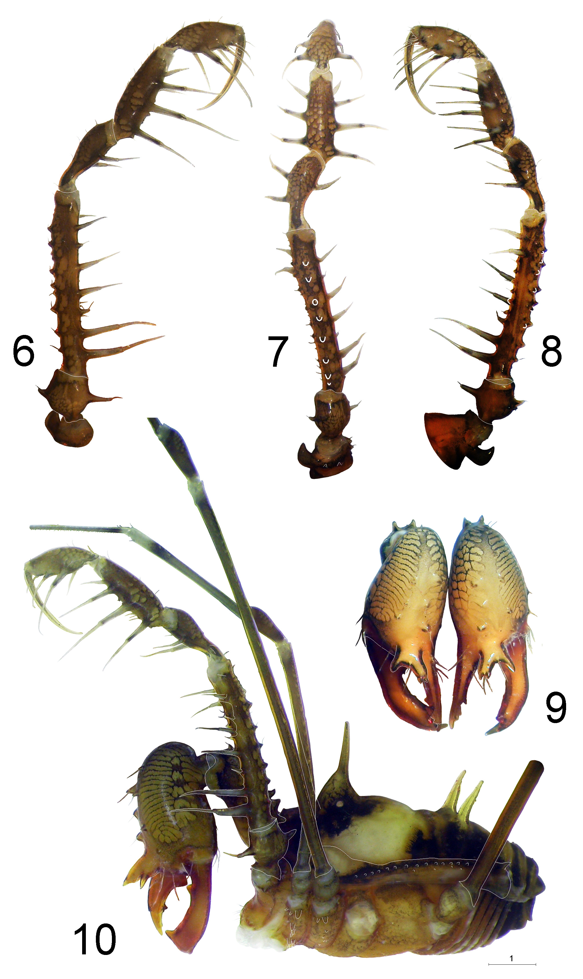
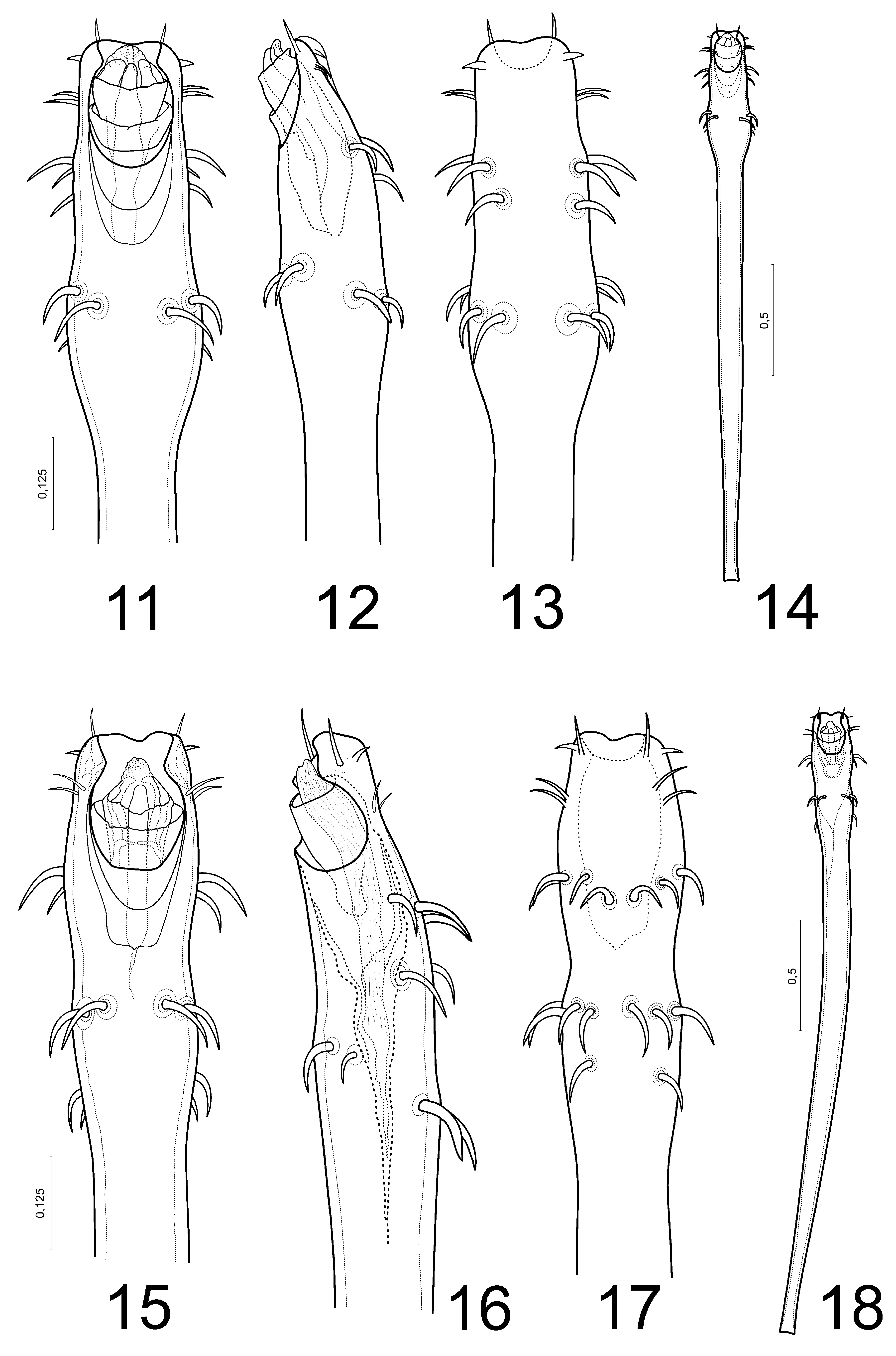
Figs. 1–18 View FIGURES 1–5 View FIGURES 6–10 View FIGURES 11–18 , 28 View FIGURE 28
Types. Holotype male ( MMUE, G7641.1 View Materials ) from Vietnam, Dong Noi Province , Cat Tien National-Park, October– December 2011, A.A. Goncharov . Paratypes: 1 male (damaged), 3 females and 3 females (damaged) ( MMUE, G7641.2 View Materials ) , 1 male, 1 female ( ISEA 001.8549 ) — idem .
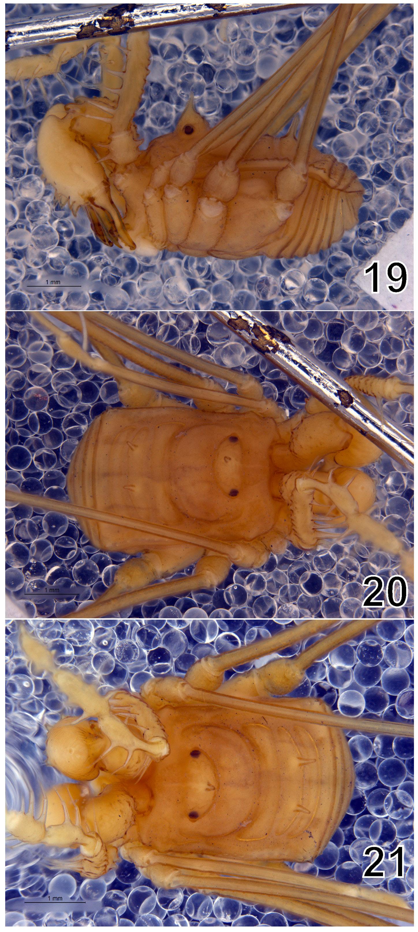
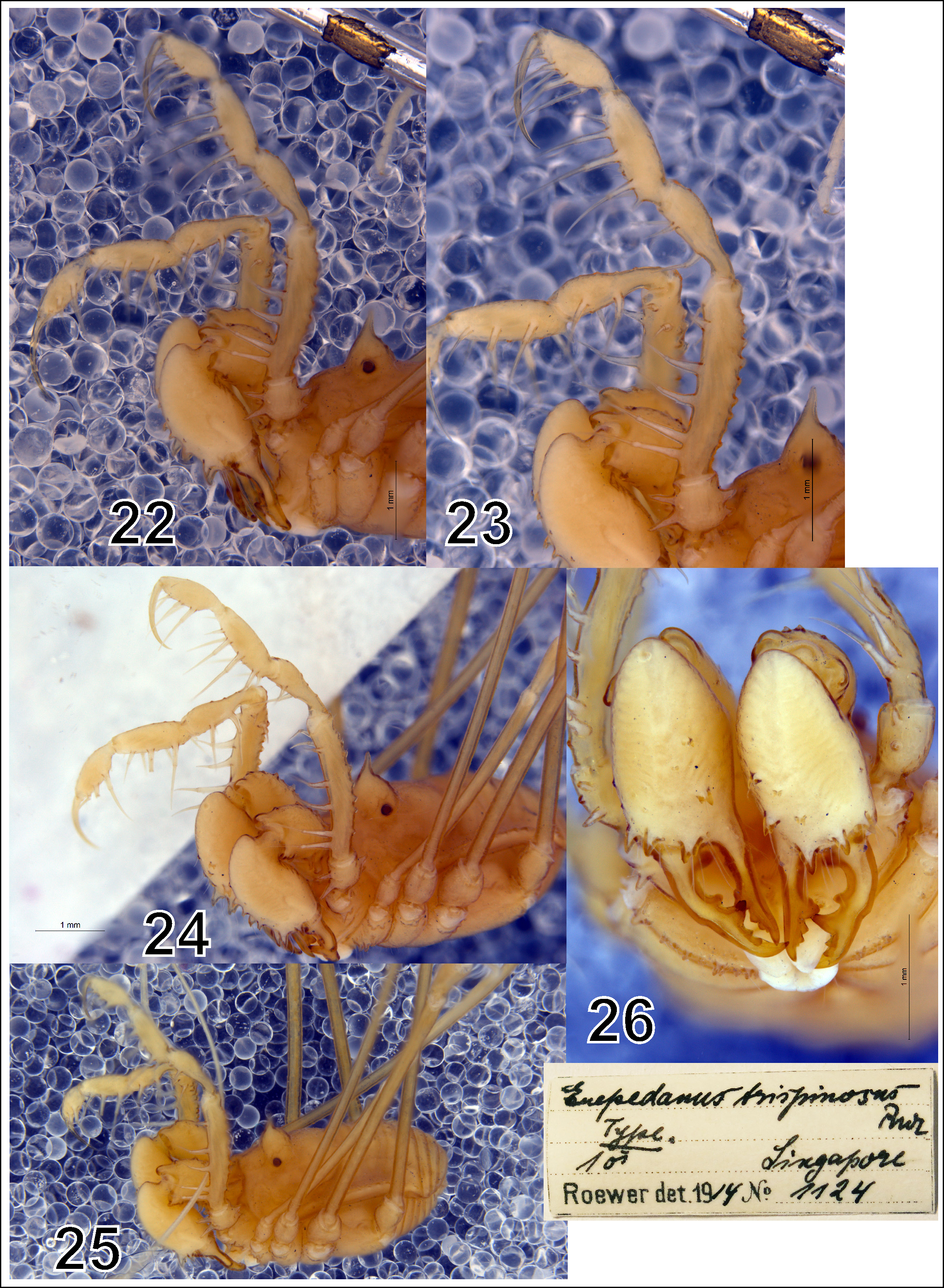
Comparative material. Euepedanus trispinosus Roewer, 1915 : Holotype ♂ ( SMF9901124 About SMF ) from Singapore, Malakka ; see Roewer (1915: 63, fig. 31 a–b), for its general appearance see Figs. 19–27 View FIGURES 19–21 View FIGURES 22–27 .
Etymology. The species epithet originates from Vietnam, the country from where the type series was collected.
Diagnosis. By the structure of body and appendages, this species is closest to Euepedanus flavimaculatus Zhu & Lian, 2006 and E. dividuus Suzuki, 1969 . The main diagnostic characters are as follows: 1) penis apex truncate, not trapeziform ( Figs. 11–18 View FIGURES 11–18 ); 2) cheliceral basal segment relatively long, somewhat curved and armed with numerous large and robust setae-tipped tubercles (but not strongly drawn), except for ectal and ventral surfaces ( Figs. 1, 3–5 View FIGURES 1–5 ); 3) distal cheliceral segment markedly swollen, with an apophysis-like structure near chelae formed by several long spines, protruding forward beyond the segment limit ( Figs. 2–4 View FIGURES 1–5 , 9–10 View FIGURES 6–10 ); 4) fingers of chelae widened and long ( Figs. 2–5 View FIGURES 1–5 , 9–10 View FIGURES 6–10 ); 5) palpal armament ( Table 1; Figs. 6–8, 10 View FIGURES 6–10 ). Morphological differences between the new species and three other Euepedanus species, including E. trispinosus ( Figs. 19–27 View FIGURES 19–21 View FIGURES 22–27 ), are shown in Table 1.
Description. MALE. Measurements. Body: length 5.92; maximal width 4.03. Scutum length: 4.90; width in the middle 3.58. ‘Clypeus’ length: 0.68. Eye tubercle width: 1.58; height (from the base of eye tubercle to top) 2.0. Chelicera: basal segment 2.57 long; distal segment 2.81 long; chela 2.35 long. Length of palp segments: 1.08 + 3.53 + 1.62 + 2.05 + 1.80 + claw 1.93 = 12.01. Length of leg segments: I: 4.92 + 1.13 + 3.40 + 6.02 + 1.32 = 16.79; II: 8.82 + 1.63 + 7.58 + 11.12 + 5.09 = 34.24; III: 6.76 + 1.53 + 3.63 + 8.16 + 3.28 = 23.36. IV: 9.28 + 1.78 + 4.65 + 10.95 + 4.12 = 30.78. Penis 2.58 long. Body. Almost rectangular, slightly trapezoid-like as shown in Fig. 1 View FIGURES 1–5 . Scutum with a wide portion consisting of four sclerites, of which the most narrow one is situated near the rear end. Abdomen widely rounded behind. Anterior carapace margin with 6–8 granules. Eye tubercle separated from the anterior carapace margin and armed with a long median spine on its top. Carapace region with small dome-shaped hump situated around odoriferous glands and a big dome-shaped hump on both sides beloweye tubercle. Opisthosomal region of scutum with four scuta; scutal area II with a pair of median sharp relatively long spines, others areas smooth; fourth area in corners with short spine; each lateral margin of the scutum with a longitudinal row of conical and distinctly transparent tiny tubercles. Each of the free tergites with a row of granules spread out across its entire width. Last granule from a longitudinal row modified to short spine. Venter. Palpal coxae distally armed with 2–3 blunt tubercles. Leg coxae I–II considerably narrower than the others. I—armed with a row of irregular blunt tubercles, IIwith a row of setae-tipped granules and 2–3 distal blunt tubercles, III—on its sides with a row of pointed tubercles, IV—with 1–2 distal blunt tubercles situated at its corners. Tracheal stigma clearly visible; it is equal to about half length of coxa IV. Each of the free sternites with a row of tiny translucentsetae. Chelicerae ( Figs. 1–5 View FIGURES 1–5 , 9–10 View FIGURES 6–10 ) strong and robust. Basal segment relatively long, not swollen, somewhat curved and armed with numerous large and robust setae-tipped tubercles, except for ectal and ventral surfaces. Distal segment large and swollen, frontally armed with two irregular rows of setae-tipped tubercles, the three lowermost long setae-tipped tubercles at the base of chelae are fused, forming a prominent apophysis resembling a crown. Chela fingers relatively long, widened and strong, movable finger curved with strong and robust cutting teeth, their cutting edges toothed as in Figs. 2–5 View FIGURES 1–5 , 9–10 View FIGURES 6–10 . Palps ( Figs. 1–2 View FIGURES 1–5 , 6–8, 10 View FIGURES 6–10 ) relatively long, slightly swollen, armed with strong spines. Coxa DE—with strong crescent -shaped tubercle, D and M with rare LCT. Palpal spinulation formula: Tr: D—2 LCT, V—1 SLT; Fm: D—7–8 LCT, V—5 SLT + 2 SMT, E—2 distal SLT, DE and VE—5–6 TCT; Pt: VE—2 SLT, VM—1 SLT; Tb: VE—3 SLT, VM—4 SLT; Tr: VE and VM—3 SLT. Tarsal claw almost equal to tarsus long, slightly curved. Legs ( Figs. 1–2 View FIGURES 1–5 , 10 View FIGURES 6–10 ) slender and relatively long, III–IV thicker than others. Femora I slightly curved, II–IV straight, femoral bases of all legs with 1–2 pseudosegments. All metatarsi with numerous pseudosegments. Tarsi III–IV with a pair of claws, no scopulae. Distitarsus I leg with two tarsomeres. Tarsal articulations: I: 9, II: 25–28, III: 8, IV: 9. Penis ( Figs. 11–18 View FIGURES 11–18 ) long and slender, its truncus is gradually widened distad. Distal area with broad deepening along the middle line and rows of spicules, glans (capsules externa and interna) are located inside the depression. Apex of the ventral plate emarginate in the middle. Base very narrow.
FEMALE. Measurements. Body: length 5.50; maximal width 4.11. Scutum length 4.42; width in middle 4.12. ‘Clypeus’ length: 0.60. Eye tubercle width 0.97, height (from the base of eye tubercle to its top) 1.47. Chelicera: basal segment 1.42 long; distal segment 1.48 long; chela 1.22 long. Length of palp segments: 0.60 + 2.91 + 1.48 + 1.70 + 1.58 + claw 1.68 = 9.95. Length of leg segments: I: 4.36 + 0.91 + 2.98 + 5.47 + 1.98 = 15.70; II: 7.44 + 1.12 + 6.35 + 9.82 + 3.85 = 28.58; III: 5.68 + 1.28 + 3.10 + 6.82 + 2.73 = 19.61; IV: 7.95 + 1.27 + 4.07 + 9.36 + 3.43 = 26.08. Female general appearance as in the male, but body trapezoid and smaller. Chelicerae smaller, not swollen, basal segment without any apophysis-like structure as in the male. Females darker than the males, their prevailing colour—dirty yellowish, on the dorsal surface with a pattern resembling the Latin letter X. Carapace region, in front and eye tubercle, with a dark brown reticulation. Opistosomal region and each lateral margin of the scutum with the similar reticulation and dark brown spots and patches. Dome-shaped humps yellowish, situated on both sides below eye tubercle. Chelicerae and palps yellowish, with the same reticulation. Distal cheliceral segment with a zebra-pattern. Legs yellowish, with an ochre-brown reticulation, distal segments lighter, grey-milky. Venter yellowish, darkest at its distal end.
Distribution. Vietnam: the type locality only ( Fig. 28 View FIGURE 28 ).
| MMUE |
Museum of Manchester University |
No known copyright restrictions apply. See Agosti, D., Egloff, W., 2009. Taxonomic information exchange and copyright: the Plazi approach. BMC Research Notes 2009, 2:53 for further explanation.
|
Kingdom |
|
|
Phylum |
|
|
Class |
|
|
Order |
|
|
Family |
|
|
Genus |