
Uropoda lichenicola, Kontschán & Starý, 2011
|
publication ID |
https://doi.org/10.5281/zenodo.202067 |
|
DOI |
https://doi.org/10.5281/zenodo.5619566 |
|
persistent identifier |
https://treatment.plazi.org/id/9C3B2D30-FF86-FFC0-D4CE-F794B70CFABA |
|
treatment provided by |
Plazi |
|
scientific name |
Uropoda lichenicola |
| status |
sp. nov. |
Uropoda lichenicola sp. nov.
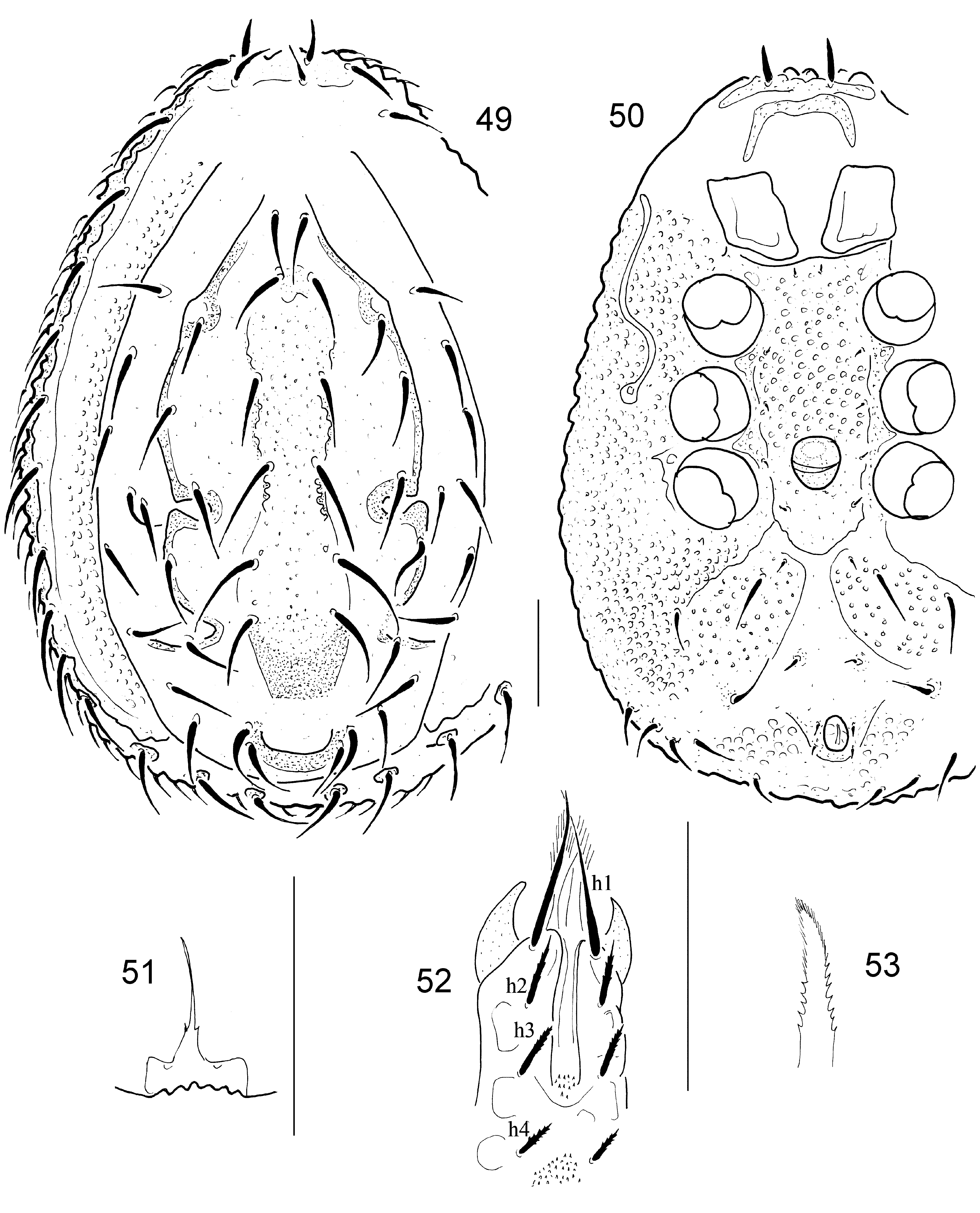
( Figs 49–57 View FIGURES 49 – 53 View FIGURES 54 – 57 )
Material examined. Holotype. Male ( HNHM), Vietnam, Lao Cai Province, Sín Chai, Hoáng Liên N.P., Tram Ton, along and over a rivulet, 1936 m, 22°20.941’N 103°46.197’E, from lichens, 12 December 2008, L. Peregovits coll. Paratypes. One male ( ISB) and one deutonymph ( HNHM). Locality and date same as holotype.
Description. Male. Length of idiosoma 710–730 µm, width 470–500 µm (n=2). Shape oval, posterior margin rounded.
Dorsal idiosoma ( Fig. 49 View FIGURES 49 – 53 ). Marginal and dorsal shields fused anteriorly. Central region of dorsal shield elevated from the other parts of dorsum, covered by small, oval pits, with six pairs of smooth (ca 60–100 µm long) needle-like setae. Lateral regions of dorsal shield with setae similar in shape and length to those in central region. Laterocentral part delimited by a strongly sclerotised line with three pairs of loops: first pair small loops at level of coxae II, second pair of larger loops at level of coxae IV, third pair at the end of the strongly sclerotised dorsal line. One large, U-shaped, strongly sclerotised line situated on caudal margin of dorsal shield, bearing two pairs of setae. Marginal shield reduced, its surface covered by small, oval pits. Marginal setae ca 60–65 µm long, smooth and needle-like. Four pairs of caudal setae situated on small smooth platelets, similar in shape and length to marginal setae.
Ventral idiosoma ( Fig. 50 View FIGURES 49 – 53 ). Ventral idiosoma covered by oval pits, except a smooth area anterior to anal opening. Sternal setae short (ca 10–12), smooth and needle-like. St1 situated near anterior margin of sternal shield, St2 at level of posterior margin of coxae II, St3 at mid-level of coxae III, St4 at anterior level of coxa IV, and St5 at level of posterior margin of coxae IV. Two pairs of ventral setae anteriorly to anal opening short (ca 12–15 µm) and needle-like, three pairs anterolateral pairs long (ca 50–60 µm) and needle-like. Four pairs of setae inserted in small protuberences near posterior margin of ventral idiosoma. Adanal setae smooth and short (ca 10 µm). Stigmata situated at level of coxae III. Peritremes s-shaped. Genital shield rounded, subdivided into two plates, without setae. Base of tritosternum wide, tritosternal laciniae subdivided into two very short, spine-like lateral branches and one long central point ( Fig. 51 View FIGURES 49 – 53 ).
Legs with smooth and needle-like setae, leg I without ambulacral claws.
Gnathosoma ( Fig. 52 View FIGURES 49 – 53 ). Corniculi horn-like, internal malae longer than corniculi, their margins pilose. Hypostomal setae h1 smooth, long (ca 57 µm), situated near anterior margins of gnathosoma, h2 marginally serrate, ca 25 µm, h3 and h4 similar in shape to h2, h3 ca 20 µm and h4 ca 15 µm long. Base of epistome marginally serrate, apical part pilose ( Fig. 53 View FIGURES 49 – 53 ). Chelicerae not clearly visible.
Deutonymph. Length of idiosoma 640 μm, width 460 μm (n = 1). Idiosoma oval, posterior margin rounded. Dorsal idisoma ( Fig. 54 View FIGURES 54 – 57 ) covered by oval pits and bearing long (ca 50–75 μm) needle-like setae, three pairs near caudal margin inserted on small protuberances. Ventral idisoma ( Fig. 55 View FIGURES 54 – 57 ) with sternal setae smooth and very short (7–9 μm), sternal shield covered by web-like sculptural pattern. Other ventral shields covered by oval pits, excluding anterolateral area of coxae IV and lateral to stigmata, with web-like structure. Ventrianal shield with two pairs of long setae (ca 42–45 μm), and base of anal pedicel. Peritremes straight, with one small lateral branch in central region ( Fig. 56 View FIGURES 54 – 57 ). Tritosternum ( Fig. 57 View FIGURES 54 – 57 ) with narrow base, laciniae with one distinct basal branch. Gnathosoma ( Fig. 57 View FIGURES 54 – 57 ) with corniculi horn-like, internal malae smooth. Hypostomal setae h1 long (ca 31 μm), smooth, h2 not clearly visible, h3 smooth, ca 13 μm long. Epistome and chelicerae not clearly visible.
Etymology. The name of the new species refers to the habitat (lichens) from where the specimens were collected.
Remarks. The new species belongs to the Uropoda difoveolata -group ( Hirschmann, 1972a), on the basis of the well-sclerotised lines on dorsal shield, reduced marginal shield and the absence of pedofossae. Thirty species is known from this species group, but the elevated central region, the U-shaped, strongly sclerotised line on the caudal margin, and the third loop on the end of the well-sclerotised line are unique characters in the Uropoda difoveolata group.

No known copyright restrictions apply. See Agosti, D., Egloff, W., 2009. Taxonomic information exchange and copyright: the Plazi approach. BMC Research Notes 2009, 2:53 for further explanation.
|
Kingdom |
|
|
Phylum |
|
|
Class |
|
|
Order |
|
|
InfraOrder |
Uropodina |
|
Family |
|
|
Genus |