
Cylapocoris tiquiensis Carvalho
|
publication ID |
https://doi.org/10.11646/zootaxa.3721.6.1 |
|
publication LSID |
lsid:zoobank.org:pub:05FE4F3C-3FB7-4BBB-91BF-A28E04064ABA |
|
DOI |
https://doi.org/10.5281/zenodo.6151243 |
|
persistent identifier |
https://treatment.plazi.org/id/9D251F73-9A0D-FFC6-FF16-FCDB43F49A6C |
|
treatment provided by |
Plazi |
|
scientific name |
Cylapocoris tiquiensis Carvalho |
| status |
|
Cylapocoris tiquiensis Carvalho View in CoL
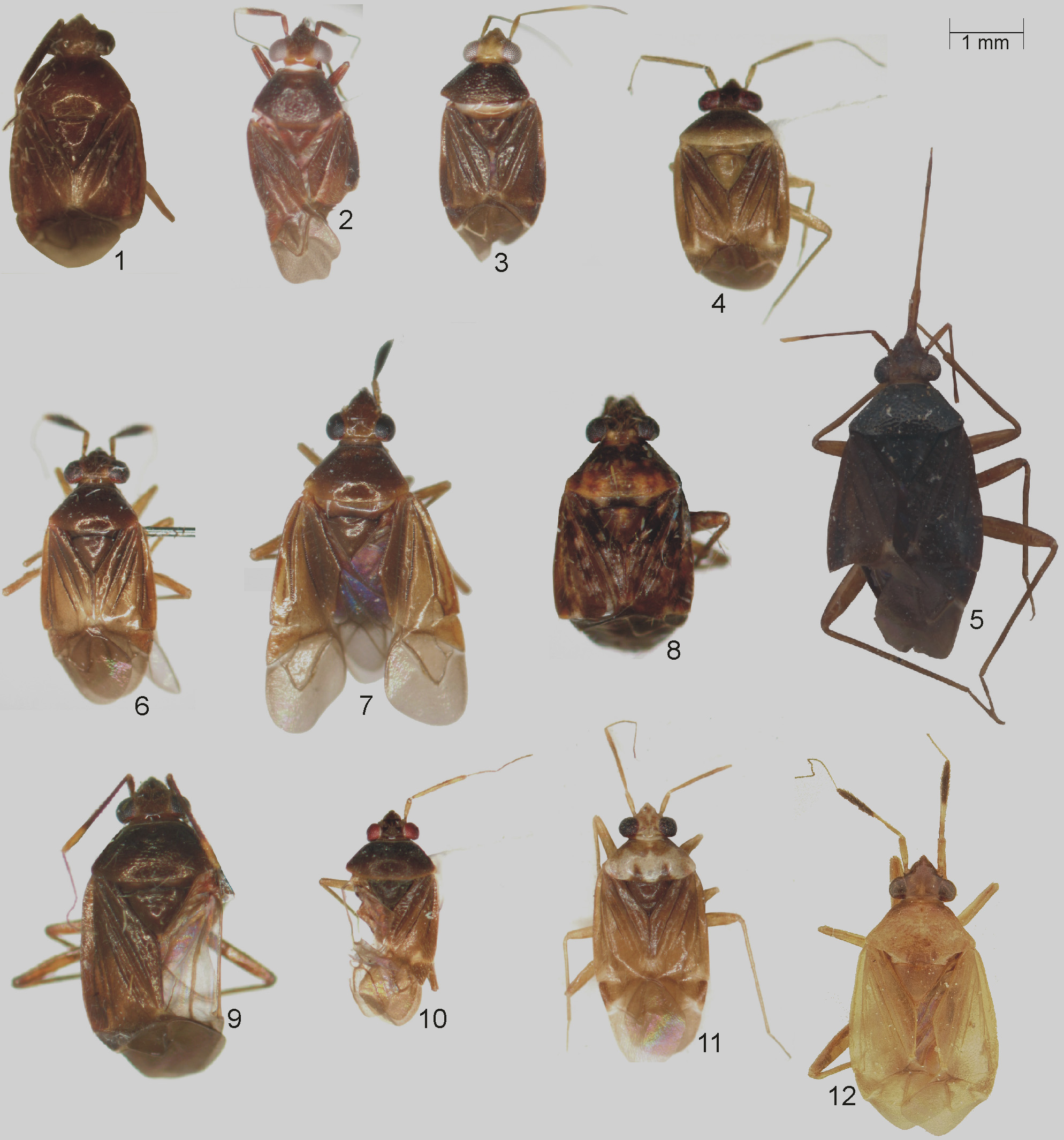
( Figures 12 View FIGURES 1 – 12 , 22 View FIGURES 13 – 22 , 36 View FIGURES 33 – 39 , 61 View FIGURE 61 , Table 1)
Cylapocoris tiquiensis Carvalho 1954: 508 , Pl. I, Fig. 5 View FIGURES 1 – 12 , Pl. 2, Figs. l–2, 5 (n. sp.); Carvalho 1957: 28 (catalog); Carvalho & Gomes 1971: 485, 487 (diag., key); Carvalho & Froeschner 1987: 154 (list); Schuh 1995: 22 (catalog); Cherot & Pauwels 2000: 7 (list); Gorczyca 2006b: 28 (catalog).
Diagnosis. Recognized by the dorsal coloration pale, yellowish brown ( Fig. 12 View FIGURES 1 – 12 ), the antennal segment II strongly thickened and contrastingly black at apical half ( Figs. 12 View FIGURES 1 – 12 , 22 View FIGURES 13 – 22 ), and the endosoma spinose (Carvalho 1954: Pl. I, Fig. 5 View FIGURES 1 – 12 ).
Most similar to C. sulinus in sharing similar body size and pale coloration of dorsum ( Fig. 11 View FIGURES 1 – 12 ). C. tiquiensis can, however, be easily distinguished by the uniformly yellowish brown dorsal coloration and the thickened and blackened apical half of antennal segment II ( Figs. 12 View FIGURES 1 – 12 , 22 View FIGURES 13 – 22 ).
Redescription. Male. COLORATION ( Figs. 12 View FIGURES 1 – 12 , 22 View FIGURES 13 – 22 ). Dorsal surface yellowish brown with castaneous areas. Head. Mostly blackish to fuscous with indistinct, yellowish tinges; antennal segment I and basal half of antennal segment II dirty yellowish, remainder of segment II black; segment III and IV dirty yellowish fuscous; labium dirty yellowish. Thorax. Pronotum. Yellowish brown tinged with fuscous at anterior angles and lateral portion of pronotal calli. Mesoscutum and scutellum. Yellowish brown. Thoracic pleura. Yellowish brown. Hemelytron. Yellowish brown. Legs. Yellowish with brownish areas. Abdomen. Yellowish brown. STRUCTURE, TEXTURE, AND VESTITURE ( Figs. 12 View FIGURES 1 – 12 , 22 View FIGURES 13 – 22 , 36 View FIGURES 33 – 39 ). Head. Shiny, almost glabrous; antennal segment I somewhat narrowed at basal one fourth, remainder of segment I cylindrical; segment II covered with dense, semierect setae, with basal half almost cylindrical, apical half distinctly thickened; segment III and IV thin, covered with long, protruding setae; labium thin and long, reaching apex of abdomen. Thorax. Pronotum. Punctation indistinct, shallow. Mesoscutum and scutellum. Scutellum moderately convex. Hemelytron. Rows of punctures along R+M vein, medial fracture, and vein on clavus indistinct, reaching beyond half of hemelytron. Legs. Tarsi two-segmented; pretarsal claw not toothed subapically.
Male genitalia (from Carvalho 1954). Aedeagus (Carvalho 1954, Pl. I, Fig. 5 View FIGURES 1 – 12 ). Endosoma spinose, theca present. Left paramere (Carvalho 1954, Pl. II, Fig. 2 View FIGURES 1 – 12 ). Curved; ended by a hook-like process, beset with setae on dorsal surface.
Measurements. ♀/♂ (based on Carvalho 1954 and the AMNH specimen, holotype measurements second): Body. Length 4.0/3.6, width 1.5/1.5. Head. Length 0.7/0.6, width 0.8, diameter of eye in dorsal view 0.18. Antenna. Length of segment I 0.5/0.4, II 1.1/1.0, III 0.7, IV 0.8. Labium. Length of labium 2.4. Pronotum. Length 0.7, width of posterior margin 1.2.
Biology. Unknown.
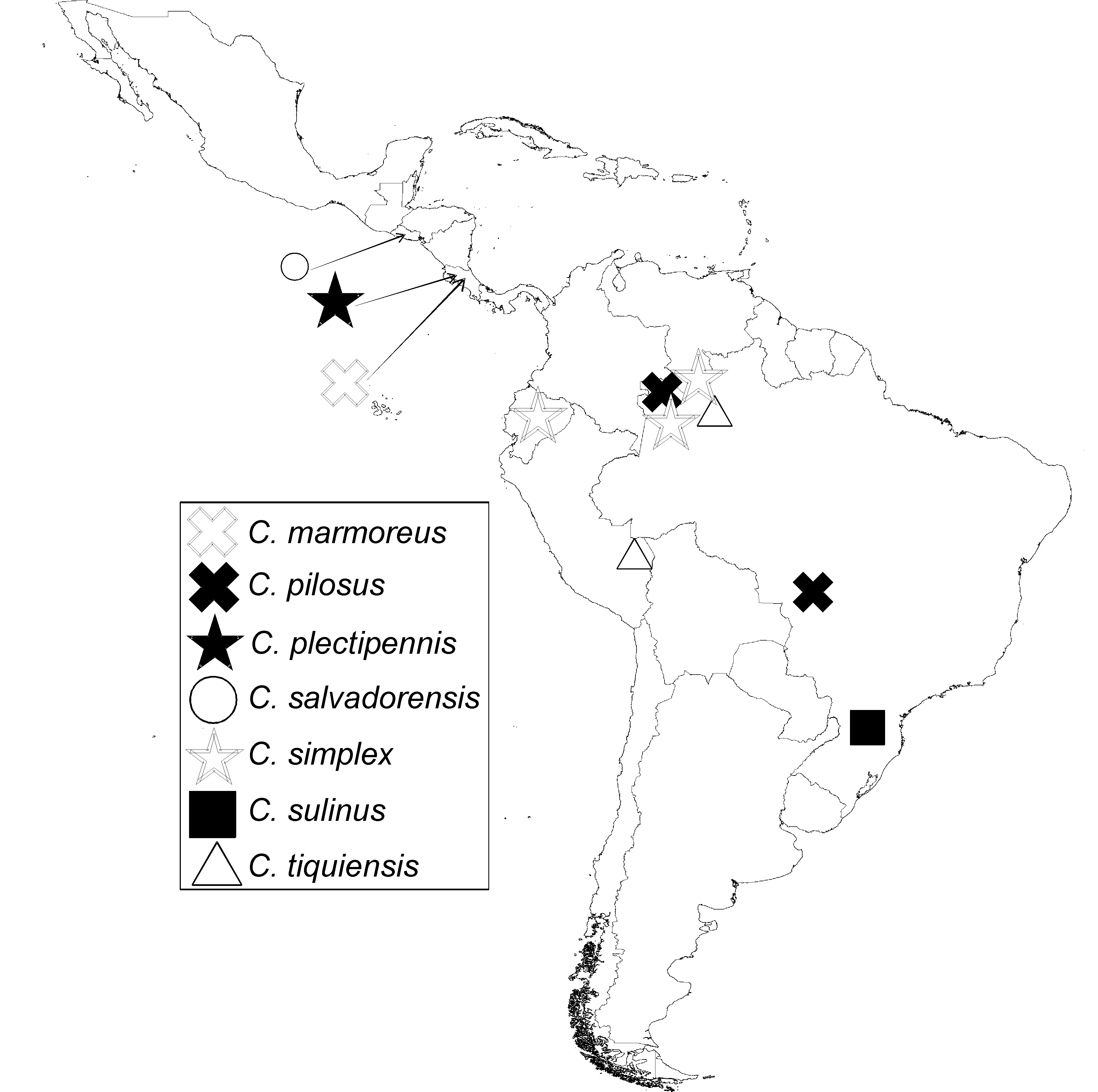
Distribution. Brasil (Amazonas), Peru (Tambopata) ( Fig. 61 View FIGURE 61 ).
Type material. Allotype ♀: Amazonas, Brasil, Taracua, 6–49, JCM Carvalho col; Allotype [reddish label]; Carvalho to Drake Coll. 1993 ( USNM) ( Figs. 12 View FIGURES 1 – 12 , 22 View FIGURES 13 – 22 ).
Additional examined specimens. 1 ♀: Peru: Tambopata Reserve, 30 km SW Puerto Maldonado, IX/19-X/10/ 84; 12º12’S, 69º16’W Trop[ical] Moist F[o]r[e]st., D.A. Grimaldi; reared from fungus: Auricularia ( AMNH); 1 ♀: Tiquié, Amazonas, JCM Carvalho Col. 1949 ( USNM).
Acknowledgments
I thank the following people for kindly offering me specimens used in this study: Randall T. Schuh (AMNH), Mick D. Webb (BMNH), Gunvi Lindberg (NHRS), Thomas J. Henry (USNM), and Klaus Schönitzer (ZSM). I’m also grateful to Magdalena Gawlak (Institute of Plant Protection–National Research Institute, Poznań, Poland) for her kind assistance in taking SEM microphotographs. Many thanks are also due to Michael D. Schwartz (Agriculture and Agri-Food Canada, Ottawa, Canada) for his very useful comments and suggestions on an earlier version of the manuscript. I also thank Mick D. Webb and Jing Sun (BMNH) and Michele A. Touchet (USNM) for the color photographs of C. funebris and C. tiquiensis .


No known copyright restrictions apply. See Agosti, D., Egloff, W., 2009. Taxonomic information exchange and copyright: the Plazi approach. BMC Research Notes 2009, 2:53 for further explanation.
|
Kingdom |
|
|
Phylum |
|
|
Class |
|
|
Order |
|
|
Family |
|
|
Genus |