
Pseudorhyncomyia
|
publication ID |
https://doi.org/10.11646/zootaxa.3736.3.3 |
|
publication LSID |
lsid:zoobank.org:pub:3ADF5D1D-20EE-4F0B-AE33-B8C9554E8ED5 |
|
DOI |
https://doi.org/10.5281/zenodo.6149435 |
|
persistent identifier |
https://treatment.plazi.org/id/A340878F-8915-FFA7-FF4D-8B57FA34A5B2 |
|
treatment provided by |
Plazi |
|
scientific name |
Pseudorhyncomyia |
| status |
|
Key to species of Pseudorhyncomyia View in CoL
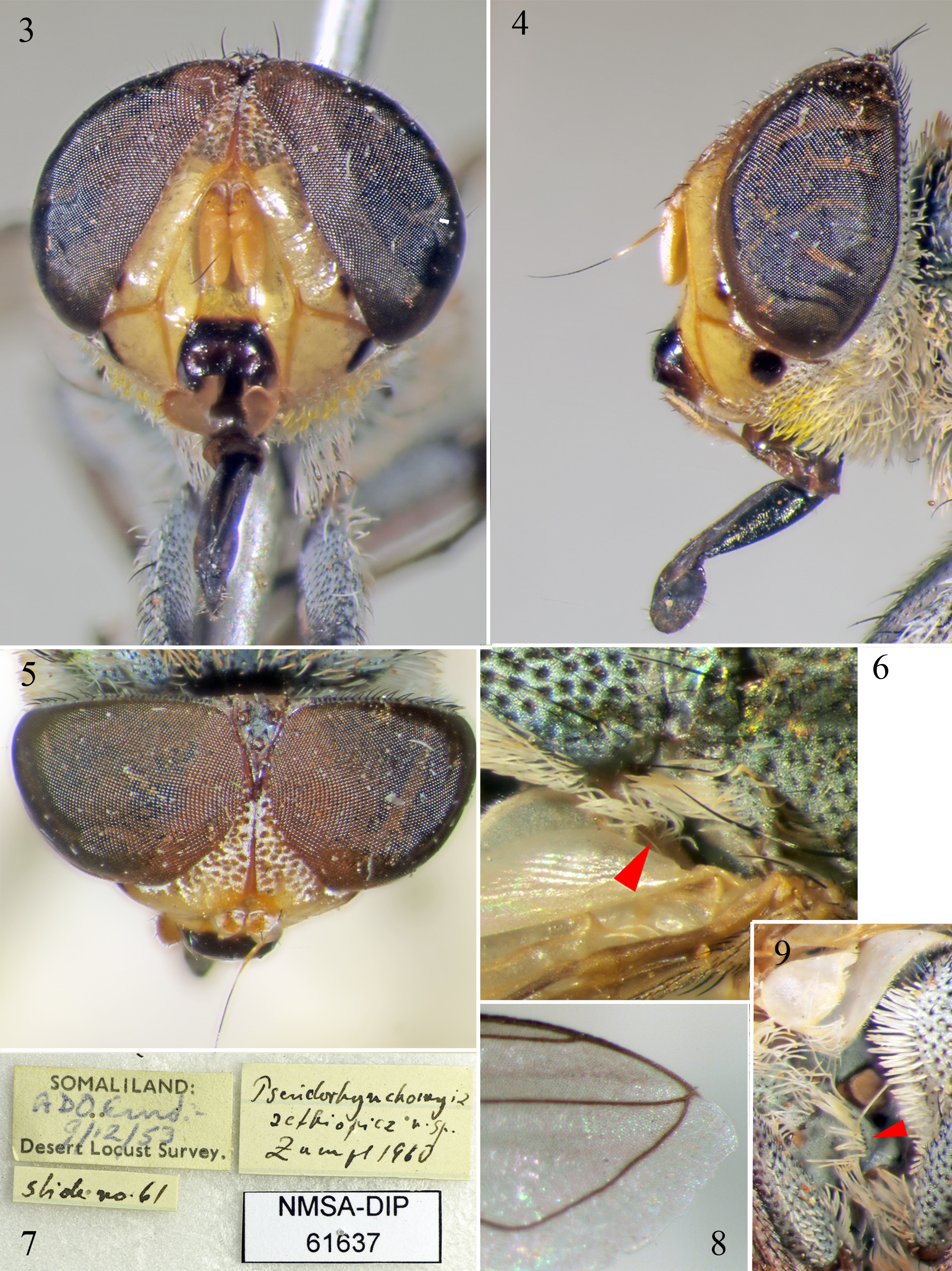
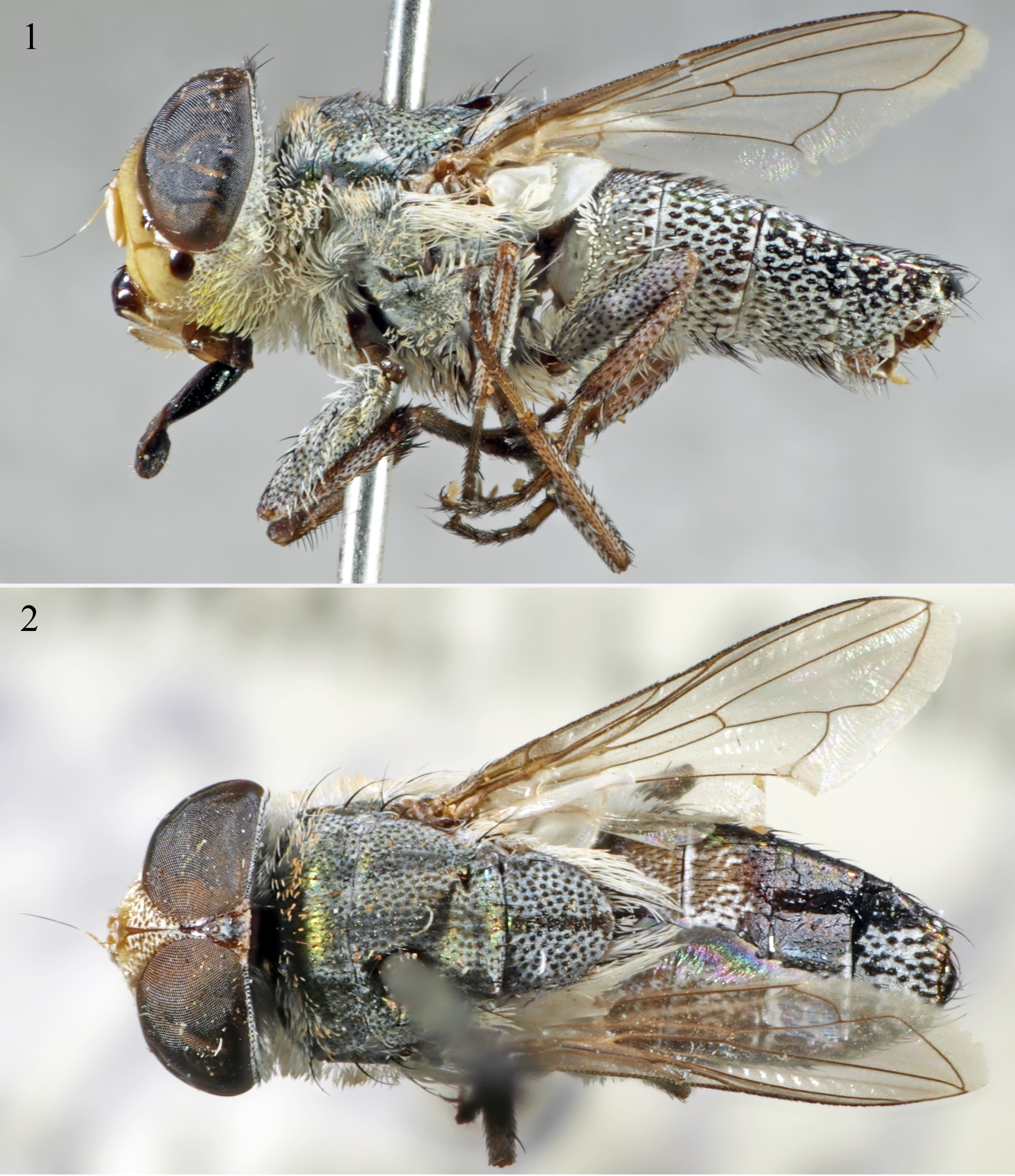
1. Wing cell r4+5 petiolate ( Fig. 8 View FIGURES 3 – 9 ). Vein M with a very shallow curve ( Fig. 2 View FIGURES 1 – 2 ). Parafacial and anterior half of genal dilation bright waxy yellow without piliferous dots ( Figs. 3–4 View FIGURES 3 – 9 ). Black spot in lower part of parafacial much smaller than black spot on gena ( Fig. 1 View FIGURES 1 – 2 ). Facial membrane without tomentum ( Fig. 3 View FIGURES 3 – 9 ). Lower facial margin projecting forward slightly beyond a vertical line through antennal pedicel in profile view; without microtomentum on anterodorsal surface ( Fig. 3 View FIGURES 3 – 9 ). Yellow area dorsally on each side of base of abdomen affecting T1+2 and T3 ( Fig. 2 View FIGURES 1 – 2 ). White setae usually rather thick and short, sometimes yellowish distally ( Figs. 1, 2 View FIGURES 1 – 2 , 4 View FIGURES 3 – 9 , 12 View FIGURES 10 – 13 )................................................. Pseudorhyncomyia aethiopica sp. nov.
– Wing cell r4+5 open ( Fig. 15 View FIGURES 14 – 16 ). Vein M angulate ( Fig. 15 View FIGURES 14 – 16 ). Parafacial and anterior half of genal dilation with white microtomentum over yellow ground colour and with distinct piliferous dots ( Figs. 17–20 View FIGURES 17 – 22 ). Black spot in lower half of parafacial larger than black spot in anterior half of gena ( Figs. 17–20 View FIGURES 17 – 22 ). Facial membrane with greyish-white tomentum ( Figs. 17, 18 View FIGURES 17 – 22 ). Lower facial margin projecting forward far beyond a vertical line through antennal pedicel in profile view, with a triangular area of white microtrichosity on anterodorsal surface ( Fig. 22 View FIGURES 17 – 22 ). Yellow area dorsally on each side of base of abdomen affecting T1+2 only ( Fig. 15 View FIGURES 14 – 16 ). White setae thin and slender, not yellowish distally................... Pseudorhyncomyia braunsi (Villeneuve) View in CoL


No known copyright restrictions apply. See Agosti, D., Egloff, W., 2009. Taxonomic information exchange and copyright: the Plazi approach. BMC Research Notes 2009, 2:53 for further explanation.