
Simulium sheilae Takaoka and Davies, 1995
|
publication ID |
https://doi.org/10.5281/zenodo.184626 |
|
DOI |
https://doi.org/10.5281/zenodo.5658683 |
|
persistent identifier |
https://treatment.plazi.org/id/A36387DA-FF96-FFB6-9CB1-8A6C4A087B35 |
|
treatment provided by |
Plazi |
|
scientific name |
Simulium sheilae Takaoka and Davies, 1995 |
| status |
|
Simulium sheilae Takaoka and Davies, 1995 View in CoL
(Figs. 6G, 7F, 9F, 11F)
Simulium sheilae Takaoka and Davies, 1995: 60 View in CoL –65 (female, male, pupa, larva) Simulium View in CoL sp. g, Kuvangkadilok et al., 2003: 74 (chromosomes)
All stages, except the egg, were described originally by Takaoka and Davies (1995) from West Malaysia. Morphological features of the adults, pupae, and larvae from site 42, are nearly identical to those in the original description, except for minor differences in the pattern of leg coloration in both sexes and the presence of hairs on the basal half of the female subcosta, as opposed to a fully haired subcosta in the original description.
Diagnosis: The larva can be distinguished morphologically from other cytoforms by having greenish transverse bands on abdominal segments I–IV and by bearing dark setae with 2–4 branches (rarely unbranched) on abdominal segments V–VIII. The pupa cannot be distinguished from that of other cytoforms.
This species differs morphologically from S. asakoae as follows:
Female. Maxillary palp with enlarged sensory vesicle, ca. 0.6 x as long as 3rd segment; fore and mid trochanters darker; hair tuft at base of costa and on stem vein black; sternite 8 of female genitalia bare medially, with ca. 18 dark macrosetae on each side. Male. Scutum entirely covered with pale recumbent pile; subcosta bare; posterior margin of ventral plate slightly convex medially or straight. Pupa. Gill ( Fig. 11 View FIGURE 11 F) with slender filaments almost subequal in thickness, arranged (1 + 2) + (1 + 2) + 2 from dorsal to ventral; dorsal and middle triplets composed of 1 individual and 2 paired filaments with stalks of moderate length; terminal hooks each with serrated outer margin. Larva. Postgenal cleft ( Fig. 9 View FIGURE 9 F) deep, ca. 6.3–6.8 x as long as postgenal bridge; abdominal segments I–IV with greenish transverse bands; abdominal segments V–VIII bearing dark setae with 2–4 branches (rarely unbranched).
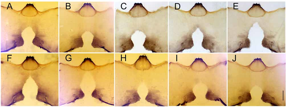
Chromosomes. The faintly staining chromosomes of 37 larvae from three populations (sites 40, 42, and 44) were studied. The centromere region of chromosome I was expanded but not of chromosomes II or III. Simulium sheilae was characterized by fixed inversion IIS-10 (Fig. 4A) and a complex set of inversions in IIIL, designated IIIL-complex 8 (Fig. 6G). Polymorphisms were not observed.
Bionomics. Larvae and pupae of this cytoform were on the surface of fallen leaves and trailing plants in warm (25.7 °C), sandy or muddy streams with low conductivity (31.2 µs/cm), at low altitudes (55–75 m) in southern Thailand ( Table 3 View TABLE 3 ). This cytoform was collected with S. trangense n. sp.
Remarks. Our results suggest that S. sp. g, previously reported by Kuvangkadilok et al. (2003), is conspecific with S. sheilae in our study, based on morphological and cytological similarity. This hypothesis also agrees with molecular (ITS2) evidence ( Thanwisai et al. 2006).

No known copyright restrictions apply. See Agosti, D., Egloff, W., 2009. Taxonomic information exchange and copyright: the Plazi approach. BMC Research Notes 2009, 2:53 for further explanation.
|
Kingdom |
|
|
Phylum |
|
|
Class |
|
|
Order |
|
|
Family |
|
|
Genus |
Simulium sheilae Takaoka and Davies, 1995
| Jitklang, Sanae, Kuvangkadilok, Chaliow, Baimai, Visut, Takaoka, Hiroyuki & Adler, Peter H. 2008 |
Simulium sheilae
| Kuvangkadilok 2003: 74 |
| Takaoka 1995: 60 |